Advertisement

Advertisement

Advertisement

Advertisement

Developmental Dysplasia of the Hip

Developmental dysplasia of the hip refers to a range of disorders of the hip joint Hip joint The hip joint is a ball-and-socket joint formed by the head of the femur and the acetabulum of the pelvis. The hip joint is the most stable joint in the body and is supported by a very strong capsule and several ligaments, allowing the joint to sustain forces that can be multiple times the total body weight. Hip Joint: Anatomy characterized by hip instability and resulting in subluxation Subluxation Radial Head Subluxation (Nursemaid’s Elbow) or dislocation that mostly presents during the first few months of life. The condition is often first recognized due to hip laxity on newborn Newborn An infant during the first 28 days after birth. Physical Examination of the Newborn exam. Developmental dysplasia of the hip occurs most commonly in otherwise healthy girls and often does not have an identifiable cause. Treatment is imperative to avoid complications, such as avascular necrosis Avascular Necrosis Hip Fractures of the femoral head and pain Pain An unpleasant sensation induced by noxious stimuli which are detected by nerve endings of nociceptive neurons. Pain: Types and Pathways with mobility. Management is dependent on the severity and age at diagnosis, with infants often treated with a Pavlik harness and children > 6 months of age often requiring open or closed surgical reduction.

Last updated: Feb 21, 2023

Editorial responsibility: Stanley Oiseth, Lindsay Jones, Evelin Maza

Advertisement

Advertisement

Advertisement

Advertisement

Advertisement

Advertisement

Overview

Definitions

  • Developmental dysplasia of the hip (DDH): a spectrum of developmental disorders of the acetabulum and proximal femur, characterized by instability of the hip joint Hip joint The hip joint is a ball-and-socket joint formed by the head of the femur and the acetabulum of the pelvis. The hip joint is the most stable joint in the body and is supported by a very strong capsule and several ligaments, allowing the joint to sustain forces that can be multiple times the total body weight. Hip Joint: Anatomy (formerly known as congenital Congenital Chorioretinitis dysplasia of the hip)
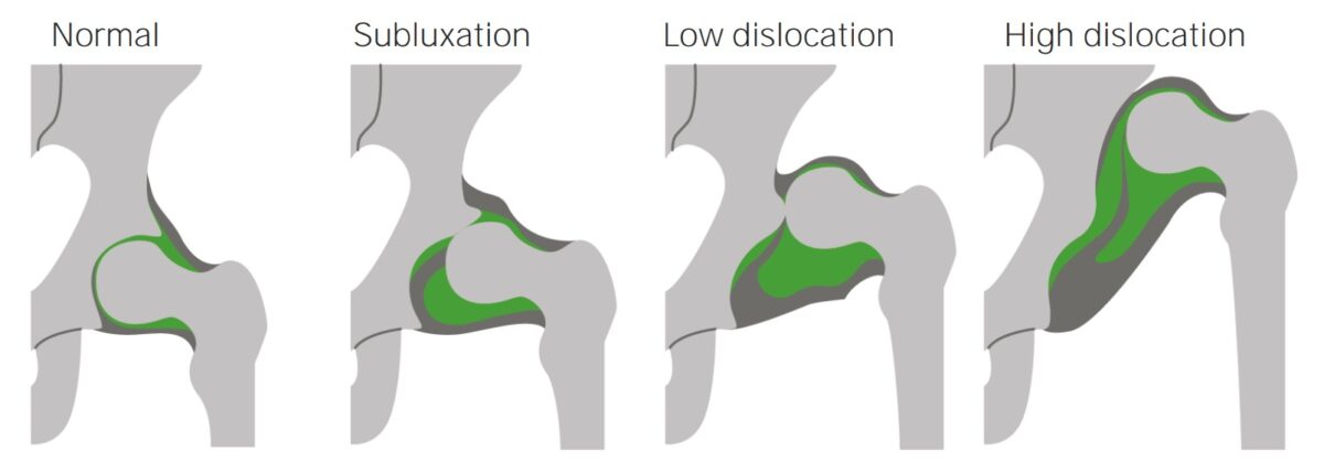
  • Subluxation Subluxation Radial Head Subluxation (Nursemaid’s Elbow): partial loss of contact between the head of the femur Head of the femur The hemispheric articular surface at the upper extremity of the thigh bone. Hip Joint: Anatomy and the acetabulum
  • Dislocation: total loss of contact

Classification

  • Typical DDH: hip dysplasia in otherwise healthy children (this is the most common form)
  • Teratologic DDH: hip dysplasia with an identifiable cause (e.g., arthrogryposis, genetic syndromes such as Down syndrome Down syndrome Down syndrome, or trisomy 21, is the most common chromosomal aberration and the most frequent genetic cause of developmental delay. Both boys and girls are affected and have characteristic craniofacial and musculoskeletal features, as well as multiple medical anomalies involving the cardiac, gastrointestinal, ocular, and auditory systems. Down syndrome (Trisomy 21))
  • Neuromuscular DDH: hip dysplasia due to weakness or spasticity Spasticity Spinal Disk Herniation in lower-extremity muscle groups (e.g., spina bifida or cerebral palsy Palsy paralysis of an area of the body, thus incapable of voluntary movement Cranial Nerve Palsies)

Epidemiology

  • Prevalence Prevalence The total number of cases of a given disease in a specified population at a designated time. It is differentiated from incidence, which refers to the number of new cases in the population at a given time. Measures of Disease Frequency: 35 in 1,000 children
  • More common in children of Caucasian descent
  • 80% of cases are girls
  • 63% of cases are unilateral, with the left hip most commonly affected.
  • Mild hip instability is reported in up to 40% of neonates; these cases usually self-resolve.

Risk factors

  • Single greatest risk factor: breech position, especially in late pregnancy Pregnancy The status during which female mammals carry their developing young (embryos or fetuses) in utero before birth, beginning from fertilization to birth. Pregnancy: Diagnosis, Physiology, and Care (≥ 34 weeks gestation)
  • Female sex Sex The totality of characteristics of reproductive structure, functions, phenotype, and genotype, differentiating the male from the female organism. Gender Dysphoria
  • Family history Family History Adult Health Maintenance of DDH
  • Tight lower-extremity swaddling
Normal hip joint vs ddh

Normal hip joint and hip joints with progressively more severe dislocations

Image by Lecturio.

Etiology and Pathophysiology

Etiology

Developmental dysplasia of the hip is caused by an inadequate reduction of the femoral head in the acetabulum. This can occur due to several factors.

  • Extrauterine factors:
    • Hypertrophy Hypertrophy General increase in bulk of a part or organ due to cell enlargement and accumulation of fluids and secretions, not due to tumor formation, nor to an increase in the number of cells (hyperplasia). Cellular Adaptation of the ligamentum teres Ligamentum teres A cord-like remnant structure formed from the closed left fetal umbilical vein. It is located along the lower edge of the falciform ligament. Liver: Anatomy
    • Fatty tissue within the socket
    • Thickening of the transverse acetabular ligament
    • Iliopsoas tendon interfering with access to the acetabulum
  • Intrauterine factors:
    • Limited fetal motility Motility The motor activity of the gastrointestinal tract. Gastrointestinal Motility in utero
    • Oligohydramnios Oligohydramnios Oligohydramnios refers to amniotic fluid volume less than expected for the current gestational age. Oligohydramnios is diagnosed by ultrasound and defined as an amniotic fluid index (AFI) of ‰¤ 5 cm or a single deep pocket (SDP) of < 2 cm in the 2nd or 3rd trimester. Oligohydramnios

Pathophysiology

Normal development of the hip is dependent on contact between the acetabulum and femoral head. However, in DDH:

  • There is interrupted contact between the femoral head and acetabulum.
  • Because the femoral head gives depth to the acetabulum during normal fetal development, this interrupted contact causes the acetabulum to become shallow.
  • A shallow acetabulum results in decreased coverage of the femoral head, which results in hip instability.

Clinical Presentation

  • The clinical presentation of DDH depends on the child’s age and the severity of the pathology.
  • For this reason, routine hip evaluation should be performed at all well-child exams until 9 months of age.
  • DDH shows a progression through time, manifesting differently and more severely as time passes.
Table: Key clinical exam findings by age group
Neonates aged 0–2 months Hip instability
Children 2–3 months old
Children who can walk

Diagnosis

The diagnosis of DDH is made clinically by demonstrating hip instability, asymmetry Asymmetry Examination of the Upper Limbs, and limited abduction Abduction Examination of the Upper Limbs of the hip.

Physical exam

Special tests have been devised to evaluate hip joint Hip joint The hip joint is a ball-and-socket joint formed by the head of the femur and the acetabulum of the pelvis. The hip joint is the most stable joint in the body and is supported by a very strong capsule and several ligaments, allowing the joint to sustain forces that can be multiple times the total body weight. Hip Joint: Anatomy structure, range of motion Range of motion The distance and direction to which a bone joint can be extended. Range of motion is a function of the condition of the joints, muscles, and connective tissues involved. Joint flexibility can be improved through appropriate muscle strength exercises. Examination of the Upper Limbs, and strength. The choice of which to employ is age-based.

Neonates aged 02 months

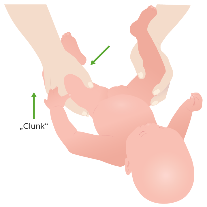
  • Ortolani maneuver (reduction of hips dislocated at rest)
    • High false-positive rate, especially in younger children
    • Remember that hip laxity may be present in up to 40% of newborns.
    • Steps:
      1. Child is placed in the supine position on a stable surface.
      2. Examiner grasps the child’s thigh Thigh The thigh is the region of the lower limb found between the hip and the knee joint. There is a single bone in the thigh called the femur, which is surrounded by large muscles grouped into 3 fascial compartments. Thigh: Anatomy with the thumb and index finger. 
      3. From an adducted position, the child’s hip is abducted while the trochanter is pushed anteriorly.
      4. Positive when a “hip clunk” is felt or if the maneuver reduces the hip. This hip clunk is the sound or sensation of the femoral head slipping back into the acetabulum.
  • Barlow maneuver (dislocation of hips reduced at rest)
    1. Child is placed in the supine position on a stable surface.
    2. Examiner grasps the child’s thighs with the thumb and index finger. 
    3. The child’s hips are adducted.
    4. Examiner palpates the femoral head for movement out of the acetabulum.
    5. Positive when the femoral head is felt slipping, posteriorly, out of the acetabulum. Either a “palpable clunk” or subluxation Subluxation Radial Head Subluxation (Nursemaid’s Elbow) may be felt on positive exams.

Children 23 months old

  • Galeazzi test
    1. Child lays supine with hips flexed to 45° and knees flexed to 90°.
    2. Child’s feet are placed flat and level on a surface beside each other.
    3. Positive if one of the knees appears lower than the other knee. This occurs because the head of the femur Head of the femur The hemispheric articular surface at the upper extremity of the thigh bone. Hip Joint: Anatomy is displaced posteriorly, shortening the length of the thigh Thigh The thigh is the region of the lower limb found between the hip and the knee joint. There is a single bone in the thigh called the femur, which is surrounded by large muscles grouped into 3 fascial compartments. Thigh: Anatomy
    4. Important: A positive test indicates a lower leg Leg The lower leg, or just “leg” in anatomical terms, is the part of the lower limb between the knee and the ankle joint. The bony structure is composed of the tibia and fibula bones, and the muscles of the leg are grouped into the anterior, lateral, and posterior compartments by extensions of fascia. Leg: Anatomy length discrepancy Length Discrepancy Blount’s Disease and is not specific to DDH.
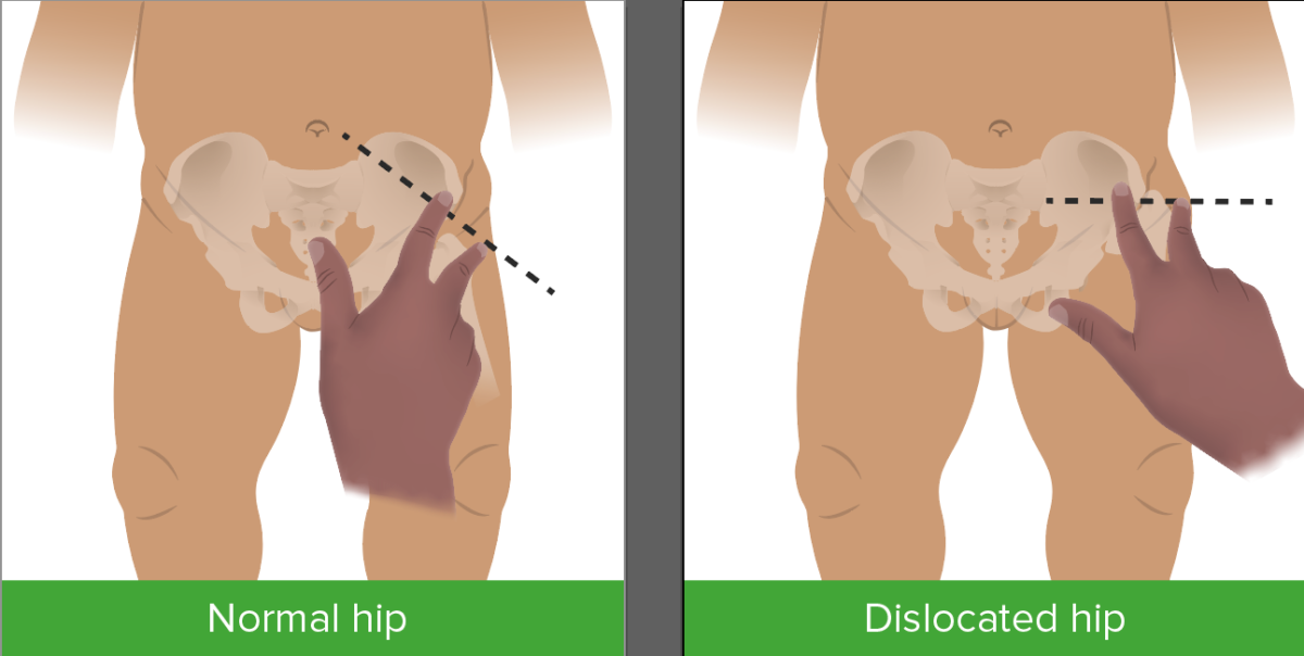
  • Klisic test
    1. Examiner places a finger on the greater trochanter and a finger of the same hand Hand The hand constitutes the distal part of the upper limb and provides the fine, precise movements needed in activities of daily living. It consists of 5 metacarpal bones and 14 phalanges, as well as numerous muscles innervated by the median and ulnar nerves. Hand: Anatomy on the anterior-superior iliac spine Spine The human spine, or vertebral column, is the most important anatomical and functional axis of the human body. It consists of 7 cervical vertebrae, 12 thoracic vertebrae, and 5 lumbar vertebrae and is limited cranially by the skull and caudally by the sacrum. Vertebral Column: Anatomy
    2. A line is drawn through the tips of both fingers.
    3. If the line passes through or above the umbilicus, the hip is adequately reduced (i.e., normal hip exam).
    4. If the line is below the umbilicus, the test is positive for a displaced hip.

Children who can walk

  • Trendelenburg sign
    • Patient is unable to maintain their pelvis Pelvis The pelvis consists of the bony pelvic girdle, the muscular and ligamentous pelvic floor, and the pelvic cavity, which contains viscera, vessels, and multiple nerves and muscles. The pelvic girdle, composed of 2 “hip” bones and the sacrum, is a ring-like bony structure of the axial skeleton that links the vertebral column with the lower extremities. Pelvis: Anatomy in line horizontally while standing on the ipsilateral leg Leg The lower leg, or just “leg” in anatomical terms, is the part of the lower limb between the knee and the ankle joint. The bony structure is composed of the tibia and fibula bones, and the muscles of the leg are grouped into the anterior, lateral, and posterior compartments by extensions of fascia. Leg: Anatomy 
    • Indicates hip abductor weakness

Imaging

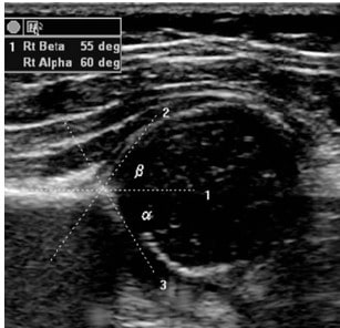
Imaging can be used to confirm the diagnosis in children with risk factors and a normal physical examination, or in children with inconclusive physical examination findings.

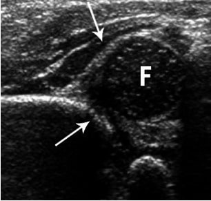
  • Hip ultrasound:
    • Useful as an adjunct to the physical exam until 46 months of age 
    • Specificity is low and usually operator-dependent.
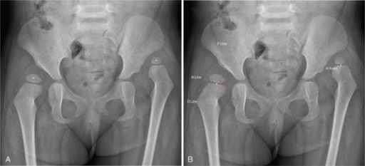
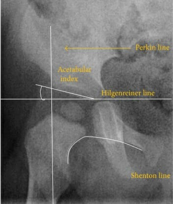
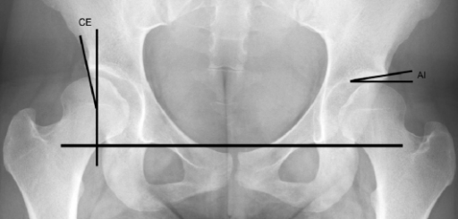
  • Hip X-ray X-ray Penetrating electromagnetic radiation emitted when the inner orbital electrons of an atom are excited and release radiant energy. X-ray wavelengths range from 1 pm to 10 nm. Hard x-rays are the higher energy, shorter wavelength x-rays. Soft x-rays or grenz rays are less energetic and longer in wavelength. The short wavelength end of the x-ray spectrum overlaps the gamma rays wavelength range. The distinction between gamma rays and x-rays is based on their radiation source. Pulmonary Function Tests: used from 4–6 months of age when the acetabulum and femoral head begin to ossify
Ddh hip ultrasound window

Window of opportunity for an optimal hip ultrasound

Image by Lecturio.
Management

Main goals

Management by age group

Newborns 0–4 weeks of age
  • Hip instability and laxity may be normal findings, so generally patients Patients Individuals participating in the health care system for the purpose of receiving therapeutic, diagnostic, or preventive procedures. Clinician–Patient Relationship are observed only.
  • Any ultrasound with abnormal findings should be repeated at 6 weeks.
Infants 4 weeks to 6 months of age
  • Often are initially observed, with reassessment every 4–6 weeks
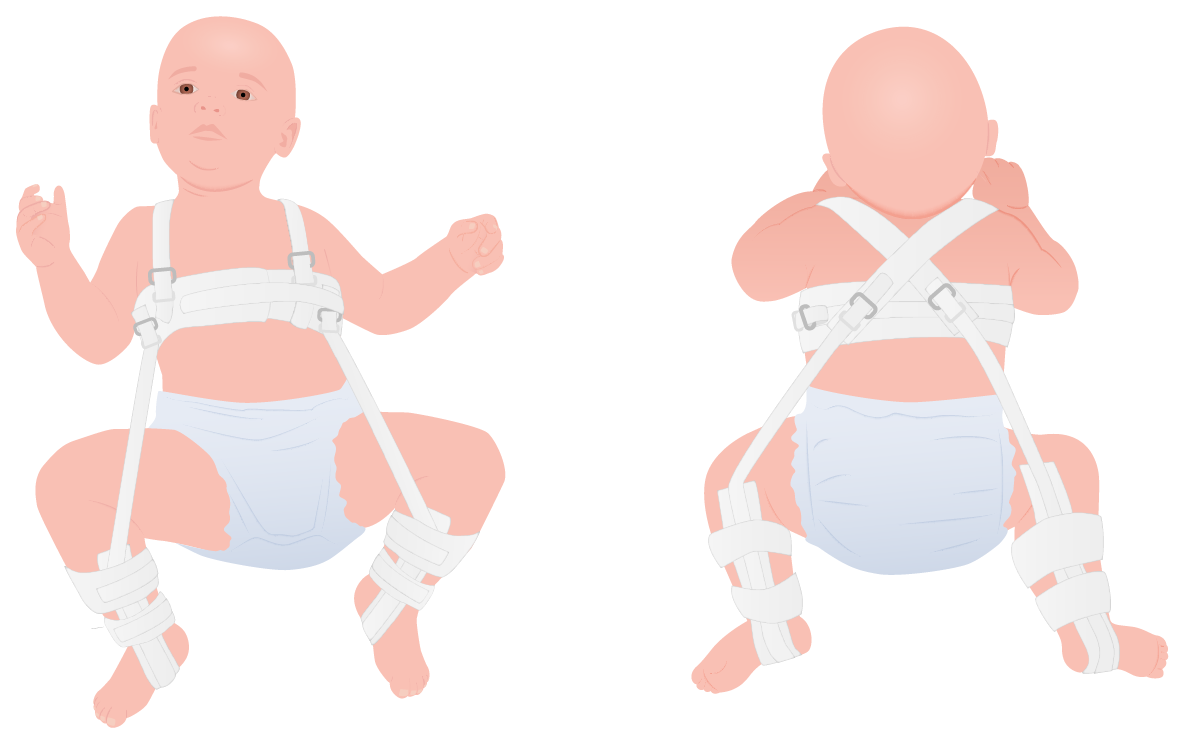
  • If symptoms continue, a Pavlik harness is most often utilized: treatment for 2–3 months, until a physical exam or ultrasound demonstrates hip stability
Infants between the ages of 6 months and 2 years
Children from 2–6 years of age Open reductions are generally required.
Pavlik harness

Pavlik harness

Image by Lecturio.

Complications

  • Most important complication: avascular necrosis Avascular Necrosis Hip Fractures of the femoral epiphysis Epiphysis The head of a long bone that is separated from the shaft by the epiphyseal plate until bone growth stops. At that time, the plate disappears and the head and shaft are united. Bones: Structure and Types (orthopedic emergency!
  • Osteoarthritis Osteoarthritis Osteoarthritis (OA) is the most common form of arthritis, and is due to cartilage destruction and changes of the subchondral bone. The risk of developing this disorder increases with age, obesity, and repetitive joint use or trauma. Patients develop gradual joint pain, stiffness lasting < 30 minutes, and decreased range of motion. Osteoarthritis ( OA OA Osteoarthritis (OA) is the most common form of arthritis, and is due to cartilage destruction and changes of the subchondral bone. The risk of developing this disorder increases with age, obesity, and repetitive joint use or trauma. Patients develop gradual joint pain, stiffness lasting < 30 minutes, and decreased range of motion. Osteoarthritis):
    • A form of arthritis Arthritis Acute or chronic inflammation of joints. Osteoarthritis caused by progressive wasting of articular cartilage Cartilage Cartilage is a type of connective tissue derived from embryonic mesenchyme that is responsible for structural support, resilience, and the smoothness of physical actions. Perichondrium (connective tissue membrane surrounding cartilage) compensates for the absence of vasculature in cartilage by providing nutrition and support. Cartilage: Histology and the surrounding joint structures
    • Patients Patients Individuals participating in the health care system for the purpose of receiving therapeutic, diagnostic, or preventive procedures. Clinician–Patient Relationship with DDH are at an increased risk of developing OA OA Osteoarthritis (OA) is the most common form of arthritis, and is due to cartilage destruction and changes of the subchondral bone. The risk of developing this disorder increases with age, obesity, and repetitive joint use or trauma. Patients develop gradual joint pain, stiffness lasting < 30 minutes, and decreased range of motion. Osteoarthritis.
  • Pain Pain An unpleasant sensation induced by noxious stimuli which are detected by nerve endings of nociceptive neurons. Pain: Types and Pathways
  • Abnormal gait Gait Manner or style of walking. Neurological Examination
  • Reduced mobility
  • Redislocation
  • Residual subluxation Subluxation Radial Head Subluxation (Nursemaid’s Elbow)
  • Dysplasia of the acetabulum

Prognosis Prognosis A prediction of the probable outcome of a disease based on a individual’s condition and the usual course of the disease as seen in similar situations. Non-Hodgkin Lymphomas

  • After reduction, the developmental potential of the acetabulum is excellent.
    • The earlier the treatment, the better the prognosis Prognosis A prediction of the probable outcome of a disease based on a individual’s condition and the usual course of the disease as seen in similar situations. Non-Hodgkin Lymphomas
    • 95% of cases treated with a Pavlik harness are successfully reduced.
  • 90% of mild instability/dysplasia cases resolve spontaneously between months 2 and 6.

Differential Diagnosis

  • Transient synovitis Synovitis Inflammation of the synovial membrane. Rheumatoid Arthritis: transient inflammation Inflammation Inflammation is a complex set of responses to infection and injury involving leukocytes as the principal cellular mediators in the body’s defense against pathogenic organisms. Inflammation is also seen as a response to tissue injury in the process of wound healing. The 5 cardinal signs of inflammation are pain, heat, redness, swelling, and loss of function. Inflammation of the hip joint Hip joint The hip joint is a ball-and-socket joint formed by the head of the femur and the acetabulum of the pelvis. The hip joint is the most stable joint in the body and is supported by a very strong capsule and several ligaments, allowing the joint to sustain forces that can be multiple times the total body weight. Hip Joint: Anatomy characterized by limited mobility and pain Pain An unpleasant sensation induced by noxious stimuli which are detected by nerve endings of nociceptive neurons. Pain: Types and Pathways. Magnetic resonance imaging (MRI) or ultrasound shows hip effusions. Management is conservative with rest and nonsteroidal anti-inflammatory drugs ( NSAIDs NSAIDS Primary vs Secondary Headaches) only. Unlike DDH, transient synovitis Synovitis Inflammation of the synovial membrane. Rheumatoid Arthritis occurs later in childhood and generally has an infectious prodrome Prodrome Symptoms that appear 24–48 hours prior to migraine onset. Migraine Headache
  • Septic arthritis Arthritis Acute or chronic inflammation of joints. Osteoarthritis: an infection of the joint causing acute, asymmetric arthritis Arthritis Acute or chronic inflammation of joints. Osteoarthritis. Most commonly caused by hematogenous Hematogenous Hepatocellular Carcinoma (HCC) and Liver Metastases spread, direct inoculation through medical procedures, or trauma. Infants present with high fevers and are unable to bear weight. Diagnosis is through ultrasound-guided needle aspiration Needle aspiration Using fine needles (finer than 22-gauge) to remove tissue or fluid specimens from the living body for examination in the pathology laboratory and for disease diagnosis. Peritonsillar Abscess, which is also the treatment of choice. 
  • Slipped capital femoral epiphysis Epiphysis The head of a long bone that is separated from the shaft by the epiphyseal plate until bone growth stops. At that time, the plate disappears and the head and shaft are united. Bones: Structure and Typesan orthopedic disorder of childhood characterized by the pathological “slipping” of the femoral head. Risk factors include obesity Obesity Obesity is a condition associated with excess body weight, specifically with the deposition of excessive adipose tissue. Obesity is considered a global epidemic. Major influences come from the western diet and sedentary lifestyles, but the exact mechanisms likely include a mixture of genetic and environmental factors. Obesity, trauma, and genetic predisposition. Patients Patients Individuals participating in the health care system for the purpose of receiving therapeutic, diagnostic, or preventive procedures. Clinician–Patient Relationship present with changes in gait Gait Manner or style of walking. Neurological Examination and pain Pain An unpleasant sensation induced by noxious stimuli which are detected by nerve endings of nociceptive neurons. Pain: Types and Pathways in the hip, knee, or groin Groin The external junctural region between the lower part of the abdomen and the thigh. Male Genitourinary Examination that is worsened by activity. Plain radiographs confirm the diagnosis. Treatment is surgical.
  • Legg-Calve-Perthes disease: a syndrome of unknown etiology characterized by idiopathic Idiopathic Dermatomyositis avascular necrosis Avascular Necrosis Hip Fractures of the femoral head.  Patients Patients Individuals participating in the health care system for the purpose of receiving therapeutic, diagnostic, or preventive procedures. Clinician–Patient Relationship present with a gradual onset of hip pain Pain An unpleasant sensation induced by noxious stimuli which are detected by nerve endings of nociceptive neurons. Pain: Types and Pathways and limp. Diagnosis involves imaging, including X-ray X-ray Penetrating electromagnetic radiation emitted when the inner orbital electrons of an atom are excited and release radiant energy. X-ray wavelengths range from 1 pm to 10 nm. Hard x-rays are the higher energy, shorter wavelength x-rays. Soft x-rays or grenz rays are less energetic and longer in wavelength. The short wavelength end of the x-ray spectrum overlaps the gamma rays wavelength range. The distinction between gamma rays and x-rays is based on their radiation source. Pulmonary Function Tests and MRI. Treatment is conservative with optional surgery in severe cases.

References

  1. Sankar, W. N., Winell, J. J., Horn, B. D., & Wells, L. (2020). The hip. In R. M. Kliegman MD, J. W. St Geme MD, N. J. Blum MD, Shah, Samir S., MD, MSCE, Tasker, Robert C., MBBS, MD & Wilson, Karen M., MD, MPH (Eds.), Nelson textbook of pediatrics (pp. 362-3633.e1). https://www.clinicalkey.es/#!/content/3-s2.0-B9780323529501006982

Create your free account or log in to continue reading!

Sign up now and get free access to Lecturio with concept pages, medical videos, and questions for your medical education.

User Reviews

Details