Dermatophytes/Tinea Infections

Tinea infections Infections Invasion of the host organism by microorganisms or their toxins or by parasites that can cause pathological conditions or diseases. Chronic Granulomatous Disease are a group of diseases caused by fungi Fungi A kingdom of eukaryotic, heterotrophic organisms that live parasitically as saprobes, including mushrooms; yeasts; smuts, molds, etc. They reproduce either sexually or asexually, and have life cycles that range from simple to complex. Filamentous fungi, commonly known as molds, refer to those that grow as multicellular colonies. Mycology infecting keratinized tissue (hair, nails, and skin Skin The skin, also referred to as the integumentary system, is the largest organ of the body. The skin is primarily composed of the epidermis (outer layer) and dermis (deep layer). The epidermis is primarily composed of keratinocytes that undergo rapid turnover, while the dermis contains dense layers of connective tissue. Skin: Structure and Functions). These infections Infections Invasion of the host organism by microorganisms or their toxins or by parasites that can cause pathological conditions or diseases. Chronic Granulomatous Disease are termed dermatomycoses and are caused by the dermatophyte fungi Fungi A kingdom of eukaryotic, heterotrophic organisms that live parasitically as saprobes, including mushrooms; yeasts; smuts, molds, etc. They reproduce either sexually or asexually, and have life cycles that range from simple to complex. Filamentous fungi, commonly known as molds, refer to those that grow as multicellular colonies. Mycology. There are approximately 40 dermatophyte fungi Fungi A kingdom of eukaryotic, heterotrophic organisms that live parasitically as saprobes, including mushrooms; yeasts; smuts, molds, etc. They reproduce either sexually or asexually, and have life cycles that range from simple to complex. Filamentous fungi, commonly known as molds, refer to those that grow as multicellular colonies. Mycology that are part of 3 genera, including Trichophyton, Epidermophyton, and Microsporum. These infections Infections Invasion of the host organism by microorganisms or their toxins or by parasites that can cause pathological conditions or diseases. Chronic Granulomatous Disease can affect any part of the body but occur most often in warm, moist regions like the groin Groin The external junctural region between the lower part of the abdomen and the thigh. Male Genitourinary Examination and the feet. The diagnosis is clinical with characteristic skin Skin The skin, also referred to as the integumentary system, is the largest organ of the body. The skin is primarily composed of the epidermis (outer layer) and dermis (deep layer). The epidermis is primarily composed of keratinocytes that undergo rapid turnover, while the dermis contains dense layers of connective tissue. Skin: Structure and Functions findings, but it can be confirmed with microscopy of skin Skin The skin, also referred to as the integumentary system, is the largest organ of the body. The skin is primarily composed of the epidermis (outer layer) and dermis (deep layer). The epidermis is primarily composed of keratinocytes that undergo rapid turnover, while the dermis contains dense layers of connective tissue. Skin: Structure and Functions scrapings. The treatment depends on the site and magnitude of infection but typically begins with topical antifungals like the -azole drugs and terbinafine Terbinafine In addition to the 3 other major classes of antifungal agents (azoles, polyenes, and echinocandins), several other clinically important antifungal agents are used, including flucytosine, griseofulvin, and terbinafine. Terbinafine acts within the stratum corneum of the skin and are used to treat dermatophyte infections of the skin, hair, and nails. Flucytosine, Griseofulvin, and Terbinafine, and it may progress to oral versions of these medications if topical treatment fails.

Last updated: May 17, 2024

Editorial responsibility: Stanley Oiseth, Lindsay Jones, Evelin Maza

Overview

General characteristics of dermatophytes

  • Dermatophytes are filamentous fungi Fungi A kingdom of eukaryotic, heterotrophic organisms that live parasitically as saprobes, including mushrooms; yeasts; smuts, molds, etc. They reproduce either sexually or asexually, and have life cycles that range from simple to complex. Filamentous fungi, commonly known as molds, refer to those that grow as multicellular colonies. Mycology that belong to the genera Trichophyton, Microsporum, and Epidermophyton.
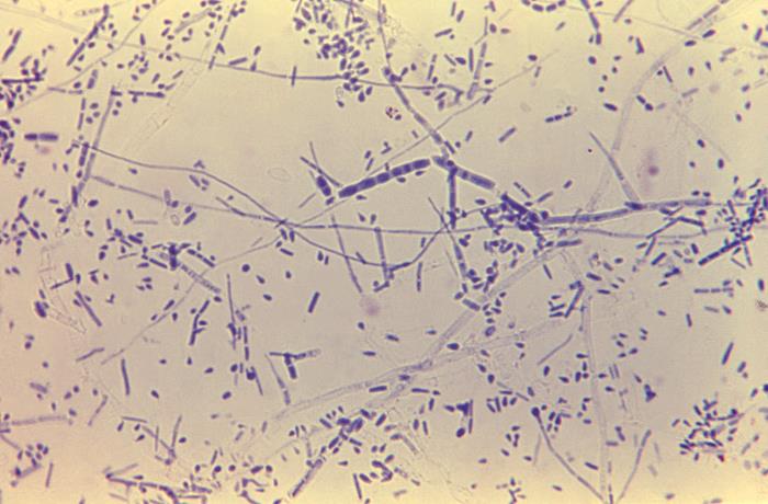
  • Dermatophytes have septate hyphae Hyphae Microscopic threadlike filaments in fungi that are filled with a layer of protoplasm. Collectively, the hyphae make up the mycelium. Mycology with chains of conidia Conidia Mycology.

Classification of tinea infections Infections Invasion of the host organism by microorganisms or their toxins or by parasites that can cause pathological conditions or diseases. Chronic Granulomatous Disease

Tinea infections Infections Invasion of the host organism by microorganisms or their toxins or by parasites that can cause pathological conditions or diseases. Chronic Granulomatous Disease are classified and named by the body region affected. 

  • Tinea pedis: 
    • “Athlete’s foot Foot The foot is the terminal portion of the lower limb, whose primary function is to bear weight and facilitate locomotion. The foot comprises 26 bones, including the tarsal bones, metatarsal bones, and phalanges. The bones of the foot form longitudinal and transverse arches and are supported by various muscles, ligaments, and tendons. Foot: Anatomy” 
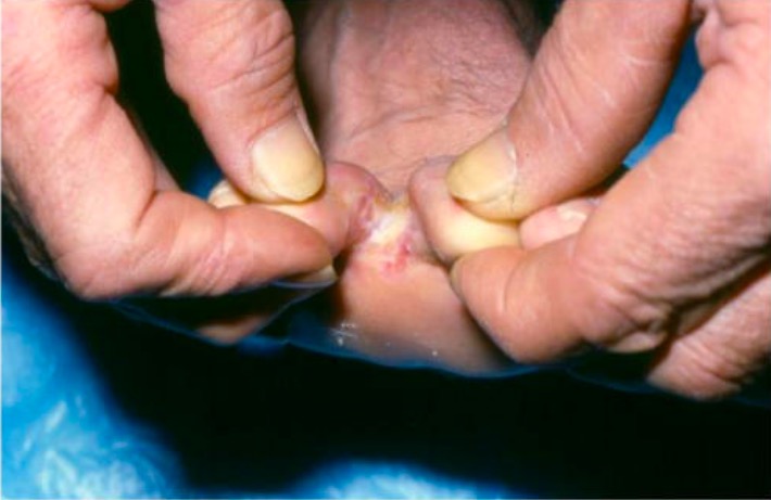
    • Infection of the interdigital webs of the feet
  • Tinea corporis: 
    • “Ringworm” 
    • Infection of the trunk or extremities (excluding hands/feet)
  • Tinea cruris: 
    • “Jock itch” 
    • Infection of the inguinal folds of the groin Groin The external junctural region between the lower part of the abdomen and the thigh. Male Genitourinary Examination
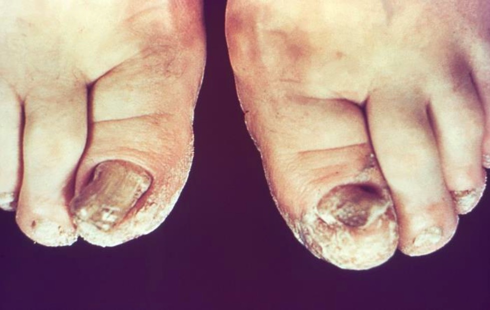
  • Tinea unguium (onychomycosis): infection of the nails
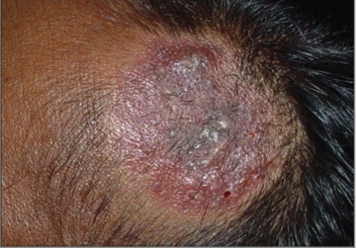
  • Tinea capitis: infection of the hair and scalp
  • Tinea manuum: infection of the hands

Pathogenesis

Reservoir Reservoir Animate or inanimate sources which normally harbor disease-causing organisms and thus serve as potential sources of disease outbreaks. Reservoirs are distinguished from vectors (disease vectors) and carriers, which are agents of disease transmission rather than continuing sources of potential disease outbreaks. Humans may serve both as disease reservoirs and carriers. Escherichia coli

  • Humans
  • Animals Animals Unicellular or multicellular, heterotrophic organisms, that have sensation and the power of voluntary movement. Under the older five kingdom paradigm, animalia was one of the kingdoms. Under the modern three domain model, animalia represents one of the many groups in the domain eukaryota. Cell Types: Eukaryotic versus Prokaryotic
  • Soil

Transmission

  • Direct contact with infected people or animals Animals Unicellular or multicellular, heterotrophic organisms, that have sensation and the power of voluntary movement. Under the older five kingdom paradigm, animalia was one of the kingdoms. Under the modern three domain model, animalia represents one of the many groups in the domain eukaryota. Cell Types: Eukaryotic versus Prokaryotic
  • Indirect contact through a fomite

Host risk factors

  • Walking barefoot in shared locker room
  • Diabetes Diabetes Diabetes mellitus (DM) is a metabolic disease characterized by hyperglycemia and dysfunction of the regulation of glucose metabolism by insulin. Type 1 DM is diagnosed mostly in children and young adults as the result of autoimmune destruction of β cells in the pancreas and the resulting lack of insulin. Type 2 DM has a significant association with obesity and is characterized by insulin resistance. Diabetes Mellitus mellitus
  • Obesity Obesity Obesity is a condition associated with excess body weight, specifically with the deposition of excessive adipose tissue. Obesity is considered a global epidemic. Major influences come from the western diet and sedentary lifestyles, but the exact mechanisms likely include a mixture of genetic and environmental factors. Obesity
  • Immunodeficiency Immunodeficiency Chédiak-Higashi Syndrome
  • Copious sweating
  • Occlusive footwear
  • Trauma
  • Shaving
  • Contact with animals Animals Unicellular or multicellular, heterotrophic organisms, that have sensation and the power of voluntary movement. Under the older five kingdom paradigm, animalia was one of the kingdoms. Under the modern three domain model, animalia represents one of the many groups in the domain eukaryota. Cell Types: Eukaryotic versus Prokaryotic

Pathophysiology

  • Infect keratinized human tissues (e.g., hair, skin Skin The skin, also referred to as the integumentary system, is the largest organ of the body. The skin is primarily composed of the epidermis (outer layer) and dermis (deep layer). The epidermis is primarily composed of keratinocytes that undergo rapid turnover, while the dermis contains dense layers of connective tissue. Skin: Structure and Functions, and nails) 
  • Possess proteases Proteases Proteins and Peptides that allow for penetration Penetration X-rays of the stratum corneum Stratum corneum Skin: Structure and Functions
  • Typically remain superficial, involving only the epidermis Epidermis The external, nonvascular layer of the skin. It is made up, from within outward, of five layers of epithelium: (1) basal layer (stratum basale epidermidis); (2) spinous layer (stratum spinosum epidermidis); (3) granular layer (stratum granulosum epidermidis); (4) clear layer (stratum lucidum epidermidis); and (5) horny layer (stratum corneum epidermidis). Skin: Structure and Functions, because they cannot survive at the normal body temperature Body Temperature The measure of the level of heat of a human or animal. Heatstroke of 37℃
  • Metabolize keratin Keratin A class of fibrous proteins or scleroproteins that represents the principal constituent of epidermis; hair; nails; horny tissues, and the organic matrix of tooth enamel. Two major conformational groups have been characterized, alpha-keratin, whose peptide backbone forms a coiled-coil alpha helical structure consisting of type I keratin and a type II keratin, and beta-keratin, whose backbone forms a zigzag or pleated sheet structure. Alpha-keratins have been classified into at least 20 subtypes. In addition multiple isoforms of subtypes have been found which may be due to gene duplication. Seborrheic Keratosis as a fuel source

Clinical Presentation

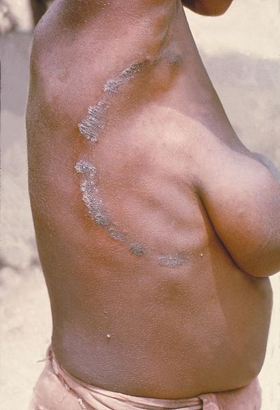
Signs and symptoms are similar between the tinea infections Infections Invasion of the host organism by microorganisms or their toxins or by parasites that can cause pathological conditions or diseases. Chronic Granulomatous Disease, with some minor differences depending on the body region affected. Lesions tend to be well-demarcated, annular Annular Dermatologic Examination, peripheral plaques with a rim of scale Scale Dermatologic Examination. They may also have associated erythema Erythema Redness of the skin produced by congestion of the capillaries. This condition may result from a variety of disease processes. Chalazion and/or maceration.

Table: Comparison of common dermatophyte infections Infections Invasion of the host organism by microorganisms or their toxins or by parasites that can cause pathological conditions or diseases. Chronic Granulomatous Disease
Name Body region Etiology Epidemiology and risk factors Clinical features
Tinea pedis (“athlete’s foot Foot The foot is the terminal portion of the lower limb, whose primary function is to bear weight and facilitate locomotion. The foot comprises 26 bones, including the tarsal bones, metatarsal bones, and phalanges. The bones of the foot form longitudinal and transverse arches and are supported by various muscles, ligaments, and tendons. Foot: Anatomy”) Feet, interdigital spaces of toes
  • Trichophyton rubrum
  • T. interdigitale
  • Epidermophyton floccosum
  • Most common tinea infection
  • Adolescents and adults
  • ↑ Risk from public locker rooms
  • Interdigital: pruritic maceration and scaling
  • Hyperkeratotic: diffuse scaling in “moccasin” distribution
  • Vesiculobullous (inflammatory): pruritic, erythematous, bullous eruption on feet
Tinea corporis (“ringworm”) Trunk and extremities (excluding hands and feet)
  • T. rubrum (most common)
  • T. tonsurans
  • Microsporum canis
  • T. interdigitale
  • Direct contact with infected person or animal, usually cat or dog
  • Common among athletes
  • Pruritic, ring-shaped, erythematous, or hyperpigmented plaques with central clearing
  • Spreads centrifugally
  • Extensive involvement may indicate diabetes Diabetes Diabetes mellitus (DM) is a metabolic disease characterized by hyperglycemia and dysfunction of the regulation of glucose metabolism by insulin. Type 1 DM is diagnosed mostly in children and young adults as the result of autoimmune destruction of β cells in the pancreas and the resulting lack of insulin. Type 2 DM has a significant association with obesity and is characterized by insulin resistance. Diabetes Mellitus or HIV HIV Anti-HIV Drugs infection.
Tinea cruris (“jock itch”) Groin Groin The external junctural region between the lower part of the abdomen and the thigh. Male Genitourinary Examination, inguinal folds
  • E. floccosum
  • T. rubrum
  • T. interdigitale
  • Men > women
  • Often spreads from preexisting tinea pedis
  • ↑ Risk with obesity Obesity Obesity is a condition associated with excess body weight, specifically with the deposition of excessive adipose tissue. Obesity is considered a global epidemic. Major influences come from the western diet and sedentary lifestyles, but the exact mechanisms likely include a mixture of genetic and environmental factors. Obesity, diabetes Diabetes Diabetes mellitus (DM) is a metabolic disease characterized by hyperglycemia and dysfunction of the regulation of glucose metabolism by insulin. Type 1 DM is diagnosed mostly in children and young adults as the result of autoimmune destruction of β cells in the pancreas and the resulting lack of insulin. Type 2 DM has a significant association with obesity and is characterized by insulin resistance. Diabetes Mellitus, immunodeficiency Immunodeficiency Chédiak-Higashi Syndrome, copious sweating
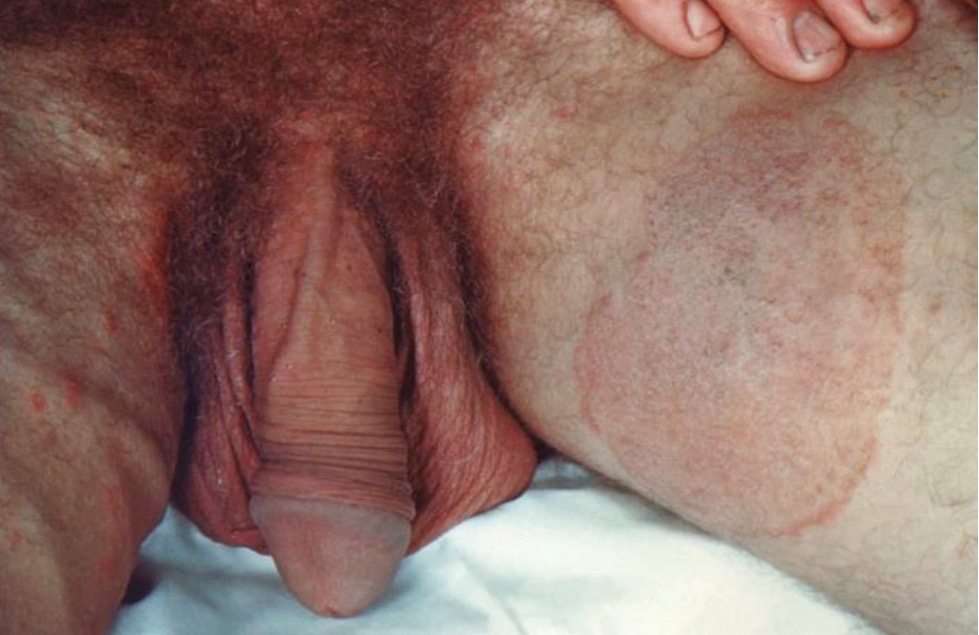
  • Involves inguinal folds
  • Erythematous patches Patches Vitiligo spread centrifugally with partial clearing.
  • May spread to perineal and perianal regions
  • Spares scrotum Scrotum A cutaneous pouch of skin containing the testicles and spermatic cords. Testicles: Anatomy
Tinea unguium (onychomycosis) Fingernails or toenails T. rubrum
  • Direct contact with dermatophytes
  • Nail injury
  • ↑ Risk with tinea pedis infection
  • Adults > children
Tinea capitis Hair follicles and scalp
  • T. tonsurans
  • T. violaceum
  • T. verrucosum
  • M. canis
  • Direct contact with dermatophytes
  • Trauma from an object (i.e., a comb)
  • Scaly plaques with alopecia Alopecia Alopecia is the loss of hair in areas anywhere on the body where hair normally grows. Alopecia may be defined as scarring or non-scarring, localized or diffuse, congenital or acquired, reversible or permanent, or confined to the scalp or universal; however, alopecia is usually classified using the 1st 3 factors. Alopecia ± black dots
  • Endothrix infection: fungal spores Spores The reproductive elements of lower organisms, such as bacteria; fungi; and cryptogamic plants. Anthrax within the hair shaft
  • Ectothrix infection: fungal spores Spores The reproductive elements of lower organisms, such as bacteria; fungi; and cryptogamic plants. Anthrax surrounding the hair shaft
  • Favus infection: fungal spores Spores The reproductive elements of lower organisms, such as bacteria; fungi; and cryptogamic plants. Anthrax and air pockets within the hair shaft
Majocchi granuloma (fungal folliculitis) Hair follicles on the body T. rubrum
  • Trauma to skin Skin The skin, also referred to as the integumentary system, is the largest organ of the body. The skin is primarily composed of the epidermis (outer layer) and dermis (deep layer). The epidermis is primarily composed of keratinocytes that undergo rapid turnover, while the dermis contains dense layers of connective tissue. Skin: Structure and Functions
  • Occlusion of hair follicles
  • Shaving hair
  • ↑ Risk with immunosuppression and corticosteroid use
  • Localized perifollicular papules
  • Erythema Erythema Redness of the skin produced by congestion of the capillaries. This condition may result from a variety of disease processes. Chalazion or hyperpigmentation Hyperpigmentation Excessive pigmentation of the skin, usually as a result of increased epidermal or dermal melanin pigmentation, hypermelanosis. Hyperpigmentation can be localized or generalized. The condition may arise from exposure to light, chemicals or other substances, or from a primary metabolic imbalance. Malassezia Fungi
  • Pustules
  • Abscesses or dissemination in immunocompromised immunocompromised A human or animal whose immunologic mechanism is deficient because of an immunodeficiency disorder or other disease or as the result of the administration of immunosuppressive drugs or radiation. Gastroenteritis patients Patients Individuals participating in the health care system for the purpose of receiving therapeutic, diagnostic, or preventive procedures. Clinician–Patient Relationship
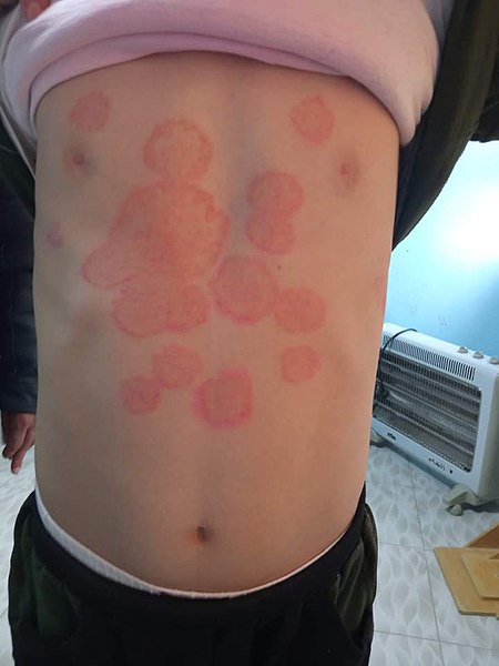
Tinea corporis

Tinea corporis, “ringworm” on a child:
Infection is most commonly due to Trichophyton rubrum, which presents with pruritic, erythematous, circular plaques with peripheral scales and central clearing.
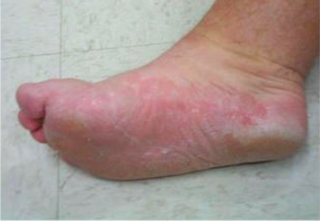
Image: “Tinea corporis” by Mohammad3021. License: CC0

Diagnosis and Management

Diagnosis

  • Primarily diagnosed clinically
  • Confirmed by KOH prep:
    • Scale Scale Dermatologic Examination from the periphery of a lesion is scraped onto a glass slide.
    • KOH is applied to the scale Scale Dermatologic Examination.
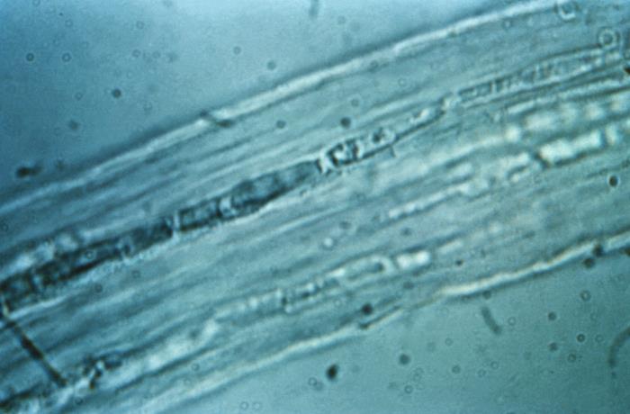
    • Dermatophytes are identified by visualizing septate hyphae Hyphae Microscopic threadlike filaments in fungi that are filled with a layer of protoplasm. Collectively, the hyphae make up the mycelium. Mycology.
  • Fungal cultures are an additional alternative for definitive diagnosis.
  • Onychomycosis can be diagnosed with a PAS stain of nail clippings.
Trichophyton rubrum

Trichophyton rubrum: This 475× magnification of T. rubrum shows centrally grouped, elongated macroconidia with septations. There are also many microconidia in the shape of teardrops.

Image: “Trichophyton rubrum” by CDC/Dr. Lucille K. Georg. License: Public Domain
Management

All variants of tinea are treated with antifungals.

  • Tinea pedis:
    • Topical terbinafine Terbinafine In addition to the 3 other major classes of antifungal agents (azoles, polyenes, and echinocandins), several other clinically important antifungal agents are used, including flucytosine, griseofulvin, and terbinafine. Terbinafine acts within the stratum corneum of the skin and are used to treat dermatophyte infections of the skin, hair, and nails. Flucytosine, Griseofulvin, and Terbinafine: Terbinafine Terbinafine In addition to the 3 other major classes of antifungal agents (azoles, polyenes, and echinocandins), several other clinically important antifungal agents are used, including flucytosine, griseofulvin, and terbinafine. Terbinafine acts within the stratum corneum of the skin and are used to treat dermatophyte infections of the skin, hair, and nails. Flucytosine, Griseofulvin, and Terbinafine is an allylamine Allylamine Possesses an unusual and selective cytotoxicity for vascular smooth muscle cells in dogs and rats. Useful for experiments dealing with arterial injury, myocardial fibrosis or cardiac decompensation. Flucytosine, Griseofulvin, and Terbinafine and works by inhibiting squalene epoxidase Squalene Epoxidase Flucytosine, Griseofulvin, and Terbinafine.
    • Oral terbinafine Terbinafine In addition to the 3 other major classes of antifungal agents (azoles, polyenes, and echinocandins), several other clinically important antifungal agents are used, including flucytosine, griseofulvin, and terbinafine. Terbinafine acts within the stratum corneum of the skin and are used to treat dermatophyte infections of the skin, hair, and nails. Flucytosine, Griseofulvin, and Terbinafine for recalcitrant cases
  • Tinea corporis:
    • Topical azoles Azoles Azoles are a widely used class of antifungal medications inhibiting the production of ergosterol, a critical component in the fungal cell membrane. The 2 primary subclasses of azoles are the imidazoles, older agents typically only used for topical applications, and the triazoles, newer agents with a wide spectrum of uses. Azoles (e.g., ketoconazole Ketoconazole Broad spectrum antifungal agent used for long periods at high doses, especially in immunosuppressed patients. Azoles) as initial therapy ( azoles Azoles Azoles are a widely used class of antifungal medications inhibiting the production of ergosterol, a critical component in the fungal cell membrane. The 2 primary subclasses of azoles are the imidazoles, older agents typically only used for topical applications, and the triazoles, newer agents with a wide spectrum of uses. Azoles work by inhibiting ergosterol Ergosterol A steroid occurring in fungi. Irradiation with ultraviolet rays results in formation of ergocalciferol (vitamin d2). Azoles synthesis Synthesis Polymerase Chain Reaction (PCR) → impair cell wall Cell wall The outermost layer of a cell in most plants; bacteria; fungi; and algae. The cell wall is usually a rigid structure that lies external to the cell membrane, and provides a protective barrier against physical or chemical agents. Cell Types: Eukaryotic versus Prokaryotic production)
    • Oral azoles Azoles Azoles are a widely used class of antifungal medications inhibiting the production of ergosterol, a critical component in the fungal cell membrane. The 2 primary subclasses of azoles are the imidazoles, older agents typically only used for topical applications, and the triazoles, newer agents with a wide spectrum of uses. Azoles (e.g., fluconazole Fluconazole Triazole antifungal agent that is used to treat oropharyngeal candidiasis and cryptococcal meningitis in aids. Azoles) for recalcitrant cases
  • Tinea cruris: topical azoles Azoles Azoles are a widely used class of antifungal medications inhibiting the production of ergosterol, a critical component in the fungal cell membrane. The 2 primary subclasses of azoles are the imidazoles, older agents typically only used for topical applications, and the triazoles, newer agents with a wide spectrum of uses. Azoles
  • Tinea unguium (onychomycosis):
    • Oral terbinafine Terbinafine In addition to the 3 other major classes of antifungal agents (azoles, polyenes, and echinocandins), several other clinically important antifungal agents are used, including flucytosine, griseofulvin, and terbinafine. Terbinafine acts within the stratum corneum of the skin and are used to treat dermatophyte infections of the skin, hair, and nails. Flucytosine, Griseofulvin, and Terbinafine
    • Oral fluconazole Fluconazole Triazole antifungal agent that is used to treat oropharyngeal candidiasis and cryptococcal meningitis in aids. Azoles
  • Tinea capitis:
    • Oral terbinafine Terbinafine In addition to the 3 other major classes of antifungal agents (azoles, polyenes, and echinocandins), several other clinically important antifungal agents are used, including flucytosine, griseofulvin, and terbinafine. Terbinafine acts within the stratum corneum of the skin and are used to treat dermatophyte infections of the skin, hair, and nails. Flucytosine, Griseofulvin, and Terbinafine for Trichophyton species
    • Oral griseofulvin Griseofulvin In addition to the 3 other major classes of antifungal agents (azoles, polyenes, and echinocandins), several other clinically important antifungal agents are used, including flucytosine, griseofulvin, and terbinafine. Griseofulvin acts within the stratum corneum of the skin and are used to treat dermatophyte infections of the skin, hair, and nails. Flucytosine, Griseofulvin, and Terbinafine (inhibition of microtubules Microtubules Slender, cylindrical filaments found in the cytoskeleton of plant and animal cells. They are composed of the protein tubulin and are influenced by tubulin modulators. The Cell: Cytosol and Cytoskeleton) for Microsporum species
  • Majocchi granuloma: oral azoles Azoles Azoles are a widely used class of antifungal medications inhibiting the production of ergosterol, a critical component in the fungal cell membrane. The 2 primary subclasses of azoles are the imidazoles, older agents typically only used for topical applications, and the triazoles, newer agents with a wide spectrum of uses. Azoles

Related videos

Differential Diagnosis

  • Erythrasma: bacterial skin Skin The skin, also referred to as the integumentary system, is the largest organ of the body. The skin is primarily composed of the epidermis (outer layer) and dermis (deep layer). The epidermis is primarily composed of keratinocytes that undergo rapid turnover, while the dermis contains dense layers of connective tissue. Skin: Structure and Functions infection caused by Corynebacterium Corynebacterium Corynebacteria are gram-positive, club-shaped bacilli. Corynebacteria are commonly isolated on tellurite or Loeffler’s media and have characteristic metachromatic granules. The major pathogenic species is Corynebacterium diphtheriae, which causes a severe respiratory infection called diphtheria. Corynebacterium minutissimum. The presentation of erythrasma is characterized by regions of scaly, pruritic, broken skin Skin The skin, also referred to as the integumentary system, is the largest organ of the body. The skin is primarily composed of the epidermis (outer layer) and dermis (deep layer). The epidermis is primarily composed of keratinocytes that undergo rapid turnover, while the dermis contains dense layers of connective tissue. Skin: Structure and Functions. The infection most commonly occurs in the spaces between the toes but may also present in intertriginous areas Intertriginous areas Malassezia Fungi. It may co-infect with Candida Candida Candida is a genus of dimorphic, opportunistic fungi. Candida albicans is part of the normal human flora and is the most common cause of candidiasis. The clinical presentation varies and can include localized mucocutaneous infections (e.g., oropharyngeal, esophageal, intertriginous, and vulvovaginal candidiasis) and invasive disease (e.g., candidemia, intraabdominal abscess, pericarditis, and meningitis). Candida/Candidiasis or dermatophytes, making the diagnosis more difficult. Areas of infection appear fluorescent red under a Wood’s lamp Wood’s lamp Malassezia Fungi. Treatment is with topical antibiotic therapy.
  • Candidal skin Skin The skin, also referred to as the integumentary system, is the largest organ of the body. The skin is primarily composed of the epidermis (outer layer) and dermis (deep layer). The epidermis is primarily composed of keratinocytes that undergo rapid turnover, while the dermis contains dense layers of connective tissue. Skin: Structure and Functions infection/intertrigo: infection and inflammation Inflammation Inflammation is a complex set of responses to infection and injury involving leukocytes as the principal cellular mediators in the body’s defense against pathogenic organisms. Inflammation is also seen as a response to tissue injury in the process of wound healing. The 5 cardinal signs of inflammation are pain, heat, redness, swelling, and loss of function. Inflammation of intertriginous regions, including the folds of an abdominal pannus Pannus A genus of cyanobacteria in the family synechococcaceae that is free-floating and occurs in various water environments attaching to algae and submersed plants. Septic Arthritis. Candidal skin Skin The skin, also referred to as the integumentary system, is the largest organ of the body. The skin is primarily composed of the epidermis (outer layer) and dermis (deep layer). The epidermis is primarily composed of keratinocytes that undergo rapid turnover, while the dermis contains dense layers of connective tissue. Skin: Structure and Functions infection may be fungal or bacterial, and it typically presents with moist, erythematous patches Patches Vitiligo within skin Skin The skin, also referred to as the integumentary system, is the largest organ of the body. The skin is primarily composed of the epidermis (outer layer) and dermis (deep layer). The epidermis is primarily composed of keratinocytes that undergo rapid turnover, while the dermis contains dense layers of connective tissue. Skin: Structure and Functions folds. This condition may eventually lead to skin Skin The skin, also referred to as the integumentary system, is the largest organ of the body. The skin is primarily composed of the epidermis (outer layer) and dermis (deep layer). The epidermis is primarily composed of keratinocytes that undergo rapid turnover, while the dermis contains dense layers of connective tissue. Skin: Structure and Functions breakdown. Diagnosis is clinical, and treatment is with topical antifungal Antifungal Azoles therapy.
  • Atopic dermatitis Dermatitis Any inflammation of the skin. Atopic Dermatitis (Eczema)/ eczema Eczema Atopic dermatitis, also known as eczema, is a chronic, relapsing, pruritic, inflammatory skin disease that occurs more frequently in children, although adults can also be affected. The condition is often associated with elevated serum levels of IgE and a personal or family history of atopy. Skin dryness, erythema, oozing, crusting, and lichenification are present. Atopic Dermatitis (Eczema): inflammatory skin Skin The skin, also referred to as the integumentary system, is the largest organ of the body. The skin is primarily composed of the epidermis (outer layer) and dermis (deep layer). The epidermis is primarily composed of keratinocytes that undergo rapid turnover, while the dermis contains dense layers of connective tissue. Skin: Structure and Functions disease that most frequently affects children but can also be present in adults. Eczema Eczema Atopic dermatitis, also known as eczema, is a chronic, relapsing, pruritic, inflammatory skin disease that occurs more frequently in children, although adults can also be affected. The condition is often associated with elevated serum levels of IgE and a personal or family history of atopy. Skin dryness, erythema, oozing, crusting, and lichenification are present. Atopic Dermatitis (Eczema) is a chronic condition that presents with dry, crusted, erythematous plaques that may be intensely pruritic. The dermatitis Dermatitis Any inflammation of the skin. Atopic Dermatitis (Eczema) most often affects the flexor surfaces and is associated with other types of atopy Atopy Atopic Dermatitis (Eczema), including asthma Asthma Asthma is a chronic inflammatory respiratory condition characterized by bronchial hyperresponsiveness and airflow obstruction. The disease is believed to result from the complex interaction of host and environmental factors that increase disease predisposition, with inflammation causing symptoms and structural changes. Patients typically present with wheezing, cough, and dyspnea. Asthma. The mainstay of therapy is gentle skin Skin The skin, also referred to as the integumentary system, is the largest organ of the body. The skin is primarily composed of the epidermis (outer layer) and dermis (deep layer). The epidermis is primarily composed of keratinocytes that undergo rapid turnover, while the dermis contains dense layers of connective tissue. Skin: Structure and Functions care and the application of topical steroids Steroids A group of polycyclic compounds closely related biochemically to terpenes. They include cholesterol, numerous hormones, precursors of certain vitamins, bile acids, alcohols (sterols), and certain natural drugs and poisons. Steroids have a common nucleus, a fused, reduced 17-carbon atom ring system, cyclopentanoperhydrophenanthrene. Most steroids also have two methyl groups and an aliphatic side-chain attached to the nucleus. Benign Liver Tumors.
  • Contact dermatitis Contact dermatitis A type of acute or chronic skin reaction in which sensitivity is manifested by reactivity to materials or substances coming in contact with the skin. It may involve allergic or non-allergic mechanisms. Male Genitourinary Examination: local inflammation Local Inflammation Surgical Site Infections of the skin Skin The skin, also referred to as the integumentary system, is the largest organ of the body. The skin is primarily composed of the epidermis (outer layer) and dermis (deep layer). The epidermis is primarily composed of keratinocytes that undergo rapid turnover, while the dermis contains dense layers of connective tissue. Skin: Structure and Functions that is a result of direct contact with an irritant or allergen. The etiologies of contact dermatitis Contact dermatitis A type of acute or chronic skin reaction in which sensitivity is manifested by reactivity to materials or substances coming in contact with the skin. It may involve allergic or non-allergic mechanisms. Male Genitourinary Examination include allergic contact dermatitis Contact dermatitis A type of acute or chronic skin reaction in which sensitivity is manifested by reactivity to materials or substances coming in contact with the skin. It may involve allergic or non-allergic mechanisms. Male Genitourinary Examination (delayed type IV hypersensitivity reaction Type IV hypersensitivity reaction Type IV hypersensitivity reaction, or delayed-type hypersensitivity, is a cell-mediated response to antigen exposure. The reaction involves T cells, not antibodies, and develops over several days. Presensitized T cells initiate the immune defense, leading to tissue damage. Type IV Hypersensitivity Reaction) or irritant dermatitis Dermatitis Any inflammation of the skin. Atopic Dermatitis (Eczema), which results from mechanical or chemical irritation of the skin Skin The skin, also referred to as the integumentary system, is the largest organ of the body. The skin is primarily composed of the epidermis (outer layer) and dermis (deep layer). The epidermis is primarily composed of keratinocytes that undergo rapid turnover, while the dermis contains dense layers of connective tissue. Skin: Structure and Functions. Lesions present with well-demarcated erythema Erythema Redness of the skin produced by congestion of the capillaries. This condition may result from a variety of disease processes. Chalazion and edema Edema Edema is a condition in which excess serous fluid accumulates in the body cavity or interstitial space of connective tissues. Edema is a symptom observed in several medical conditions. It can be categorized into 2 types, namely, peripheral (in the extremities) and internal (in an organ or body cavity). Edema and may be pruritic or develop bullae Bullae Erythema Multiforme with oozing. Treatment is with trigger Trigger The type of signal that initiates the inspiratory phase by the ventilator Invasive Mechanical Ventilation avoidance and topical corticosteroids Corticosteroids Chorioretinitis.
  • Psoriasis Psoriasis Psoriasis is a common T-cell-mediated inflammatory skin condition. The etiology is unknown, but is thought to be due to genetic inheritance and environmental triggers. There are 4 major subtypes, with the most common form being chronic plaque psoriasis. Psoriasis: multisystem inflammatory disease that includes skin Skin The skin, also referred to as the integumentary system, is the largest organ of the body. The skin is primarily composed of the epidermis (outer layer) and dermis (deep layer). The epidermis is primarily composed of keratinocytes that undergo rapid turnover, while the dermis contains dense layers of connective tissue. Skin: Structure and Functions findings. There are several subtypes of psoriasis Psoriasis Psoriasis is a common T-cell-mediated inflammatory skin condition. The etiology is unknown, but is thought to be due to genetic inheritance and environmental triggers. There are 4 major subtypes, with the most common form being chronic plaque psoriasis. Psoriasis. The most common subtype, chronic plaque Plaque Primary Skin Lesions psoriasis Psoriasis Psoriasis is a common T-cell-mediated inflammatory skin condition. The etiology is unknown, but is thought to be due to genetic inheritance and environmental triggers. There are 4 major subtypes, with the most common form being chronic plaque psoriasis. Psoriasis, presents with scaly, erythematous plaques that are intensely pruritic. These plaques most commonly affect extensor surfaces, which can distinguish it from other papulosquamous disorders. Treatment is focused on managing symptoms.
  • Scabies Scabies Scabies is an infestation of the skin by the Sarcoptes scabiei mite, which presents most commonly with intense pruritus, characteristic linear burrows, and erythematous papules, particularly in the interdigital folds and the flexor aspects of the wrists. Scabies: local skin Skin The skin, also referred to as the integumentary system, is the largest organ of the body. The skin is primarily composed of the epidermis (outer layer) and dermis (deep layer). The epidermis is primarily composed of keratinocytes that undergo rapid turnover, while the dermis contains dense layers of connective tissue. Skin: Structure and Functions infection caused by the mite Sarcoptes scabiei Sarcoptes scabiei A species of mite that causes scabies in humans and sarcoptic mange in other animals. Specific variants of s. Scabiei exist for humans and animals, but many have the ability to cross species and cause disease. Scabies. The clinical presentation of scabies Scabies Scabies is an infestation of the skin by the Sarcoptes scabiei mite, which presents most commonly with intense pruritus, characteristic linear burrows, and erythematous papules, particularly in the interdigital folds and the flexor aspects of the wrists. Scabies usually includes small erythematous papules that are very pruritic and may have a linear distribution, which is evidence of mite movement underneath the skin Skin The skin, also referred to as the integumentary system, is the largest organ of the body. The skin is primarily composed of the epidermis (outer layer) and dermis (deep layer). The epidermis is primarily composed of keratinocytes that undergo rapid turnover, while the dermis contains dense layers of connective tissue. Skin: Structure and Functions. Common sites of infection are the extremities, classically the hands, and interdigital webs. Lesions may also appear as crusted, erythematous patches Patches Vitiligo. Diagnosis is made clinically or with the detection of mites Mites Any arthropod of the subclass acari except the ticks. They are minute animals related to the spiders, usually having transparent or semitransparent bodies. They may be parasitic on humans and domestic animals, producing various irritations of the skin (mite infestations). Many mite species are important to human and veterinary medicine as both parasite and vector. Mites also infest plants. Scabies, eggs, or feces on microscopy of skin Skin The skin, also referred to as the integumentary system, is the largest organ of the body. The skin is primarily composed of the epidermis (outer layer) and dermis (deep layer). The epidermis is primarily composed of keratinocytes that undergo rapid turnover, while the dermis contains dense layers of connective tissue. Skin: Structure and Functions scrapings. Treatment is with topical permethrin Permethrin A pyrethroid insecticide commonly used in the treatment of lice infestations and scabies. Scabies.
  • Pityriasis rosea Pityriasis rosea Pityriasis rosea is an acute, self-limited skin disease. The etiology is not known, and it commonly occurs in young adults. Patients initially present with a single, ovoid “herald patch.” This is followed by diffuse, pruritic, scaly, oval lesions over the trunk (often in a “Christmas tree” distribution on the back) and extremities. Pityriasis Rosea: skin Skin The skin, also referred to as the integumentary system, is the largest organ of the body. The skin is primarily composed of the epidermis (outer layer) and dermis (deep layer). The epidermis is primarily composed of keratinocytes that undergo rapid turnover, while the dermis contains dense layers of connective tissue. Skin: Structure and Functions disease that results in oval-shaped salmon-colored patches Patches Vitiligo. An initial “herald” patch Patch Nonpalpable lesion > 1 cm in diameter Generalized and Localized Rashes is followed by the emergence of multiple additional patches Patches Vitiligo within 1–2 weeks after the initial patch Patch Nonpalpable lesion > 1 cm in diameter Generalized and Localized Rashes. Pityriasis rosea Pityriasis rosea Pityriasis rosea is an acute, self-limited skin disease. The etiology is not known, and it commonly occurs in young adults. Patients initially present with a single, ovoid “herald patch.” This is followed by diffuse, pruritic, scaly, oval lesions over the trunk (often in a “Christmas tree” distribution on the back) and extremities. Pityriasis Rosea may be associated with a prodrome Prodrome Symptoms that appear 24–48 hours prior to migraine onset. Migraine Headache of generalized malaise Malaise Tick-borne Encephalitis Virus, headache Headache The symptom of pain in the cranial region. It may be an isolated benign occurrence or manifestation of a wide variety of headache disorders. Brain Abscess, and pharyngitis Pharyngitis Pharyngitis is an inflammation of the back of the throat (pharynx). Pharyngitis is usually caused by an upper respiratory tract infection, which is viral in most cases. It typically results in a sore throat and fever. Other symptoms may include a runny nose, cough, headache, and hoarseness. Pharyngitis. The disease course is typically mild and self-limited.
  • Seborrheic dermatitis Dermatitis Any inflammation of the skin. Atopic Dermatitis (Eczema): relapsing skin disorder Relapsing Skin Disorder Seborrheic Dermatitis that has an unclear pathogenesis and presents with yellow-tinged scales Scales Dry or greasy masses of keratin that represent thickened stratum corneum. Secondary Skin Lesions on well-demarcated, pruritic, erythematous plaques. These lesions are distributed in regions of the body with numerous sebaceous glands, including the scalp, face, and chest. Seborrheic dermatitis Dermatitis Any inflammation of the skin. Atopic Dermatitis (Eczema) commonly presents as dandruff Dandruff Excessive shedding of dry scaly material from the scalp in humans. Seborrheic Dermatitis in adults and as “cradle cap” in infants. Some evidence suggests that Malassezia Malassezia Malassezia is a lipophilic yeast commonly found on the skin surfaces of many animals, including humans. In the presence of certain environments or triggers, this fungus can cause pathologic diseases ranging from superficial skin conditions (tinea versicolor and dermatitis) to invasive disease (e.g., Malassezia folliculitis, catheter-associated fungemia, meningitis, and urinary tract infections). Malassezia Fungi proliferation may be the etiology. Treatment includes topical antifungals and corticosteroids Corticosteroids Chorioretinitis.
  • Subacute cutaneous lupus erythematosus (SCLE): form of skin Skin The skin, also referred to as the integumentary system, is the largest organ of the body. The skin is primarily composed of the epidermis (outer layer) and dermis (deep layer). The epidermis is primarily composed of keratinocytes that undergo rapid turnover, while the dermis contains dense layers of connective tissue. Skin: Structure and Functions lupus that is not usually associated with systemic lupus erythematosus Systemic lupus erythematosus Systemic lupus erythematosus (SLE) is a chronic autoimmune, inflammatory condition that causes immune-complex deposition in organs, resulting in systemic manifestations. Women, particularly those of African American descent, are more commonly affected. Systemic Lupus Erythematosus ( SLE SLE Systemic lupus erythematosus (SLE) is a chronic autoimmune, inflammatory condition that causes immune-complex deposition in organs, resulting in systemic manifestations. Women, particularly those of African American descent, are more commonly affected. Systemic Lupus Erythematosus). Subacute cutaneous lupus erythematosus presents with annular Annular Dermatologic Examination, erythematous, scaly plaques and usually appears on sun-exposed skin Skin The skin, also referred to as the integumentary system, is the largest organ of the body. The skin is primarily composed of the epidermis (outer layer) and dermis (deep layer). The epidermis is primarily composed of keratinocytes that undergo rapid turnover, while the dermis contains dense layers of connective tissue. Skin: Structure and Functions, including the neck Neck The part of a human or animal body connecting the head to the rest of the body. Peritonsillar Abscess, torso, and upper extremities. Counterintuitively, the face is usually spared. Subacute cutaneous lupus erythematosus can be drug-induced, with common offenders being antihypertensive agents, antihyperlipidemic agents, proton-pump inhibitors, antifungals, and tumor Tumor Inflammation necrosis Necrosis The death of cells in an organ or tissue due to disease, injury or failure of the blood supply. Ischemic Cell Damage factor α inhibitors.
  • Granuloma annulare Granuloma Annulare Benign granulomatous disease of unknown etiology characterized by a ring of localized or disseminated papules or nodules on the skin and palisading histiocytes surrounding necrobiotic tissue resulting from altered collagen structures. Secondary Skin Lesions and erythema Erythema Redness of the skin produced by congestion of the capillaries. This condition may result from a variety of disease processes. Chalazion annulare centrifugum: benign Benign Fibroadenoma, inflammatory conditions that present with annular Annular Dermatologic Examination, erythematous plaques. Granuloma annulare Granuloma Annulare Benign granulomatous disease of unknown etiology characterized by a ring of localized or disseminated papules or nodules on the skin and palisading histiocytes surrounding necrobiotic tissue resulting from altered collagen structures. Secondary Skin Lesions and erythema Erythema Redness of the skin produced by congestion of the capillaries. This condition may result from a variety of disease processes. Chalazion annulare centrifugum often do not have scales Scales Dry or greasy masses of keratin that represent thickened stratum corneum. Secondary Skin Lesions, which can be the factor distinguishing them from tinea infections Infections Invasion of the host organism by microorganisms or their toxins or by parasites that can cause pathological conditions or diseases. Chronic Granulomatous Disease.

References

  1. Mitchel TG. (2015). Medical mycology. In Brooks JE, et al. (Eds.), Jawetz, Melnick, & Adelberg’s Medical Microbiology, 27th ed. [VitalSource Bookshelf 9.4.3]. 
  2. Goldstein AO, Goldstein BG. (2021). Dermatophyte (tinea) infections. UpToDate. Retrieved April 29, 2021, from https://www.uptodate.com/contents/dermatophyte-tinea-infections
  3. Hawkins DM, Smidt, AC. (2014). Superficial fungal infections in children. Pediatric Clinics of North America 61:443–455. https://doi-org.online.uchc.edu/10.1016/j.pcl.2013.12.003
  4. Noble SL, Forbes RC, Stamm, PL. (1998). Diagnosis and management of common tinea infections. American Family Physician 58:163–178.
  5. Smith KJ, Neafie RC, Skelton HG, Barrett TL, Graham JF, Lupton GP.  (1991). Majocchi’s granuloma. Journal of Cutaneous Pathology 18:28. https://doi-org.online.uchc.edu/10.1111/j.1600-0560.1991.tb00598.x
  6. Merola JF. (2021). Overview of cutaneous lupus erythematosus. UpToDate. Retrieved May 1, 2021, from https://www.uptodate.com/contents/overview-of-cutaneous-lupus-erythematosus
  7. van Zuuren EJ, Fedorowicz Z, El-Gohary M. (2015). Evidence-based topical treatments for tinea cruris and tinea corporis: a summary of a Cochrane systematic review. British Journal of Dermatology 172:616–641. https://doi-org.online.uchc.edu/10.1111/bjd.13441
  8. Centers for Disease Control and Prevention. Ringworm. Retrieved May 2, 2021, from https://www.cdc.gov/fungal/diseases/ringworm/index.html

Create your free account or log in to continue reading!

Sign up now and get free access to Lecturio with concept pages, medical videos, and questions for your medical education.

User Reviews

Details