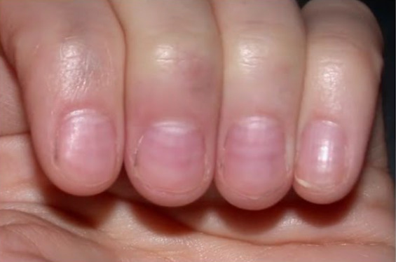
Dermatologic Examination

Examination of the skin Skin The skin, also referred to as the integumentary system, is the largest organ of the body. The skin is primarily composed of the epidermis (outer layer) and dermis (deep layer). The epidermis is primarily composed of keratinocytes that undergo rapid turnover, while the dermis contains dense layers of connective tissue. Skin: Structure and Functions is a fundamental part of the standard physical exam. This exam consists of a thorough inspection of the skin Skin The skin, also referred to as the integumentary system, is the largest organ of the body. The skin is primarily composed of the epidermis (outer layer) and dermis (deep layer). The epidermis is primarily composed of keratinocytes that undergo rapid turnover, while the dermis contains dense layers of connective tissue. Skin: Structure and Functions of the entire body. The assessment focuses on identifying abnormal signs on the skin Skin The skin, also referred to as the integumentary system, is the largest organ of the body. The skin is primarily composed of the epidermis (outer layer) and dermis (deep layer). The epidermis is primarily composed of keratinocytes that undergo rapid turnover, while the dermis contains dense layers of connective tissue. Skin: Structure and Functions, such as the scalp, orifices, nails, and mucosal surfaces. Dermatologic findings can represent localized processes or may be a sign of systemic disease.

Last updated: May 17, 2024

Editorial responsibility: Stanley Oiseth, Lindsay Jones, Evelin Maza

Terminology

Primary and secondary skin lesions Secondary Skin Lesions The identification and classification of a patient’s skin lesions are important steps in the diagnosis of any skin disorder. Secondary lesions develop from irritated or manipulated primary lesions and/or manifestations of disease progression. Secondary Skin Lesions

Primary skin lesions Primary Skin Lesions The identification and classification of skin lesions in a patient are important steps in the diagnosis of any skin disorder. Primary lesions represent the initial presentation of the disease process. Primary Skin Lesions occur as a direct result of a pathologic process, while secondary skin lesions Secondary Skin Lesions The identification and classification of a patient’s skin lesions are important steps in the diagnosis of any skin disorder. Secondary lesions develop from irritated or manipulated primary lesions and/or manifestations of disease progression. Secondary Skin Lesions are those that result from manipulation of the skin Skin The skin, also referred to as the integumentary system, is the largest organ of the body. The skin is primarily composed of the epidermis (outer layer) and dermis (deep layer). The epidermis is primarily composed of keratinocytes that undergo rapid turnover, while the dermis contains dense layers of connective tissue. Skin: Structure and Functions.

Table: Primary skin lesions Primary Skin Lesions The identification and classification of skin lesions in a patient are important steps in the diagnosis of any skin disorder. Primary lesions represent the initial presentation of the disease process. Primary Skin Lesions
Primary skin Skin The skin, also referred to as the integumentary system, is the largest organ of the body. The skin is primarily composed of the epidermis (outer layer) and dermis (deep layer). The epidermis is primarily composed of keratinocytes that undergo rapid turnover, while the dermis contains dense layers of connective tissue. Skin: Structure and Functions lesion Description
Macule Macule Nonpalpable lesion < 1 cm in diameter Generalized and Localized Rashes
  • Flat, nonpalpable skin Skin The skin, also referred to as the integumentary system, is the largest organ of the body. The skin is primarily composed of the epidermis (outer layer) and dermis (deep layer). The epidermis is primarily composed of keratinocytes that undergo rapid turnover, while the dermis contains dense layers of connective tissue. Skin: Structure and Functions lesion ≤ 1 cm in size
  • Differs in color from the surrounding skin Skin The skin, also referred to as the integumentary system, is the largest organ of the body. The skin is primarily composed of the epidermis (outer layer) and dermis (deep layer). The epidermis is primarily composed of keratinocytes that undergo rapid turnover, while the dermis contains dense layers of connective tissue. Skin: Structure and Functions
Papule Papule Elevated lesion < 1 cm in diameter Generalized and Localized Rashes Small palpable skin Skin The skin, also referred to as the integumentary system, is the largest organ of the body. The skin is primarily composed of the epidermis (outer layer) and dermis (deep layer). The epidermis is primarily composed of keratinocytes that undergo rapid turnover, while the dermis contains dense layers of connective tissue. Skin: Structure and Functions lesion ≤ 1 cm in diameter
Plaque Plaque Primary Skin Lesions Palpable, raised lesion > 1 cm
Nodule Nodule Chalazion
  • Elevated lesion
  • > 1 cm in both diameter and depth
Vesicle Vesicle Primary Skin Lesions Small fluid-containing blister Blister Bullous Pemphigoid and Pemphigus Vulgaris ≤ 1 cm in diameter
Bullae Bullae Erythema Multiforme Large fluid-containing blister Blister Bullous Pemphigoid and Pemphigus Vulgaris > 1 cm in diameter
Pustule Pustule Blister filled with pus Generalized and Localized Rashes Vesicle Vesicle Primary Skin Lesions filled with pus
Table: Secondary skin lesions Secondary Skin Lesions The identification and classification of a patient’s skin lesions are important steps in the diagnosis of any skin disorder. Secondary lesions develop from irritated or manipulated primary lesions and/or manifestations of disease progression. Secondary Skin Lesions
Secondary skin lesions Secondary Skin Lesions The identification and classification of a patient’s skin lesions are important steps in the diagnosis of any skin disorder. Secondary lesions develop from irritated or manipulated primary lesions and/or manifestations of disease progression. Secondary Skin Lesions Description
Scale
Crust Crust Dried exudate of body fluids (blood, pus, or sebum) on an area of damaged skin Secondary Skin Lesions Dried exudates, such as pus or blood
Fissure Fissure A crack or split that extends into the dermis Generalized and Localized Rashes
  • Linear crack Crack The purified, alkaloidal, extra-potent form of cocaine. It is smoked (free-based), injected intravenously, and orally ingested. Use of crack results in alterations in function of the cardiovascular system, the autonomic nervous system, the central nervous system, and the gastrointestinal system. The slang term ‘crack’ was derived from the crackling sound made upon igniting of this form of cocaine for smoking. Cocaine Use Disorder through the epidermis Epidermis The external, nonvascular layer of the skin. It is made up, from within outward, of five layers of epithelium: (1) basal layer (stratum basale epidermidis); (2) spinous layer (stratum spinosum epidermidis); (3) granular layer (stratum granulosum epidermidis); (4) clear layer (stratum lucidum epidermidis); and (5) horny layer (stratum corneum epidermidis). Skin: Structure and Functions extending to the dermis Dermis A layer of vascularized connective tissue underneath the epidermis. The surface of the dermis contains innervated papillae. Embedded in or beneath the dermis are sweat glands; hair follicles; and sebaceous glands. Skin: Structure and Functions
  • Loss of continuity of the skin Skin The skin, also referred to as the integumentary system, is the largest organ of the body. The skin is primarily composed of the epidermis (outer layer) and dermis (deep layer). The epidermis is primarily composed of keratinocytes that undergo rapid turnover, while the dermis contains dense layers of connective tissue. Skin: Structure and Functions
Ulcer
  • Break in the continuity of the epithelium Epithelium The epithelium is a complex of specialized cellular organizations arranged into sheets and lining cavities and covering the surfaces of the body. The cells exhibit polarity, having an apical and a basal pole. Structures important for the epithelial integrity and function involve the basement membrane, the semipermeable sheet on which the cells rest, and interdigitations, as well as cellular junctions. Surface Epithelium: Histology
  • Occurs owing to loss of epidermis Epidermis The external, nonvascular layer of the skin. It is made up, from within outward, of five layers of epithelium: (1) basal layer (stratum basale epidermidis); (2) spinous layer (stratum spinosum epidermidis); (3) granular layer (stratum granulosum epidermidis); (4) clear layer (stratum lucidum epidermidis); and (5) horny layer (stratum corneum epidermidis). Skin: Structure and Functions and dermis Dermis A layer of vascularized connective tissue underneath the epidermis. The surface of the dermis contains innervated papillae. Embedded in or beneath the dermis are sweat glands; hair follicles; and sebaceous glands. Skin: Structure and Functions
Erosion Erosion Partial-thickness loss of the epidermis Generalized and Localized Rashes
  • Circumscribed depressed lesion due to loss of epidermis Epidermis The external, nonvascular layer of the skin. It is made up, from within outward, of five layers of epithelium: (1) basal layer (stratum basale epidermidis); (2) spinous layer (stratum spinosum epidermidis); (3) granular layer (stratum granulosum epidermidis); (4) clear layer (stratum lucidum epidermidis); and (5) horny layer (stratum corneum epidermidis). Skin: Structure and Functions
  • Heals without scarring Scarring Inflammation
Necrosis Necrosis The death of cells in an organ or tissue due to disease, injury or failure of the blood supply. Ischemic Cell Damage Dead skin Skin The skin, also referred to as the integumentary system, is the largest organ of the body. The skin is primarily composed of the epidermis (outer layer) and dermis (deep layer). The epidermis is primarily composed of keratinocytes that undergo rapid turnover, while the dermis contains dense layers of connective tissue. Skin: Structure and Functions tissue
Atrophy Atrophy Decrease in the size of a cell, tissue, organ, or multiple organs, associated with a variety of pathological conditions such as abnormal cellular changes, ischemia, malnutrition, or hormonal changes. Cellular Adaptation
  • Thinning of the skin Skin The skin, also referred to as the integumentary system, is the largest organ of the body. The skin is primarily composed of the epidermis (outer layer) and dermis (deep layer). The epidermis is primarily composed of keratinocytes that undergo rapid turnover, while the dermis contains dense layers of connective tissue. Skin: Structure and Functions
  • Skin Skin The skin, also referred to as the integumentary system, is the largest organ of the body. The skin is primarily composed of the epidermis (outer layer) and dermis (deep layer). The epidermis is primarily composed of keratinocytes that undergo rapid turnover, while the dermis contains dense layers of connective tissue. Skin: Structure and Functions becomes shiny and wrinkled
Scar Replacement of the skin Skin The skin, also referred to as the integumentary system, is the largest organ of the body. The skin is primarily composed of the epidermis (outer layer) and dermis (deep layer). The epidermis is primarily composed of keratinocytes that undergo rapid turnover, while the dermis contains dense layers of connective tissue. Skin: Structure and Functions with fibrotic and connective tissue Connective tissue Connective tissues originate from embryonic mesenchyme and are present throughout the body except inside the brain and spinal cord. The main function of connective tissues is to provide structural support to organs. Connective tissues consist of cells and an extracellular matrix. Connective Tissue: Histology as a result of destruction
Lichenification Lichenification Atopic Dermatitis (Eczema)
  • Thickening of the skin Skin The skin, also referred to as the integumentary system, is the largest organ of the body. The skin is primarily composed of the epidermis (outer layer) and dermis (deep layer). The epidermis is primarily composed of keratinocytes that undergo rapid turnover, while the dermis contains dense layers of connective tissue. Skin: Structure and Functions
  • Leathery appearance

Special skin Skin The skin, also referred to as the integumentary system, is the largest organ of the body. The skin is primarily composed of the epidermis (outer layer) and dermis (deep layer). The epidermis is primarily composed of keratinocytes that undergo rapid turnover, while the dermis contains dense layers of connective tissue. Skin: Structure and Functions lesions

  • Comedones Comedones Acne Vulgaris: dilated hair follicles with debris such as keratin Keratin A class of fibrous proteins or scleroproteins that represents the principal constituent of epidermis; hair; nails; horny tissues, and the organic matrix of tooth enamel. Two major conformational groups have been characterized, alpha-keratin, whose peptide backbone forms a coiled-coil alpha helical structure consisting of type I keratin and a type II keratin, and beta-keratin, whose backbone forms a zigzag or pleated sheet structure. Alpha-keratins have been classified into at least 20 subtypes. In addition multiple isoforms of subtypes have been found which may be due to gene duplication. Seborrheic Keratosis, sebum Sebum The oily substance secreted by sebaceous glands. It is composed of keratin, fat, and cellular debris. Infectious Folliculitis and, bacteria Bacteria Bacteria are prokaryotic single-celled microorganisms that are metabolically active and divide by binary fission. Some of these organisms play a significant role in the pathogenesis of diseases. Bacteriology (blackhead)
  • Burrows: a tunnel noted by raised lines, suggesting scabies Scabies Scabies is an infestation of the skin by the Sarcoptes scabiei mite, which presents most commonly with intense pruritus, characteristic linear burrows, and erythematous papules, particularly in the interdigital folds and the flexor aspects of the wrists. Scabies infection
  • Alopecia Alopecia Alopecia is the loss of hair in areas anywhere on the body where hair normally grows. Alopecia may be defined as scarring or non-scarring, localized or diffuse, congenital or acquired, reversible or permanent, or confined to the scalp or universal; however, alopecia is usually classified using the 1st 3 factors. Alopecia: hair loss
  • Telangiectasia Telangiectasia Permanent dilation of preexisting blood vessels creating small focal red lesions, most commonly in the skin or mucous membranes. It is characterized by the prominence of skin blood vessels, such as vascular spiders. Chronic Venous Insufficiency: small, superficial blood vessels
  • Petechia: bruise < 3 mm
  • Purpura: bruise between 3 mm and 10 mm
  • Ecchymosis Ecchymosis Extravasation of blood into the skin, resulting in a nonelevated, rounded or irregular, blue or purplish patch, larger than a petechia. Orbital Fractures: bruise > 1 cm

Clinical Examination

Initial steps

  • Obtain informed consent Informed consent Informed consent is a medicolegal term describing the documented conversation between a patient and their physician wherein the physician discloses all relevant and necessary information to a patient who is competent to make an informed and voluntary decision regarding their care. Competency, disclosure, and voluntariness are the key elements upon which IC is based. Informed Consent:
    • Explain the need for the examination 
    • Describe the steps of the examination 
    • Ensure that the individual has a good understanding of the exam and agrees to proceed
  • Ask the individual to undress to their undergarments; have a chaperone present.

Components

  • Examination of skin Skin The skin, also referred to as the integumentary system, is the largest organ of the body. The skin is primarily composed of the epidermis (outer layer) and dermis (deep layer). The epidermis is primarily composed of keratinocytes that undergo rapid turnover, while the dermis contains dense layers of connective tissue. Skin: Structure and Functions and mucosal surfaces:
    • Inspect
    • Palpate
    • Special tests
  • Examination of hair:
    • Inspection
    • Hair-pull test
  • Examination of nails:
    • Inspection
    • Palpation

Examination of Skin and Mucosal Surfaces

All cutaneous and mucosal surfaces should be examined in good light. Wear gloves, especially if the skin Skin The skin, also referred to as the integumentary system, is the largest organ of the body. The skin is primarily composed of the epidermis (outer layer) and dermis (deep layer). The epidermis is primarily composed of keratinocytes that undergo rapid turnover, while the dermis contains dense layers of connective tissue. Skin: Structure and Functions is broken.
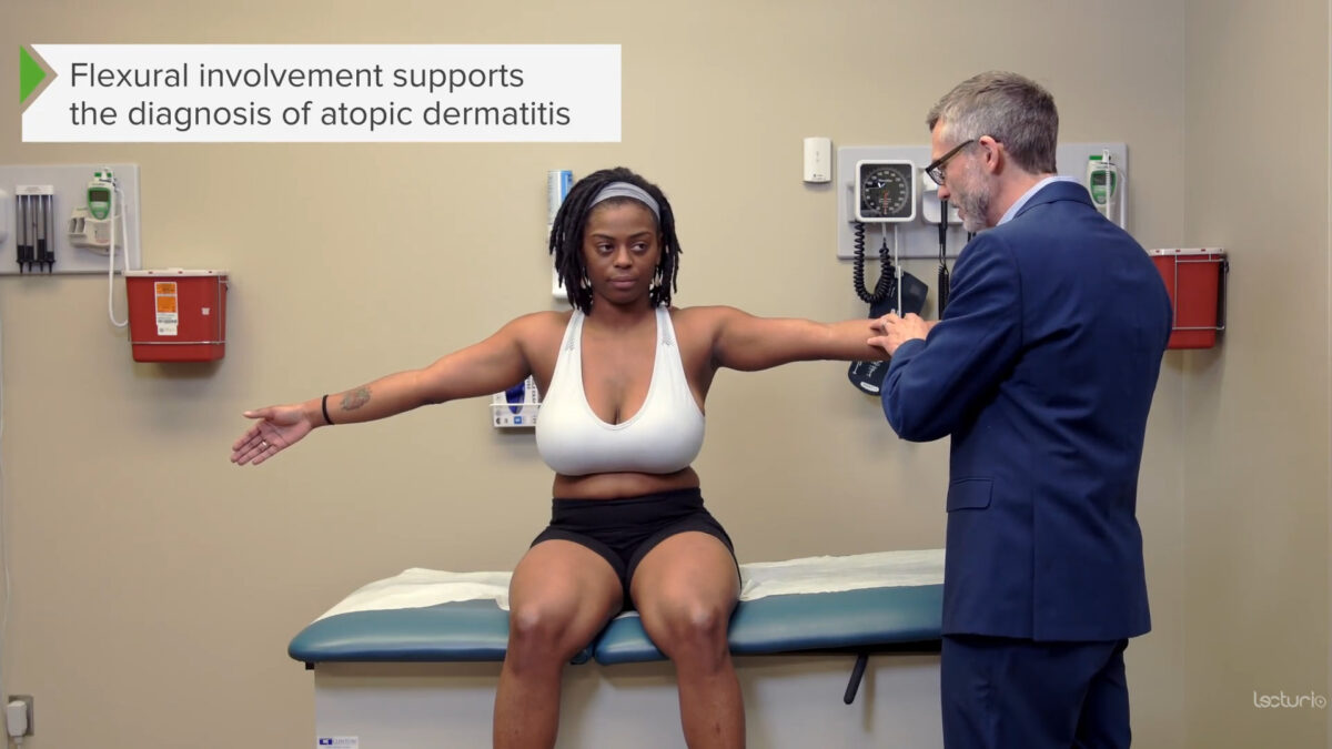
Inspection

  • Appearance:
    • Site of lesion 
    • Number of lesions
    • Size of lesion 
    • Configuration
      • Linear 
      • Annular (circular with central clearing)
      • Grouped 
    • Morphology
      • Macular 
      • Papular 
      • Maculopapular 
      • Vesicular 
    • Color
    • Presence of scale or crust Crust Dried exudate of body fluids (blood, pus, or sebum) on an area of damaged skin Secondary Skin Lesions
  • Distribution:
    • Diffuse or localized
    • Symmetrical or asymmetrical
    • Dermatomal
    • Clustered or grouped
    • Mucosal involvement
    • Atypical sites (e.g., palms and soles)

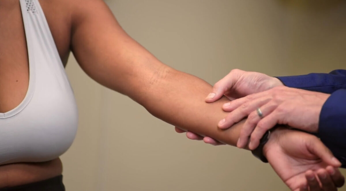
Palpation

  • Lesional:
    • Tenderness 
    • Warmth 
    • Consistency
      • Hard 
      • Firm 
      • Fluctuant 
    • Raised
    • Flat
    • Texture changes
    • Induration
    • Blanching with pressure
  • Regional lymph Lymph The interstitial fluid that is in the lymphatic system. Secondary Lymphatic Organs node examination

Examination of mucosal surfaces

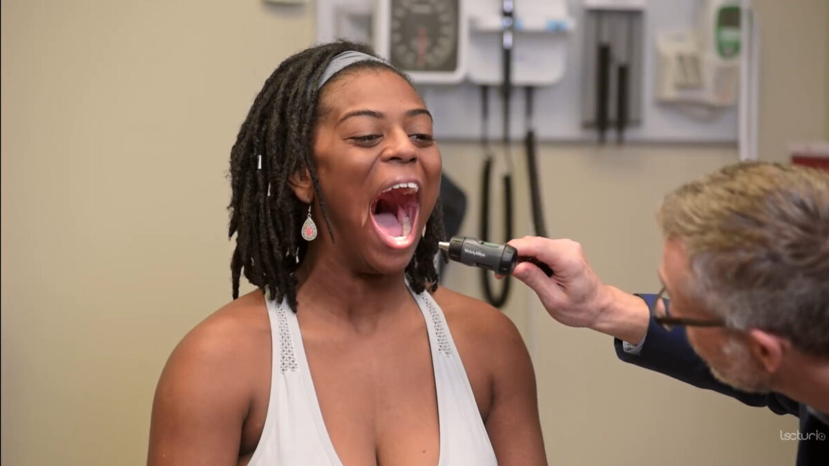
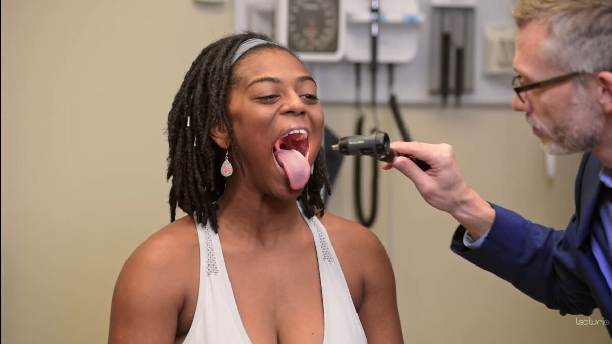
  • Oral cavity: buccal surface, palate Palate The palate is the structure that forms the roof of the mouth and floor of the nasal cavity. This structure is divided into soft and hard palates. Palate: Anatomy, and tongue Tongue The tongue, on the other hand, is a complex muscular structure that permits tasting and facilitates the process of mastication and communication. The blood supply of the tongue originates from the external carotid artery, and the innervation is through cranial nerves. Lips and Tongue: Anatomy 
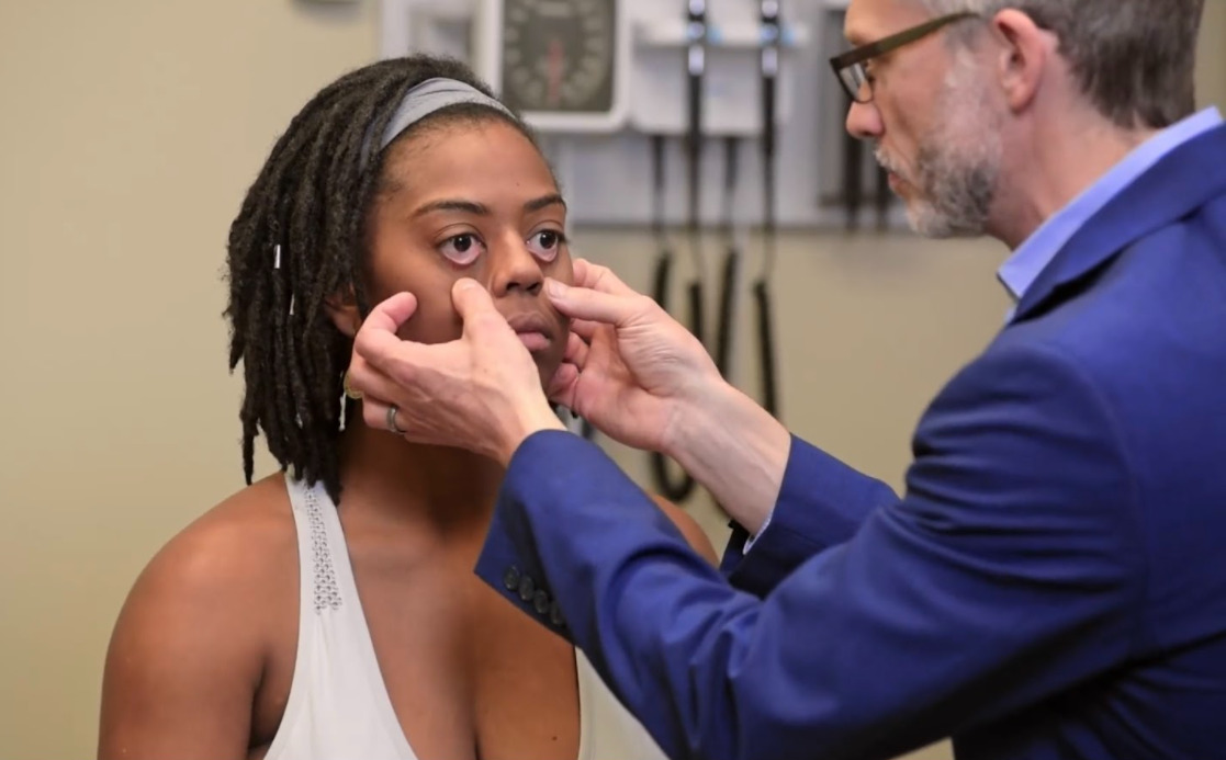
  • Conjunctiva Conjunctiva The mucous membrane that covers the posterior surface of the eyelids and the anterior pericorneal surface of the eyeball. Eye: Anatomy 
  • If sexually history is positive, examine the genital system.

Special tests

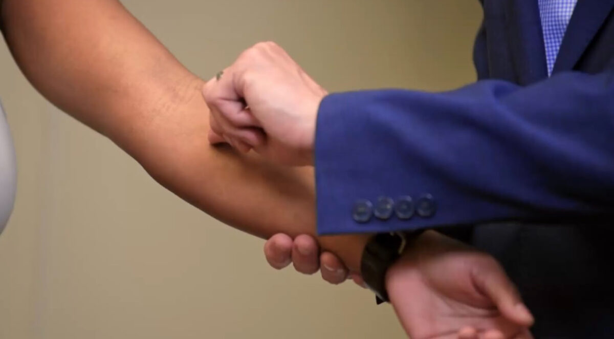
  • Nikolsky sign:
    • Performed by rubbing across the surface of the skin Skin The skin, also referred to as the integumentary system, is the largest organ of the body. The skin is primarily composed of the epidermis (outer layer) and dermis (deep layer). The epidermis is primarily composed of keratinocytes that undergo rapid turnover, while the dermis contains dense layers of connective tissue. Skin: Structure and Functions
    • If positive, the top thin layer of the skin Skin The skin, also referred to as the integumentary system, is the largest organ of the body. The skin is primarily composed of the epidermis (outer layer) and dermis (deep layer). The epidermis is primarily composed of keratinocytes that undergo rapid turnover, while the dermis contains dense layers of connective tissue. Skin: Structure and Functions shears off, leaving tender, pink  skin Skin The skin, also referred to as the integumentary system, is the largest organ of the body. The skin is primarily composed of the epidermis (outer layer) and dermis (deep layer). The epidermis is primarily composed of keratinocytes that undergo rapid turnover, while the dermis contains dense layers of connective tissue. Skin: Structure and Functions.
    • Positive sign indicates a blistering condition (e.g., pemphigus vulgaris Pemphigus vulgaris Bullous pemphigoid and pemphigus vulgaris are two different blistering autoimmune diseases. In pemphigus vulgaris, autoantibodies attack the desmosomal proteins, which connect the keratinocytes to one another. This attack results in a more severe, potentially fatal condition with fragile, flaccid blisters, usually with significant mucosal involvement. Bullous Pemphigoid and Pemphigus Vulgaris).
  • Dermographism:
    • Involves stroking the surface of the skin Skin The skin, also referred to as the integumentary system, is the largest organ of the body. The skin is primarily composed of the epidermis (outer layer) and dermis (deep layer). The epidermis is primarily composed of keratinocytes that undergo rapid turnover, while the dermis contains dense layers of connective tissue. Skin: Structure and Functions, resulting in hives Hives Urticaria is raised, well-circumscribed areas (wheals) of edema (swelling) and erythema (redness) involving the dermis and epidermis with associated pruritus (itch). Urticaria is not a single disease but rather is a reaction pattern representing cutaneous mast cell degranulation. Urticaria (Hives)
    • Often positive in conditions such as urticaria Urticaria Urticaria is raised, well-circumscribed areas (wheals) of edema (swelling) and erythema (redness) involving the dermis and epidermis with associated pruritus (itch). Urticaria is not a single disease but rather is a reaction pattern representing cutaneous mast cell degranulation. Urticaria (Hives)

Examination of Hair

Evaluate the hair carefully, looking for any changes from the individual’s baseline.

  • Texture
  • Color 
  • Quantity 
  • Distribution
  • Patches Patches Vitiligo/ skin Skin The skin, also referred to as the integumentary system, is the largest organ of the body. The skin is primarily composed of the epidermis (outer layer) and dermis (deep layer). The epidermis is primarily composed of keratinocytes that undergo rapid turnover, while the dermis contains dense layers of connective tissue. Skin: Structure and Functions lesions of the scalp
  • Hair loss
    • Diffuse 
    • Patchy 
    • Pattern thinning 
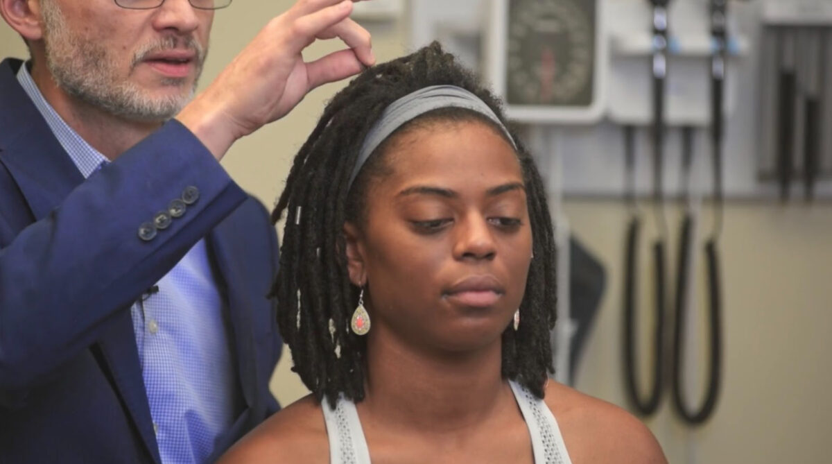
  • Hair-pull test
    • Done to demonstrate the severity of hair loss 
    • Grasp around 60 hair strands, gently tug; > 3–5 hair strands coming out indicates active hair loss.
Table: Common hair loss conditions
Disease Description
Alopecia areata Alopecia Areata Loss of scalp and body hair involving microscopically inflammatory patchy areas. Alopecia
Androgenetic alopecia Alopecia Alopecia is the loss of hair in areas anywhere on the body where hair normally grows. Alopecia may be defined as scarring or non-scarring, localized or diffuse, congenital or acquired, reversible or permanent, or confined to the scalp or universal; however, alopecia is usually classified using the 1st 3 factors. Alopecia
  • Male or female pattern baldness Baldness Alopecia is the loss of hair in areas anywhere on the body where hair normally grows. Alopecia may be defined as scarring or non-scarring, localized or diffuse, congenital or acquired, reversible or permanent, or confined to the scalp or universal; however, alopecia is usually classified using the 1st 3 factors. Alopecia
  • Due to a combination of genetic and hormonal factors
Telogen effluvium Telogen Effluvium Alopecia
  • Temporary hair loss due to excessive shedding of resting hairs after acute stressful events
  • Nonscarring form of diffuse hair loss

Examination of Nails

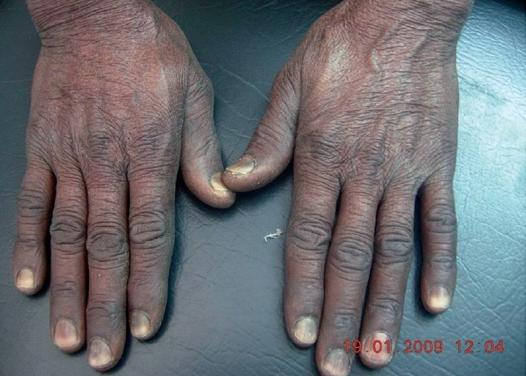
Inspect and palpate the nails carefully, looking for any changes.

  • Discoloration 
  • Symmetry 
  • Abnormal shape 
  • Abnormalities in the nail plate surface 
  • Complete loss of nails
  • Separation of the distal nail ( onycholysis Onycholysis Separation of nail plate from the underlying nail bed. It can be a sign of skin disease, infection (such as onychomycosis) or tissue injury. Psoriasis
  • Lesions around nails
Table: Common nail findings
Clinical findings Description Possible underlying etiology
Clubbing Clubbing Cardiovascular Examination Loss of angle between the nail fold and nail plate
  • Cardiac: chronic heart failure Heart Failure A heterogeneous condition in which the heart is unable to pump out sufficient blood to meet the metabolic need of the body. Heart failure can be caused by structural defects, functional abnormalities (ventricular dysfunction), or a sudden overload beyond its capacity. Chronic heart failure is more common than acute heart failure which results from sudden insult to cardiac function, such as myocardial infarction. Total Anomalous Pulmonary Venous Return (TAPVR)
  • Inflammatory bowel disease
  • Liver Liver The liver is the largest gland in the human body. The liver is found in the superior right quadrant of the abdomen and weighs approximately 1.5 kilograms. Its main functions are detoxification, metabolism, nutrient storage (e.g., iron and vitamins), synthesis of coagulation factors, formation of bile, filtration, and storage of blood. Liver: Anatomy diseases
  • Pulmonary fibrosis Fibrosis Any pathological condition where fibrous connective tissue invades any organ, usually as a consequence of inflammation or other injury. Bronchiolitis Obliterans
Onycholysis Onycholysis Separation of nail plate from the underlying nail bed. It can be a sign of skin disease, infection (such as onychomycosis) or tissue injury. Psoriasis Distal nail separation
  • Psoriasis Psoriasis Psoriasis is a common T-cell-mediated inflammatory skin condition. The etiology is unknown, but is thought to be due to genetic inheritance and environmental triggers. There are 4 major subtypes, with the most common form being chronic plaque psoriasis. Psoriasis
  • Fungal infection
  • Trauma
Nail pitting
  • Psoriasis Psoriasis Psoriasis is a common T-cell-mediated inflammatory skin condition. The etiology is unknown, but is thought to be due to genetic inheritance and environmental triggers. There are 4 major subtypes, with the most common form being chronic plaque psoriasis. Psoriasis
  • Eczema Eczema Atopic dermatitis, also known as eczema, is a chronic, relapsing, pruritic, inflammatory skin disease that occurs more frequently in children, although adults can also be affected. The condition is often associated with elevated serum levels of IgE and a personal or family history of atopy. Skin dryness, erythema, oozing, crusting, and lichenification are present. Atopic Dermatitis (Eczema)
Koilonychia Koilonychia Iron Deficiency Anemia Spoon-shaped depression Iron Iron A metallic element with atomic symbol fe, atomic number 26, and atomic weight 55. 85. It is an essential constituent of hemoglobins; cytochromes; and iron-binding proteins. It plays a role in cellular redox reactions and in the transport of oxygen. Trace Elements deficiency anemia Anemia Anemia is a condition in which individuals have low Hb levels, which can arise from various causes. Anemia is accompanied by a reduced number of RBCs and may manifest with fatigue, shortness of breath, pallor, and weakness. Subtypes are classified by the size of RBCs, chronicity, and etiology. Anemia: Overview and Types
Mees lines Mees Lines Metal Poisoning (Lead, Arsenic, Iron) White streaks
  • Heavy-metal poisoning
  • Renal failure Renal failure Conditions in which the kidneys perform below the normal level in the ability to remove wastes, concentrate urine, and maintain electrolyte balance; blood pressure; and calcium metabolism. Renal insufficiency can be classified by the degree of kidney damage (as measured by the level of proteinuria) and reduction in glomerular filtration rate. Crush Syndrome
  • Chemotherapy Chemotherapy Osteosarcoma
Brittle nails Hypothyroidism Hypothyroidism Hypothyroidism is a condition characterized by a deficiency of thyroid hormones. Iodine deficiency is the most common cause worldwide, but Hashimoto’s disease (autoimmune thyroiditis) is the leading cause in non-iodine-deficient regions. Hypothyroidism
Yellow nail syndrome
Terry nails Proximal red/brown band on the nail plate
  • Heart failure Heart Failure A heterogeneous condition in which the heart is unable to pump out sufficient blood to meet the metabolic need of the body. Heart failure can be caused by structural defects, functional abnormalities (ventricular dysfunction), or a sudden overload beyond its capacity. Chronic heart failure is more common than acute heart failure which results from sudden insult to cardiac function, such as myocardial infarction. Total Anomalous Pulmonary Venous Return (TAPVR)
  • Liver Liver The liver is the largest gland in the human body. The liver is found in the superior right quadrant of the abdomen and weighs approximately 1.5 kilograms. Its main functions are detoxification, metabolism, nutrient storage (e.g., iron and vitamins), synthesis of coagulation factors, formation of bile, filtration, and storage of blood. Liver: Anatomy disease
Half-and-half nails Kidney disease
Muehrcke lines Double white lines Hypoalbuminemia Hypoalbuminemia A condition in which albumin level in blood (serum albumin) is below the normal range. Hypoalbuminemia may be due to decreased hepatic albumin synthesis, increased albumin catabolism, altered albumin distribution, or albumin loss through the urine (albuminuria). Nephrotic Syndrome in Children

Clinical Relevance

Table: Common dermatologic conditions
Psoriasis Psoriasis Psoriasis is a common T-cell-mediated inflammatory skin condition. The etiology is unknown, but is thought to be due to genetic inheritance and environmental triggers. There are 4 major subtypes, with the most common form being chronic plaque psoriasis. Psoriasis Atopic dermatitis Dermatitis Any inflammation of the skin. Atopic Dermatitis (Eczema) Acne vulgaris Acne vulgaris Acne vulgaris, also known as acne, is a common disorder of the pilosebaceous units in adolescents and young adults. The condition occurs due to follicular hyperkeratinization, excess sebum production, follicular colonization by Cutibacterium acnes, and inflammation. Acne Vulgaris Fungal infections Infections Invasion of the host organism by microorganisms or their toxins or by parasites that can cause pathological conditions or diseases. Chronic Granulomatous Disease
Appearance
  • Salmon-colored plaques
  • Thick, silvery scales Scales Dry or greasy masses of keratin that represent thickened stratum corneum. Secondary Skin Lesions
  • Pruritic
  • Lichenified (thickening of skin Skin The skin, also referred to as the integumentary system, is the largest organ of the body. The skin is primarily composed of the epidermis (outer layer) and dermis (deep layer). The epidermis is primarily composed of keratinocytes that undergo rapid turnover, while the dermis contains dense layers of connective tissue. Skin: Structure and Functions)
  • Pink scaly plaques
  • Intensely pruritic
  • Pruritic
  • Erythematous plaques
  • Leading edge of scale
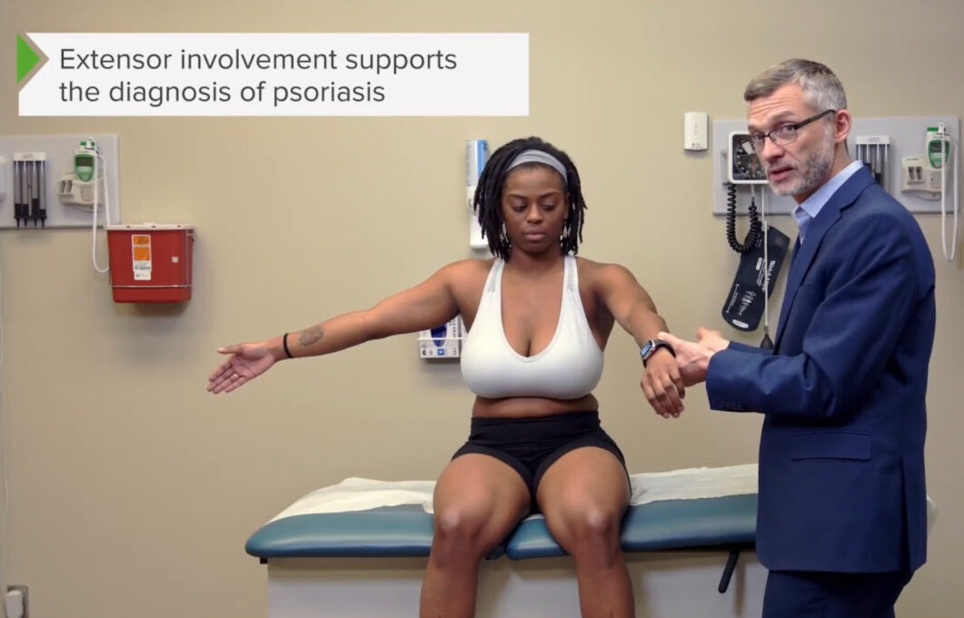
Distribution Symmetrical, extensor surfaces Flexor surfaces Face, neck Neck The part of a human or animal body connecting the head to the rest of the body. Peritonsillar Abscess, chest, and back
Associated findings Auspitz sign Auspitz Sign Psoriasis: pinpoint bleeding with removal of scale History of atopy Atopy Atopic Dermatitis (Eczema) ( asthma Asthma Asthma is a chronic inflammatory respiratory condition characterized by bronchial hyperresponsiveness and airflow obstruction. The disease is believed to result from the complex interaction of host and environmental factors that increase disease predisposition, with inflammation causing symptoms and structural changes. Patients typically present with wheezing, cough, and dyspnea. Asthma and seasonal allergies Allergies A medical specialty concerned with the hypersensitivity of the individual to foreign substances and protection from the resultant infection or disorder. Selective IgA Deficiency) Scarring Scarring Inflammation/ hyperpigmentation Hyperpigmentation Excessive pigmentation of the skin, usually as a result of increased epidermal or dermal melanin pigmentation, hypermelanosis. Hyperpigmentation can be localized or generalized. The condition may arise from exposure to light, chemicals or other substances, or from a primary metabolic imbalance. Malassezia Fungi Confirmed by KOH prep KOH prep Dermatophytes/Tinea Infections

References

  1. Cole, J.M., Gray-Miceli, D. (2002). Necessary elements of a dermatologic history and physical evaluation. Dermatology Nursing. 14(6). Retrieved October 23, 2021, from https://www.medscape.com/viewarticle/447729
  2. DermNet NZ. (2008). Principles of dermatological practice—examination of the skin. Retrieved October 23, 2021, from https://www.dermnetnz.org/cme/principles/examination-of-the-skin/
  3. Armstrong, C.A. (2021). Approach to the clinical dermatologic diagnosis. UpToDate. Retrieved October 23, 2021, from https://www.uptodate.com/contents/approach-to-the-clinical-dermatologic-diagnosis
  4. Stanford Medicine. (n.a.) The hand examination. Retrieved October 23, 2021, from https://stanfordmedicine25.stanford.edu/the25/hand.html

Create your free account or log in to continue reading!

Sign up now and get free access to Lecturio with concept pages, medical videos, and questions for your medical education.

User Reviews

Details