Advertisement

Advertisement

Advertisement

Advertisement

Decompression Sickness

Decompression sickness (DCS), known informally as “the bends,” is a condition caused by compression Compression Blunt Chest Trauma and decompression of gases contained in the body during descent and rapid ascent while diving. Clinical presentation of DCS may be nonspecific and variable Variable Variables represent information about something that can change. The design of the measurement scales, or of the methods for obtaining information, will determine the data gathered and the characteristics of that data. As a result, a variable can be qualitative or quantitative, and may be further classified into subgroups. Types of Variables, with a time of onset that can vary from immediately to 12 hours after surfacing. Diagnosis is made clinically. Management is early supportive therapy and hyperbaric recompression treatment carried out in a specialized facility.

Last updated: Jan 24, 2023

Editorial responsibility: Stanley Oiseth, Lindsay Jones, Evelin Maza

Advertisement

Advertisement

Advertisement

Advertisement

Advertisement

Advertisement

Overview

Epidemiology

  • Population statistics:
    • Sports diving: 3 cases per 10,000 dives
    • Commercial diving: 1.5–10 cases per 10,000 dives
    • 2.5 times more likely in men than women 
  • Risk factors:
    • Dive details:
      • Depth
      • Duration
      • Number of dives
      • Interval surface time between dives
      • Water conditions
    • Diver:
      • Weight ( obesity Obesity Obesity is a condition associated with excess body weight, specifically with the deposition of excessive adipose tissue. Obesity is considered a global epidemic. Major influences come from the western diet and sedentary lifestyles, but the exact mechanisms likely include a mixture of genetic and environmental factors. Obesity is predisposing)
      • History of lung or cardiac disease
      • Right-to-left cardiac shunts (e.g., patent foramen ovale Foramen ovale An opening in the wall between the right and the left upper chambers (heart atria) of a fetal heart. Oval foramen normally closes soon after birth; when it fails to close the condition is called patent oval foramen. Patent Foramen Ovale)
      • Inexperienced divers are more commonly affected.
      • Rate of ascent
      • Length of time between low altitude (scuba dive) and high altitude (air travel or ground ascent)

Etiology

Decompression sickness (DCS) comprises varied symptoms caused by gas bubbles that come out of solution in the body after ascending from a deep dive.

  • Most likely to occur when:
    • Scuba diving is followed closely by travel to high altitudes 
    • Divers do not adhere to dive protocols 
  • Panic can make divers ascend too quickly.

Classification

Based on severity of symptoms and location of gas bubbles:
  • Type I: mild bubbles form in:
    • Lymphatic system Lymphatic system A system of organs and tissues that process and transport immune cells and lymph. Primary Lymphatic Organs 
    • Skin Skin The skin, also referred to as the integumentary system, is the largest organ of the body. The skin is primarily composed of the epidermis (outer layer) and dermis (deep layer). The epidermis is primarily composed of keratinocytes that undergo rapid turnover, while the dermis contains dense layers of connective tissue. Skin: Structure and Functions
    • Muscles and joints
  • Type II: severe/lethal bubbles form in:

Pathophysiology

  • Descent: gas in body under higher atmospheric pressure Atmospheric pressure The pressure at any point in an atmosphere due solely to the weight of the atmospheric gases above the point concerned. Ventilation: Mechanics of Breathing → dissolves into tissues and blood
  • Rapid ascent: partial pressure Partial pressure The pressure that would be exerted by one component of a mixture of gases if it were present alone in a container. Gas Exchange of gas > ambient pressure → gas comes out of solution → formation of bubbles
  • Bubbles affect:
    • Organ tissue
      • Mechanical disruption/damage to tissue 
      • Alteration of the functionality of important structures
    • Venous circulation Circulation The movement of the blood as it is pumped through the cardiovascular system. ABCDE Assessment → picks gas up from tissues
      • In ↓ quantities → bubbles are asymptomatic and filtered in pulmonary capillaries Capillaries Capillaries are the primary structures in the circulatory system that allow the exchange of gas, nutrients, and other materials between the blood and the extracellular fluid (ECF). Capillaries are the smallest of the blood vessels. Because a capillary diameter is so small, only 1 RBC may pass through at a time. Capillaries: Histology
      • In ↑ quantities, bubbles can cause:
        • Inflammatory response
        • Activation of coagulation cascades
        • Damage to endothelium Endothelium A layer of epithelium that lines the heart, blood vessels (vascular endothelium), lymph vessels (lymphatic endothelium), and the serous cavities of the body. Arteries: Histology
        • Activation of platelet aggregation Platelet aggregation The attachment of platelets to one another. This clumping together can be induced by a number of agents (e.g., thrombin; collagen) and is part of the mechanism leading to the formation of a thrombus. Hemostasis
        • Occlusion of blood flow Blood flow Blood flow refers to the movement of a certain volume of blood through the vasculature over a given unit of time (e.g., mL per minute). Vascular Resistance, Flow, and Mean Arterial Pressure
        • Capillary leakage
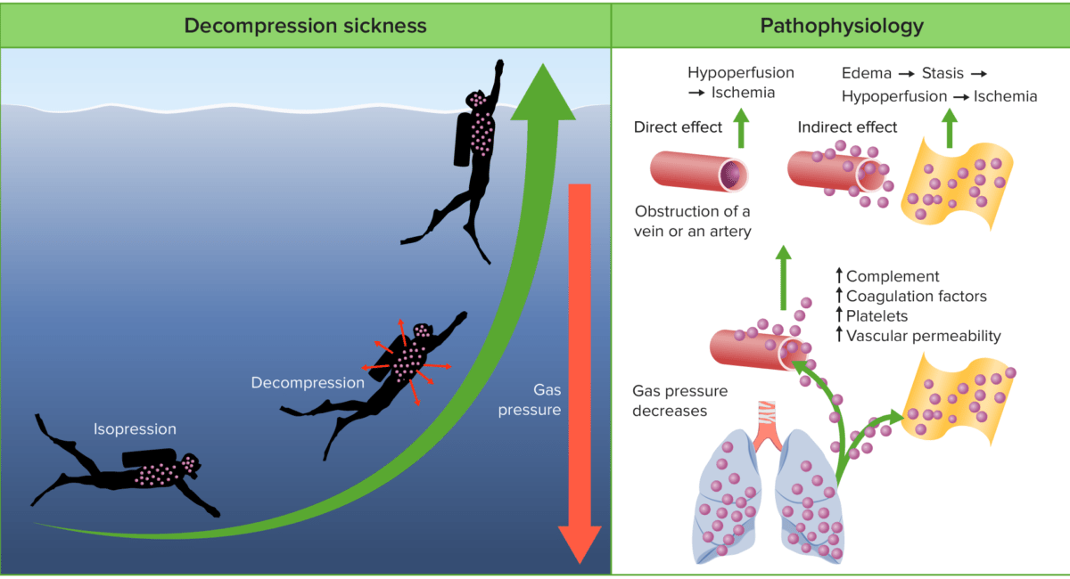
Decompression sickness (dcs)

Pathophysiology of decompression sickness:
During descent, the increased pressure results in gases dissolving into the body’s tissues and blood. During decompression, the tissues become supersaturated with these gases, resulting in the formation of free gas. These bubbles of gas can be filtered in the pulmonary capillaries in small quantities. However, in large quantities, the gases can cause direct vascular obstruction, the activation of platelets and coagulation cascades, capillary leakage, and inflammatory responses.

Image by Lecturio.

Clinical Presentation

  • Nonspecific, depending on:
    • Location of gas bubbles
    • Compressibility of gases in body
  • Quicker onset of symptoms = ↑ severity
    • Severe cases start < 30 minutes after surfacing.
    • Mild symptoms can take 6 hours to appear.
    • 75% of cases show symptoms in < 1 hour.
  • Most cases are mild.
  • Pulmonary/cardiovascular manifestations can be lethal.
Table: Clinical manifestations of DCS
Neurologic: cerebral
  • Confusion
  • Visual and speech disturbances
Neurologic: spinal
Neurologic: vestibulocochlear
  • Labyrinthine decompression illness (“the staggers”): central vertigo Central Vertigo Vertigo
  • Hearing loss Hearing loss Hearing loss, also known as hearing impairment, is any degree of impairment in the ability to apprehend sound as determined by audiometry to be below normal hearing thresholds. Clinical presentation may occur at birth or as a gradual loss of hearing with age, including a short-term or sudden loss at any point. Hearing Loss
  • Nausea Nausea An unpleasant sensation in the stomach usually accompanied by the urge to vomit. Common causes are early pregnancy, sea and motion sickness, emotional stress, intense pain, food poisoning, and various enteroviruses. Antiemetics and vomiting Vomiting The forcible expulsion of the contents of the stomach through the mouth. Hypokalemia
Neurologic: peripheral Patchy nondermatomal sensory Sensory Neurons which conduct nerve impulses to the central nervous system. Nervous System: Histology disturbance
Musculoskeletal Joint pain Pain An unpleasant sensation induced by noxious stimuli which are detected by nerve endings of nociceptive neurons. Pain: Types and Pathways (“the bends”)
Ocular
  • Uveitis Uveitis Uveitis is the inflammation of the uvea, the pigmented middle layer of the eye, which comprises the iris, ciliary body, and choroid. The condition is categorized based on the site of disease; anterior uveitis is the most common. Diseases of the Uvea
  • Conjunctivitis Conjunctivitis Conjunctivitis is a common inflammation of the bulbar and/or palpebral conjunctiva. It can be classified into infectious (mostly viral) and noninfectious conjunctivitis, which includes allergic causes. Patients commonly present with red eyes, increased tearing, burning, foreign body sensation, and photophobia. Conjunctivitis
Pulmonary
  • Cough
  • Dyspnea Dyspnea Dyspnea is the subjective sensation of breathing discomfort. Dyspnea is a normal manifestation of heavy physical or psychological exertion, but also may be caused by underlying conditions (both pulmonary and extrapulmonary). Dyspnea
Cardiovascular
  • Hemoconcentration Hemoconcentration Neonatal Polycythemia
  • Coagulopathy
  • Hypotension Hypotension Hypotension is defined as low blood pressure, specifically < 90/60 mm Hg, and is most commonly a physiologic response. Hypotension may be mild, serious, or life threatening, depending on the cause. Hypotension
  • Acute coronary syndrome
Cutaneous
Lymphatic Soft tissue Soft Tissue Soft Tissue Abscess edema Edema Edema is a condition in which excess serous fluid accumulates in the body cavity or interstitial space of connective tissues. Edema is a symptom observed in several medical conditions. It can be categorized into 2 types, namely, peripheral (in the extremities) and internal (in an organ or body cavity). Edema
Constitutional Fatigue Fatigue The state of weariness following a period of exertion, mental or physical, characterized by a decreased capacity for work and reduced efficiency to respond to stimuli. Fibromyalgia and malaise Malaise Tick-borne Encephalitis Virus

Diagnosis

Diagnosis is based on: 

  • History: relationship of symptoms to a diving event
    • DCS should be considered in any diver manifesting symptoms that cannot be explained by other mechanisms.
  • Imaging:
    • Computed tomography (CT) scan can be used to detect vacuum phenomenon of trapped gas.
    • Magnetic resonance imaging (MRI): most accurate for detection of brain Brain The part of central nervous system that is contained within the skull (cranium). Arising from the neural tube, the embryonic brain is comprised of three major parts including prosencephalon (the forebrain); mesencephalon (the midbrain); and rhombencephalon (the hindbrain). The developed brain consists of cerebrum; cerebellum; and other structures in the brain stem. Nervous System: Anatomy, Structure, and Classification and spinal cord Spinal cord The spinal cord is the major conduction pathway connecting the brain to the body; it is part of the CNS. In cross section, the spinal cord is divided into an H-shaped area of gray matter (consisting of synapsing neuronal cell bodies) and a surrounding area of white matter (consisting of ascending and descending tracts of myelinated axons). Spinal Cord: Anatomy lesions
Decompression sickness diagnostic imaging

A 61‐year‐old experienced male diver presenting with a diagnosis of DCS:
(A) MRI of the head showing multiple cerebral thromboembolisms.
(B) CT scan of the chest 6 hours after the first symptoms showing multiple pulmonary thromboembolisms of the segmental arteries.
Follow‐up CT scans of the chest 9 hours later: no pulmonary thromboembolism of the same segmental arteries.

Image: “Decompression illness” by Sebastian Klapa et al. License: CC BY 4.0

Management and Prognosis

The main goal of therapy is to dissolve bubbles and recompress gas in body fluids.

First aid

  • Trendelenburg positioning → puts right ventricular outflow tract below right ventricular cavity → air migrates up and out of way of flow Flow Blood flows through the heart, arteries, capillaries, and veins in a closed, continuous circuit. Flow is the movement of volume per unit of time. Flow is affected by the pressure gradient and the resistance fluid encounters between 2 points. Vascular resistance is the opposition to flow, which is caused primarily by blood friction against vessel walls. Vascular Resistance, Flow, and Mean Arterial Pressure of blood
  • Intravenous (IV) fluids
  • Supplementary oxygen → accelerates inert gas washout

Definitive therapy

  • Recompression therapy in hyperbaric treatment facility
    • Hyperbaric oxygen
    • Should never be withheld, even if initiation is delayed 
  • IV fluids IV fluids Intravenous fluids are one of the most common interventions administered in medicine to approximate physiologic bodily fluids. Intravenous fluids are divided into 2 categories: crystalloid and colloid solutions. Intravenous fluids have a wide variety of indications, including intravascular volume expansion, electrolyte manipulation, and maintenance fluids. Intravenous Fluids
  • A consult with diving medicine/hyperbaric oxygen specialist is required, even if symptoms resolve.

Prevention

  • Diver education
  • Pre-dive medical screening Screening Preoperative Care and dive planning
  • Strict adherence to dive course, timing, and depths
  • Slow and controlled ascent (decompression stops: Experienced divers control their ascent using algorithms that indicate when ascent has to stop at different depths to allow for gas washout.)
  • Recommendation to avoid high altitudes for 24 hours after a dive

Contraindications Contraindications A condition or factor associated with a recipient that makes the use of a drug, procedure, or physical agent improper or inadvisable. Contraindications may be absolute (life threatening) or relative (higher risk of complications in which benefits may outweigh risks). Noninvasive Ventilation to deep-sea diving

  • Active asthma Asthma Asthma is a chronic inflammatory respiratory condition characterized by bronchial hyperresponsiveness and airflow obstruction. The disease is believed to result from the complex interaction of host and environmental factors that increase disease predisposition, with inflammation causing symptoms and structural changes. Patients typically present with wheezing, cough, and dyspnea. Asthma
  • Reduced pulmonary function
  • Lung cysts Cysts Any fluid-filled closed cavity or sac that is lined by an epithelium. Cysts can be of normal, abnormal, non-neoplastic, or neoplastic tissues. Fibrocystic Change
  • Recent thoracic trauma or pneumothorax Pneumothorax A pneumothorax is a life-threatening condition in which air collects in the pleural space, causing partial or full collapse of the lung. A pneumothorax can be traumatic or spontaneous. Patients present with a sudden onset of sharp chest pain, dyspnea, and diminished breath sounds on exam. Pneumothorax
  • Cardiovascular disease
  • History of bowel obstruction Bowel obstruction Any impairment, arrest, or reversal of the normal flow of intestinal contents toward the anal canal. Ascaris/Ascariasis
  • Recent brain Brain The part of central nervous system that is contained within the skull (cranium). Arising from the neural tube, the embryonic brain is comprised of three major parts including prosencephalon (the forebrain); mesencephalon (the midbrain); and rhombencephalon (the hindbrain). The developed brain consists of cerebrum; cerebellum; and other structures in the brain stem. Nervous System: Anatomy, Structure, and Classification or eye surgery
  • Seizures Seizures A seizure is abnormal electrical activity of the neurons in the cerebral cortex that can manifest in numerous ways depending on the region of the brain affected. Seizures consist of a sudden imbalance that occurs between the excitatory and inhibitory signals in cortical neurons, creating a net excitation. The 2 major classes of seizures are focal and generalized. Seizures
  • Diabetes Diabetes Diabetes mellitus (DM) is a metabolic disease characterized by hyperglycemia and dysfunction of the regulation of glucose metabolism by insulin. Type 1 DM is diagnosed mostly in children and young adults as the result of autoimmune destruction of β cells in the pancreas and the resulting lack of insulin. Type 2 DM has a significant association with obesity and is characterized by insulin resistance. Diabetes Mellitus mellitus and hypoglycemic episodes
  • History of syncope Syncope Syncope is a short-term loss of consciousness and loss of postural stability followed by spontaneous return of consciousness to the previous neurologic baseline without the need for resuscitation. The condition is caused by transient interruption of cerebral blood flow that may be benign or related to a underlying life-threatening condition. Syncope

Prognosis Prognosis A prediction of the probable outcome of a disease based on a individual’s condition and the usual course of the disease as seen in similar situations. Non-Hodgkin Lymphomas

  • 75% of cases completely resolve.
  • 16% of cases may have residual symptoms for up to 3 months.
  • Spinal cord Spinal cord The spinal cord is the major conduction pathway connecting the brain to the body; it is part of the CNS. In cross section, the spinal cord is divided into an H-shaped area of gray matter (consisting of synapsing neuronal cell bodies) and a surrounding area of white matter (consisting of ascending and descending tracts of myelinated axons). Spinal Cord: Anatomy involvement often causes permanent damage.

Clinical Relevance

The following conditions greatly increase the likelihood of developing decompression sickness:

  • Asthma Asthma Asthma is a chronic inflammatory respiratory condition characterized by bronchial hyperresponsiveness and airflow obstruction. The disease is believed to result from the complex interaction of host and environmental factors that increase disease predisposition, with inflammation causing symptoms and structural changes. Patients typically present with wheezing, cough, and dyspnea. Asthma: a chronic inflammatory condition of the airways characterized by bronchial hyperreactivity Bronchial hyperreactivity Tendency of the smooth muscle of the tracheobronchial tree to contract more intensely in response to a given stimulus than it does in the response seen in normal individuals. This condition is present in virtually all symptomatic patients with asthma. The most prominent manifestation of this smooth muscle contraction is a decrease in airway caliber that can be readily measured in the pulmonary function laboratory. Asthma, which presents as wheezing Wheezing Wheezing is an abnormal breath sound characterized by a whistling noise that can be relatively high-pitched and shrill (more common) or coarse. Wheezing is produced by the movement of air through narrowed or compressed small (intrathoracic) airways. Wheezing, cough, and dyspnea Dyspnea Dyspnea is the subjective sensation of breathing discomfort. Dyspnea is a normal manifestation of heavy physical or psychological exertion, but also may be caused by underlying conditions (both pulmonary and extrapulmonary). Dyspnea. People with asthma Asthma Asthma is a chronic inflammatory respiratory condition characterized by bronchial hyperresponsiveness and airflow obstruction. The disease is believed to result from the complex interaction of host and environmental factors that increase disease predisposition, with inflammation causing symptoms and structural changes. Patients typically present with wheezing, cough, and dyspnea. Asthma have increased risk for pulmonary barotrauma Barotrauma Injury following pressure changes; includes injury to the eustachian tube, ear drum, lung and stomach. Invasive Mechanical Ventilation and decompression sickness, which can lead to a pneumothorax Pneumothorax A pneumothorax is a life-threatening condition in which air collects in the pleural space, causing partial or full collapse of the lung. A pneumothorax can be traumatic or spontaneous. Patients present with a sudden onset of sharp chest pain, dyspnea, and diminished breath sounds on exam. Pneumothorax and breathing difficulties. Treatment often requires intubation Intubation Peritonsillar Abscess
  • Pneumothorax Pneumothorax A pneumothorax is a life-threatening condition in which air collects in the pleural space, causing partial or full collapse of the lung. A pneumothorax can be traumatic or spontaneous. Patients present with a sudden onset of sharp chest pain, dyspnea, and diminished breath sounds on exam. Pneumothorax: a collection of air in the pleural space Pleural space The thin serous membrane enveloping the lungs (lung) and lining the thoracic cavity. Pleura consist of two layers, the inner visceral pleura lying next to the pulmonary parenchyma and the outer parietal pleura. Between the two layers is the pleural cavity which contains a thin film of liquid. Pleuritis that can occur due to dysbarism, causing shortness of breath Shortness of breath Dyspnea is the subjective sensation of breathing discomfort. Dyspnea is a normal manifestation of heavy physical or psychological exertion, but also may be caused by underlying conditions (both pulmonary and extrapulmonary). Dyspnea and hypoxia Hypoxia Sub-optimal oxygen levels in the ambient air of living organisms. Ischemic Cell Damage. Treatment is with thoracostomy Thoracostomy Surgical procedure involving the creation of an opening (stoma) into the chest cavity for drainage; used in the treatment of pleural effusion; pneumothorax; hemothorax; and empyema. Hemothorax (chest tube) placement and oxygen.
  • Bowel obstruction Bowel obstruction Any impairment, arrest, or reversal of the normal flow of intestinal contents toward the anal canal. Ascaris/Ascariasis: the interruption of the normal transit of intestinal contents either due to a functional decrease in peristalsis Peristalsis A movement, caused by sequential muscle contraction, that pushes the contents of the intestines or other tubular organs in one direction. Gastrointestinal Motility or mechanical obstruction Mechanical Obstruction Any impairment, arrest, or reversal of the normal flow of intestinal contents toward the anal canal. Imaging of the Intestines. Over-pressurization of the bowels can result in gastric rupture, bowel perforation Bowel perforation Perforated viscus or GI perforation represents a condition in which the integrity of the GI wall is lost with subsequent leakage of enteric contents into the peritoneal cavity, resulting in peritonitis. The causes of perforated viscus include trauma, bowel ischemia, infections, or ulcerative conditions, all of which ultimately lead to a full-thickness disruption of the intestinal wall. Perforated Viscus, or pneumoperitoneum Pneumoperitoneum A condition with trapped gas or air in the peritoneal cavity, usually secondary to perforation of the internal organs such as the lung and the gastrointestinal tract, or to recent surgery. Pneumoperitoneum may be purposely introduced to aid radiological examination. Perforated Viscus. Treatment often requires surgery.

References

  1. Chandy, D. and Weinhouse, G. Complications of SCUBA diving. (2019). UpToDate. Accessed November 13, 2020 from: https://www.uptodate.com/contents/complications-of-scuba-diving
  2. Nemer, J. A., & Juarez, M. A. (2020). Dysbarism & decompression sickness. Current medical diagnosis and treatment (2020). New York, NY: McGraw-Hill Education. accessmedicine.mhmedical.com/content.aspx?aid=1166176018
  3. Pollock NW, Buteau D. Updates in Decompression Illness. Emerg Med Clin North Am. 2017 May;35(2):301-319. doi: 10.1016/j.emc.2016.12.002. Epub 2017 Mar 15. PMID: 28411929.
  4. Bennett, M. H., & Mitchell, S. J. (2018). Hyperbaric and diving medicine. In J. L. Jameson, Harrison’s principles of internal medicine, 20e. New York, NY: McGraw-Hill Education. http://accessmedicine.mhmedical.com/content.aspx?aid=1164035738
  5. Nemer, J. A., & Juarez, M. A. (2020). Dysbarism & decompression sickness. Current medical diagnosis and treatment (2020). New York, NY: McGraw-Hill Education. https://accessmedicine.mhmedical.com/content.aspx?bookid=2683&sectionid=225057753

Create your free account or log in to continue reading!

Sign up now and get free access to Lecturio with concept pages, medical videos, and questions for your medical education.

User Reviews

Details