Cryptorchidism

Cryptorchidism is one of the most common congenital anomalies in young boys. Typically, this asymptomatic condition presents during a routine well-child examination where 1 or both testicles Testicles The testicles, also known as the testes or the male gonads, are a pair of egg-shaped glands suspended within the scrotum. The testicles have multiple layers: an outer tunica vaginalis, an intermediate tunica albuginea, and an innermost tunica vasculosa. The testicles are composed of testicular lobules and seminiferous tubules. Testicles: Anatomy are not palpable in the scrotum Scrotum A cutaneous pouch of skin containing the testicles and spermatic cords. Testicles: Anatomy. Diagnosis depends on physical exam and ultrasound is performed only when other disorders are suspected. Treatment is dependent on patient age and testis position. In boys with persistent undescended testis by age 6 months, the condition is unlikely to spontaneously resolve. If the testis is palpable but not in the dependent intrascrotal location, an orchiopexy (bringing the testicle to the scrotum Scrotum A cutaneous pouch of skin containing the testicles and spermatic cords. Testicles: Anatomy) is preferred. If the testis is non-palpable, a diagnostic laparoscopy Diagnostic Laparoscopy Laparotomy and Laparoscopy is needed to inspect for an intra-abdominal testis, which requires a staged Fowler-Stephen’s orchiopexy.

Last updated: Nov 18, 2024

Editorial responsibility: Stanley Oiseth, Lindsay Jones, Evelin Maza

Overview

Definition

Cryptorchidism is the absence of 1 or both testicles Testicles The testicles, also known as the testes or the male gonads, are a pair of egg-shaped glands suspended within the scrotum. The testicles have multiple layers: an outer tunica vaginalis, an intermediate tunica albuginea, and an innermost tunica vasculosa. The testicles are composed of testicular lobules and seminiferous tubules. Testicles: Anatomy in the scrotum Scrotum A cutaneous pouch of skin containing the testicles and spermatic cords. Testicles: Anatomy. Cryptorchid testis (“hidden testis”) may be undescended or absent (atrophied).

Epidemiology

  • Prevalence Prevalence The total number of cases of a given disease in a specified population at a designated time. It is differentiated from incidence, which refers to the number of new cases in the population at a given time. Measures of Disease Frequency at birth about 3% overall:
  • Risk factors:
    • Maternal smoking Smoking Willful or deliberate act of inhaling and exhaling smoke from burning substances or agents held by hand. Interstitial Lung Diseases and alcohol exposure 
    • Fetal exposure to paracetamol paracetamol Acetaminophen is an over-the-counter nonopioid analgesic and antipyretic medication and the most commonly used analgesic worldwide. Despite the widespread use of acetaminophen, its mechanism of action is not entirely understood. Acetaminophen/ acetaminophen Acetaminophen Acetaminophen is an over-the-counter nonopioid analgesic and antipyretic medication and the most commonly used analgesic worldwide. Despite the widespread use of acetaminophen, its mechanism of action is not entirely understood. Acetaminophen and diethylstilbestrol Diethylstilbestrol A synthetic nonsteroidal estrogen used in the treatment of menopausal and postmenopausal disorders. It was also used formerly as a growth promoter in animals. According to the fourth annual report on carcinogens, diethylstilbestrol has been listed as a known carcinogen. Noncontraceptive Estrogen and Progestins  
    • Prematurity Prematurity Neonatal Respiratory Distress Syndrome 
    • Small for gestational age Gestational age The age of the conceptus, beginning from the time of fertilization. In clinical obstetrics, the gestational age is often estimated as the time from the last day of the last menstruation which is about 2 weeks before ovulation and fertilization. Pregnancy: Diagnosis, Physiology, and Care and low birth weight
  • Cryptorchidism is usually an isolated condition, but there are related disorders:
    • Disorders of sexual differentiation (DSD) 
    • Congenital adrenal hyperplasia Hyperplasia An increase in the number of cells in a tissue or organ without tumor formation. It differs from hypertrophy, which is an increase in bulk without an increase in the number of cells. Cellular Adaptation ( CAH CAH Congenital adrenal hyperplasia (CAH) consists of a group of autosomal recessive disorders that cause a deficiency of an enzyme needed in cortisol, aldosterone, and androgen synthesis. The most common subform of cah is 21-hydroxylase deficiency, followed by 11β-hydroxylase deficiency. Congenital Adrenal Hyperplasia
    • Genetic disorders such as Klinefelter syndrome Klinefelter syndrome Klinefelter syndrome is a chromosomal aneuploidy characterized by the presence of 1 or more extra X chromosomes in a male karyotype, most commonly leading to karyotype 47,XXY. Klinefelter syndrome is associated with decreased levels of testosterone and is the most common cause of congenital hypogonadism. Klinefelter Syndrome and Prader-Willi syndrome Prader-Willi syndrome Prader-Willi syndrome (PWS) is a rare autosomal neurodevelopmental genetic disorders mapped to a specific region of chromosome 15 attributed to genomic imprinting. A paternally derived chromosome 15 with this deletion results in 15q11-13 paternal deletion syndrome, or PWS. Prader-Willi Syndrome and Angelman Syndrome

Pathophysiology

Pathogenesis overall is not well understood but is affected by hormonal and anatomical embryologic factors.

Normal mechanical descent:

  • Intra-abdominal phase: mediated by the hormone descendin
  • Inguinal phase (by the 28th week of gestation): mediated by androgen/ testosterone Testosterone A potent androgenic steroid and major product secreted by the leydig cells of the testis. Its production is stimulated by luteinizing hormone from the pituitary gland. In turn, testosterone exerts feedback control of the pituitary LH and FSH secretion. Depending on the tissues, testosterone can be further converted to dihydrotestosterone or estradiol. Androgens and Antiandrogens 
  • Gubernaculum Gubernaculum An embryonic structure that helps guide proper descent of gonads into their final positions. It attaches the caudal end of the fetal gonads to the developing scrotum in male and the labium majorum in female. It gives rise to the caudal ligaments of the gonad: the scrotal ligament in male and the uterine round and proper ovarian ligaments in female. It includes morphofunctional equivalent structures in non-mammals. Congenital Malformations of the Female Reproductive System swells and pulls the testis down into the scrotum Scrotum A cutaneous pouch of skin containing the testicles and spermatic cords. Testicles: Anatomy along the inguinal canal Inguinal canal The tunnel in the lower anterior abdominal wall through which the spermatic cord, in the male; round ligament, in the female; nerves; and vessels pass. Its internal end is at the deep inguinal ring and its external end is at the superficial inguinal ring. Inguinal Canal: Anatomy and Hernias

Defective mechanisms in the different phases of descent:
  • Alterations in hypothalamic-pituitary-adrenal (HPA) axis, especially testosterone Testosterone A potent androgenic steroid and major product secreted by the leydig cells of the testis. Its production is stimulated by luteinizing hormone from the pituitary gland. In turn, testosterone exerts feedback control of the pituitary LH and FSH secretion. Depending on the tissues, testosterone can be further converted to dihydrotestosterone or estradiol. Androgens and Antiandrogens 
  • In utero, increased expression of estradiol Estradiol The 17-beta-isomer of estradiol, an aromatized C18 steroid with hydroxyl group at 3-beta- and 17-beta-position. Estradiol-17-beta is the most potent form of mammalian estrogenic steroids. Noncontraceptive Estrogen and Progestins in the placenta Placenta A highly vascularized mammalian fetal-maternal organ and major site of transport of oxygen, nutrients, and fetal waste products. It includes a fetal portion (chorionic villi) derived from trophoblasts and a maternal portion (decidua) derived from the uterine endometrium. The placenta produces an array of steroid, protein and peptide hormones (placental hormones). Placenta, Umbilical Cord, and Amniotic Cavity 
  • Testicular agenesis Agenesis Teratogenic Birth Defects (from vascular compromise, causing absent testis)
  • Changes in abdominal pressure
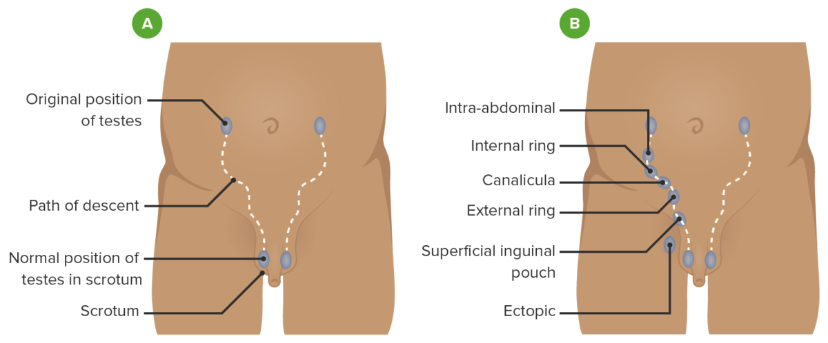
Path of descent of the testis

Path of descent of the testis
A: normal path the testis follows as it descends into the scrotum
B: possible locations where an undescended testis can be found
Image by Lecturio.

Related videos

Clinical Presentation and Diagnosis

Clinical findings

  • Palpable:
    • Suprascrotal (most common location): outside the external inguinal ring 
    • Intracanalicular: inguinal canal Inguinal canal The tunnel in the lower anterior abdominal wall through which the spermatic cord, in the male; round ligament, in the female; nerves; and vessels pass. Its internal end is at the deep inguinal ring and its external end is at the superficial inguinal ring. Inguinal Canal: Anatomy and Hernias 
  • Non-palpable:
    • Intra-abdominal 
    • Absent testis 
    • Empty or hypoplastic scrotum Scrotum A cutaneous pouch of skin containing the testicles and spermatic cords. Testicles: Anatomy

Examination

  • Diagnosis is mainly based on a comprehensive physical examination.
    • Testicular exam:
      • Position infant in the supine position.
      • Exam requires warm lubricated hands: 1 hand Hand The hand constitutes the distal part of the upper limb and provides the fine, precise movements needed in activities of daily living. It consists of 5 metacarpal bones and 14 phalanges, as well as numerous muscles innervated by the median and ulnar nerves. Hand: Anatomy near the anterior superior iliac spine Anterior Superior Iliac Spine Chronic Apophyseal Injury ( ASIS ASIS Chronic Apophyseal Injury) and the other on the scrotum Scrotum A cutaneous pouch of skin containing the testicles and spermatic cords. Testicles: Anatomy
      • Use fingertips to sweep from ASIS ASIS Chronic Apophyseal Injury along the inguinal canal Inguinal canal The tunnel in the lower anterior abdominal wall through which the spermatic cord, in the male; round ligament, in the female; nerves; and vessels pass. Its internal end is at the deep inguinal ring and its external end is at the superficial inguinal ring. Inguinal Canal: Anatomy and Hernias.
    • Genital exam:
      • Assess position of urethral meatus to rule out hypospadias Hypospadias A birth defect due to malformation of the urethra in which the urethral opening is below its normal location. In the male, the malformed urethra generally opens on the ventral surface of the penis or on the perineum. In the female, the malformed urethral opening is in the vagina. Penile Anomalies and Conditions.
      • Check phallus for size and atypical appearance. 
      • Look for hypoplastic scrotum Scrotum A cutaneous pouch of skin containing the testicles and spermatic cords. Testicles: Anatomy or poorly rugated scrotal skin Skin The skin, also referred to as the integumentary system, is the largest organ of the body. The skin is primarily composed of the epidermis (outer layer) and dermis (deep layer). The epidermis is primarily composed of keratinocytes that undergo rapid turnover, while the dermis contains dense layers of connective tissue. Skin: Structure and Functions
    • Inguinal exam: Assess for fullness; may indicate hernia Hernia Protrusion of tissue, structure, or part of an organ through the bone, muscular tissue, or the membrane by which it is normally contained. Hernia may involve tissues such as the abdominal wall or the respiratory diaphragm. Hernias may be internal, external, congenital, or acquired. Abdominal Hernias or lymphadenopathy Lymphadenopathy Lymphadenopathy is lymph node enlargement (> 1 cm) and is benign and self-limited in most patients. Etiologies include malignancy, infection, and autoimmune disorders, as well as iatrogenic causes such as the use of certain medications. Generalized lymphadenopathy often indicates underlying systemic disease. Lymphadenopathy.
  • Imaging:
    • Generally not necessary as it lacks sensitivity or specificity on treatment decision
    • Ultrasound: may be indicated when ruling out disorder of sexual differentiation or gonads Gonads The gamete-producing glands, ovary or testis. Hormones: Overview and Types/ uterus Uterus The uterus, cervix, and fallopian tubes are part of the internal female reproductive system. The uterus has a thick wall made of smooth muscle (the myometrium) and an inner mucosal layer (the endometrium). The most inferior portion of the uterus is the cervix, which connects the uterine cavity to the vagina. Uterus, Cervix, and Fallopian Tubes: Anatomy 

Management

Treatment principles

  • Goal: Fix the viable undescended testis in the normal scrotal position or remove nonviable testicular elements.
  • Timing:
    • No spontaneous descent by 6 months of age triggers the need for intervention.
    • Surgery before 2 years of age (ideally before 1 year) 
  • Treatment is recommended to:
    • Allow testicular growth and for fertility preservation
    • Reduce the risk of testicular cancer Testicular cancer Testicular cancer is the most common solid malignancy affecting men 15-35 years of age. Most of the testicular cancers are of the germ cell tumor type, and they can be classified as seminomas and nonseminomas. The most common presentation of testicular cancer is a painless testicular mass. Testicular Cancer
    • Allow for detection of testicular masses
    • Decrease the risk of testicular torsion Testicular torsion Testicular torsion is the sudden rotation of the testicle, specifically the spermatic cord, around its axis in the inguinal canal or below. The acute rotation results in compromised blood flow to and from the testicle, which puts the testicle at risk for necrosis. Testicular Torsion

Hormonal therapy

  • Less commonly used due to variable Variable Variables represent information about something that can change. The design of the measurement scales, or of the methods for obtaining information, will determine the data gathered and the characteristics of that data. As a result, a variable can be qualitative or quantitative, and may be further classified into subgroups. Types of Variables success rates
  • Involves the administration of human chorionic gonadotropin (hCG) or gonadotropin-releasing hormone Gonadotropin-releasing hormone A decapeptide that stimulates the synthesis and secretion of both pituitary gonadotropins, luteinizing hormone and follicle stimulating hormone. Gnrh is produced by neurons in the septum preoptic area of the hypothalamus and released into the pituitary portal blood, leading to stimulation of gonadotrophs in the anterior pituitary gland. Puberty (GnRH) analogs
  • May be considered in specific cases, but surgical intervention remains the standard approach

Surgery for palpable testis

  • Orchiopexy via inguinal approach: Testis and spermatic cord Spermatic Cord Either of a pair of tubular structures formed by ductus deferens; arteries; veins; lymphatic vessels; and nerves. The spermatic cord extends from the deep inguinal ring through the inguinal canal to the testis in the scrotum. Testicles: Anatomy are released from attachments and the testis is sutured in the scrotum Scrotum A cutaneous pouch of skin containing the testicles and spermatic cords. Testicles: Anatomy
  • Complications: testicular atrophy Atrophy Decrease in the size of a cell, tissue, organ, or multiple organs, associated with a variety of pathological conditions such as abnormal cellular changes, ischemia, malnutrition, or hormonal changes. Cellular Adaptation, infection, bleeding, inguinal hernia Hernia Protrusion of tissue, structure, or part of an organ through the bone, muscular tissue, or the membrane by which it is normally contained. Hernia may involve tissues such as the abdominal wall or the respiratory diaphragm. Hernias may be internal, external, congenital, or acquired. Abdominal Hernias

Surgery for nonpalpable testis

  • Diagnostic laparoscopy Diagnostic Laparoscopy Laparotomy and Laparoscopy to search for intra-abdominal testis
  • If no testis is found, must determine presence of blind-ending vessels or testicular nubbin.
  • If testis is found, can proceed with laparoscopic orchiopexy (options):
    • Single-stage orchiopexy without division of gonadal vessels 
    • 1-stage Fowler-Stephens (FS) orchiopexy with gonadal vessels divided
    • 2-stage FS orchiopexy where the gonadal vessels are clipped to allow collateral blood flow Blood flow Blood flow refers to the movement of a certain volume of blood through the vasculature over a given unit of time (e.g., mL per minute). Vascular Resistance, Flow, and Mean Arterial Pressure to testis in 6 months to mobilize testis
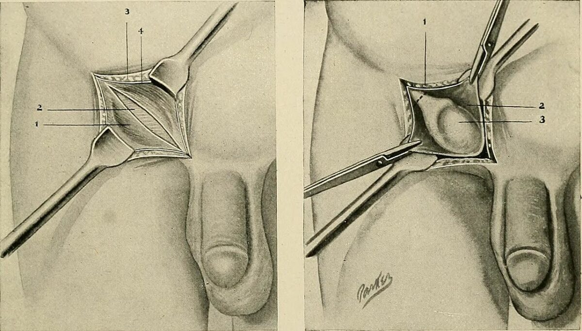
Surgery for undescended testis

Surgical correction of undescended testis
Left: inguinal region dissection
Right: inguinal approach to orchiopexy with testicle exposed in inguinal canal

Image: “The practice of surgery” by Internet Archive Book Images. License: Public Domain

Clinical Relevance

  • Testicular cancer Testicular cancer Testicular cancer is the most common solid malignancy affecting men 15-35 years of age. Most of the testicular cancers are of the germ cell tumor type, and they can be classified as seminomas and nonseminomas. The most common presentation of testicular cancer is a painless testicular mass. Testicular Cancer: Patients Patients Individuals participating in the health care system for the purpose of receiving therapeutic, diagnostic, or preventive procedures. Clinician–Patient Relationship with a history of cryptorchidism have an increased risk of testicular cancer Testicular cancer Testicular cancer is the most common solid malignancy affecting men 15-35 years of age. Most of the testicular cancers are of the germ cell tumor type, and they can be classified as seminomas and nonseminomas. The most common presentation of testicular cancer is a painless testicular mass. Testicular Cancer ( relative risk Relative risk Relative risk (RR) is the risk of a disease or condition occurring in a group or population with a particular exposure relative to a control (unexposed) group. Measures of Risk of 2.9). The most common presentation is a painless testicular nodule Nodule Chalazion. Most of the testicular cancers are germ cell tumors Germ cell tumors A true neoplasm composed of a number of different types of tissue, none of which is native to the area in which it occurs. It is composed of tissues that are derived from three germinal layers, the endoderm, mesoderm, and ectoderm. They are classified histologically as mature (benign) or immature (malignant). Ovarian Cancer. Diagnosis involves physical exam, testicular ultrasound, serum tumor Tumor Inflammation markers, and metastatic workup. Treatment is radical orchiectomy.  
  • Testicular torsion Testicular torsion Testicular torsion is the sudden rotation of the testicle, specifically the spermatic cord, around its axis in the inguinal canal or below. The acute rotation results in compromised blood flow to and from the testicle, which puts the testicle at risk for necrosis. Testicular Torsion: the most common cause of acute testicular and scrotal emergency pain Pain An unpleasant sensation induced by noxious stimuli which are detected by nerve endings of nociceptive neurons. Pain: Types and Pathways. Patients Patients Individuals participating in the health care system for the purpose of receiving therapeutic, diagnostic, or preventive procedures. Clinician–Patient Relationship have unilateral testicular pain Pain An unpleasant sensation induced by noxious stimuli which are detected by nerve endings of nociceptive neurons. Pain: Types and Pathways, most frequently caused by a high abnormal attachment of the tunica vaginalis Tunica vaginalis Testicles: Anatomy, making it prone to twisting. Scrotal ultrasound is helpful, but testicular torsion Testicular torsion Testicular torsion is the sudden rotation of the testicle, specifically the spermatic cord, around its axis in the inguinal canal or below. The acute rotation results in compromised blood flow to and from the testicle, which puts the testicle at risk for necrosis. Testicular Torsion is mostly a clinical diagnosis and requires immediate scrotal exploration and orchiopexy. 
  • Inguinal canal Inguinal canal The tunnel in the lower anterior abdominal wall through which the spermatic cord, in the male; round ligament, in the female; nerves; and vessels pass. Its internal end is at the deep inguinal ring and its external end is at the superficial inguinal ring. Inguinal Canal: Anatomy and Hernias and hernias: hernia Hernia Protrusion of tissue, structure, or part of an organ through the bone, muscular tissue, or the membrane by which it is normally contained. Hernia may involve tissues such as the abdominal wall or the respiratory diaphragm. Hernias may be internal, external, congenital, or acquired. Abdominal Hernias occurs in parts of the abdominal wall Abdominal wall The outer margins of the abdomen, extending from the osteocartilaginous thoracic cage to the pelvis. Though its major part is muscular, the abdominal wall consists of at least seven layers: the skin, subcutaneous fat, deep fascia; abdominal muscles, transversalis fascia, extraperitoneal fat, and the parietal peritoneum. Surgical Anatomy of the Abdomen where the musculature is not continuous. The increased pressure in the abdominal cavity leads to an evagination (protrusion) of the parietal Parietal One of a pair of irregularly shaped quadrilateral bones situated between the frontal bone and occipital bone, which together form the sides of the cranium. Skull: Anatomy peritoneum Peritoneum The peritoneum is a serous membrane lining the abdominopelvic cavity. This lining is formed by connective tissue and originates from the mesoderm. The membrane lines both the abdominal walls (as parietal peritoneum) and all of the visceral organs (as visceral peritoneum). Peritoneum: Anatomy and parts of organs through the surrounding muscles (in this case, the inguinal canal Inguinal canal The tunnel in the lower anterior abdominal wall through which the spermatic cord, in the male; round ligament, in the female; nerves; and vessels pass. Its internal end is at the deep inguinal ring and its external end is at the superficial inguinal ring. Inguinal Canal: Anatomy and Hernias). Diagnosis is made by physical exam and ultrasound, with manual reduction or hernia Hernia Protrusion of tissue, structure, or part of an organ through the bone, muscular tissue, or the membrane by which it is normally contained. Hernia may involve tissues such as the abdominal wall or the respiratory diaphragm. Hernias may be internal, external, congenital, or acquired. Abdominal Hernias repair as necessary. 
  • Infertility Infertility Infertility is the inability to conceive in the context of regular intercourse. The most common causes of infertility in women are related to ovulatory dysfunction or tubal obstruction, whereas, in men, abnormal sperm is a common cause. Infertility: A history of cryptorchidism will increase the incidence Incidence The number of new cases of a given disease during a given period in a specified population. It also is used for the rate at which new events occur in a defined population. It is differentiated from prevalence, which refers to all cases in the population at a given time. Measures of Disease Frequency of impaired spermatogenesis Spermatogenesis The process of germ cell development in the male from the primordial germ cells, through spermatogonia; spermatocytes; spermatids; to the mature haploid spermatozoa. Gametogenesis and lower fertility rates. Studies show that early orchiopexy and diagnosis of undescended testis will lead to increased fertility preservation. 
  • Retractile testis: when the testicles Testicles The testicles, also known as the testes or the male gonads, are a pair of egg-shaped glands suspended within the scrotum. The testicles have multiple layers: an outer tunica vaginalis, an intermediate tunica albuginea, and an innermost tunica vasculosa. The testicles are composed of testicular lobules and seminiferous tubules. Testicles: Anatomy are palpable but in an extra-scrotal position on examination and can be manipulated into the scrotum Scrotum A cutaneous pouch of skin containing the testicles and spermatic cords. Testicles: Anatomy. The testicles Testicles The testicles, also known as the testes or the male gonads, are a pair of egg-shaped glands suspended within the scrotum. The testicles have multiple layers: an outer tunica vaginalis, an intermediate tunica albuginea, and an innermost tunica vasculosa. The testicles are composed of testicular lobules and seminiferous tubules. Testicles: Anatomy will remain in a dependent scrotal location temporarily and re-ascend. Treatment is with serial exams and monitoring of descent. 

References

  1. Cooper, C.S. (2024). Undescended testes (cryptorchidism) in children: Management. UpToDate. Retrieved November 18, 2024, from https://www.uptodate.com/contents/undescended-testes-cryptorchidism-in-children-management
  2. Cooper, C.S. (2024). Undescended testes (cryptorchidism) in children: Clinical features and evaluation. UpToDate. Retrieved November 18, 2024, from https://www.uptodate.com/contents/undescended-testes-cryptorchidism-in-children-clinical-features-and-evaluation
  3. Jacobs, Micah. (2020). Undescended Testis. AUA University: AUA Core Curriculum. https://university.auanet.org/modules/webapps/core/index.cfm#/corecontent/213
  4. Sickkids staff. (2010). Undescended testicle. Retrieved January 21, 2021, from https://www.aboutkidshealth.ca/Article?contentid=884&language=English

Create your free account or log in to continue reading!

Sign up now and get free access to Lecturio with concept pages, medical videos, and questions for your medical education.

User Reviews

Details