Advertisement

Advertisement

Advertisement

Advertisement

Craniosynostosis

Craniosynostosis is the premature Premature Childbirth before 37 weeks of pregnancy (259 days from the first day of the mother's last menstrual period, or 245 days after fertilization). Necrotizing Enterocolitis fusion of 1 or more cranial sutures during the 1st year of life. Craniosynostosis is classified as simple or complex, and can be caused by environmental factors or genetic syndromes. Patients Patients Individuals participating in the health care system for the purpose of receiving therapeutic, diagnostic, or preventive procedures. Clinician–Patient Relationship are typically asymptomatic and concern may arise from caregiver observations. Diagnosis is made clinically and by imaging of the head Imaging of the head Today, CT and MRI, especially the latter, are the preferred imaging methods for the study of the cranial vault and its contents. In conditions where emergent management is decided on the basis of presentation and imaging, CT has the advantage of rapid scan time and wider availability. Imaging of the Head and Brain. Treatment is surgical and prognosis Prognosis A prediction of the probable outcome of a disease based on a individual's condition and the usual course of the disease as seen in similar situations. Non-Hodgkin Lymphomas depends on classification and the presence of genetic syndromes.

Last updated: Sep 29, 2022

Editorial responsibility: Stanley Oiseth, Lindsay Jones, Evelin Maza

Advertisement

Advertisement

Advertisement

Advertisement

Advertisement

Advertisement

Definitions and Epidemiology

Definitions

  • Craniosynostosis: premature Premature Childbirth before 37 weeks of pregnancy (259 days from the first day of the mother’s last menstrual period, or 245 days after fertilization). Necrotizing Enterocolitis fusion of 1 ⩲ cranial sutures, causing an abnormal shaping of the skull Skull The skull (cranium) is the skeletal structure of the head supporting the face and forming a protective cavity for the brain. The skull consists of 22 bones divided into the viscerocranium (facial skeleton) and the neurocranium. Skull: Anatomy 
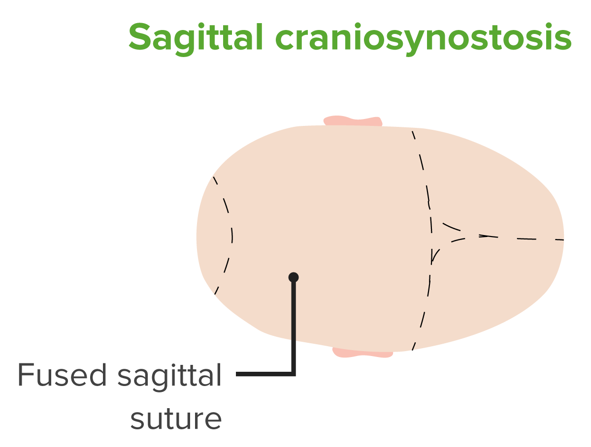
  • Scaphocephaly: elongated skull Skull The skull (cranium) is the skeletal structure of the head supporting the face and forming a protective cavity for the brain. The skull consists of 22 bones divided into the viscerocranium (facial skeleton) and the neurocranium. Skull: Anatomy with frontal bossing Frontal Bossing Hydrocephalus in Children, often due to fusion of sagittal Sagittal Computed Tomography (CT) sutures 
  • Plagiocephaly: flattening of half of the forehead Forehead The part of the face above the eyes. Melasma with raising of ipsilateral eyebrow due to unilateral coronal Coronal Computed Tomography (CT) suture fusion
  • Trigonocephaly: triangular-shaped forehead Forehead The part of the face above the eyes. Melasma with prominent midline ridge caused by fusion of the metopic suture
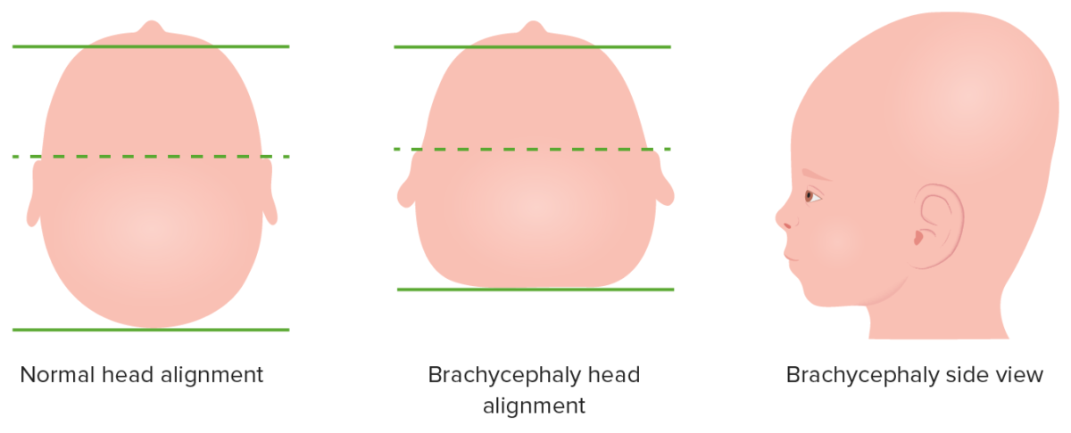
  • Brachycephaly: shortened skull Skull The skull (cranium) is the skeletal structure of the head supporting the face and forming a protective cavity for the brain. The skull consists of 22 bones divided into the viscerocranium (facial skeleton) and the neurocranium. Skull: Anatomy that is wider and taller than normal due to bilateral coronal Coronal Computed Tomography (CT) suture fusion
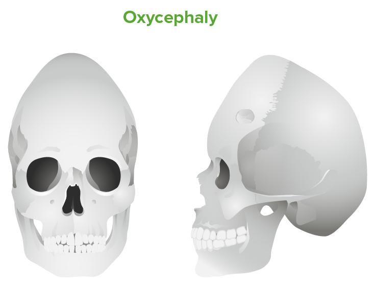
  • Oxycephaly: tall cranium Cranium The skull (cranium) is the skeletal structure of the head supporting the face and forming a protective cavity for the brain. The skull consists of 22 bones divided into the viscerocranium (facial skeleton) and the neurocranium. Skull: Anatomy due to inadequately treated brachycephaly
  • Acrocephaly: elevation of the anterior skull Skull The skull (cranium) is the skeletal structure of the head supporting the face and forming a protective cavity for the brain. The skull consists of 22 bones divided into the viscerocranium (facial skeleton) and the neurocranium. Skull: Anatomy due to combined fusion of lambdoid, sagittal Sagittal Computed Tomography (CT), and coronal Coronal Computed Tomography (CT) sutures
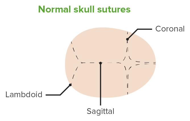
Normal cranial sutures

Normal cranial sutures

Image by Lecturio.

Classification

  • Simple versus complex:
    • Simple: A single suture fuses.
    • Complex or compound: Multiple sutures fuse.
  • Primary versus secondary:
    • Primary: premature Premature Childbirth before 37 weeks of pregnancy (259 days from the first day of the mother’s last menstrual period, or 245 days after fertilization). Necrotizing Enterocolitis closure due to abnormalities of skull Skull The skull (cranium) is the skeletal structure of the head supporting the face and forming a protective cavity for the brain. The skull consists of 22 bones divided into the viscerocranium (facial skeleton) and the neurocranium. Skull: Anatomy development
    • Secondary: closure due to failure of brain Brain The part of central nervous system that is contained within the skull (cranium). Arising from the neural tube, the embryonic brain is comprised of three major parts including prosencephalon (the forebrain); mesencephalon (the midbrain); and rhombencephalon (the hindbrain). The developed brain consists of cerebrum; cerebellum; and other structures in the brain stem. Nervous System: Anatomy, Structure, and Classification growth and expansion

Epidemiology

  • Incidence Incidence The number of new cases of a given disease during a given period in a specified population. It also is used for the rate at which new events occur in a defined population. It is differentiated from prevalence, which refers to all cases in the population at a given time. Measures of Disease Frequency:
    • 1 in 2,000 live births in the United States
    • 1 in 2,500 live births worldwide
    • No gender Gender Gender Dysphoria or racial predilection
  • Scaphocephaly ( sagittal Sagittal Computed Tomography (CT) suture) is the most common.
  • 85% of cases are simple.
  • 10%–20% of cases are associated with genetic syndromes.
  • Complex cases tend to be associated with other malformations.

Etiology and Pathophysiology

Etiology

Nonsyndromic:

  • Genetic mutations Genetic Mutations Carcinogenesis:
    • Caused by gene Gene A category of nucleic acid sequences that function as units of heredity and which code for the basic instructions for the development, reproduction, and maintenance of organisms. Basic Terms of Genetics mutations of fibroblast growth factor Fibroblast growth factor A family of small polypeptide growth factors that share several common features including a strong affinity for heparin, and a central barrel-shaped core region of 140 amino acids that is highly homologous between family members. Although originally studied as proteins that stimulate the growth of fibroblasts this distinction is no longer a requirement for membership in the fibroblast growth factor family. X-linked Hypophosphatemic Rickets (FGF), FGF receptor Receptor Receptors are proteins located either on the surface of or within a cell that can bind to signaling molecules known as ligands (e.g., hormones) and cause some type of response within the cell. Receptors, or transforming growth factor beta Transforming growth factor beta Cell-surface proteins that bind transforming growth factor beta and trigger changes influencing the behavior of cells. Two types of transforming growth factor receptors have been recognized. They differ in affinity for different members of the transforming growth factor beta family and in cellular mechanisms of action. Pulmonary Fibrosis (TGF-β)
    • Alters communication Communication The exchange or transmission of ideas, attitudes, or beliefs between individuals or groups. Decision-making Capacity and Legal Competence between the dura mater Dura mater The outermost of the three meninges, a fibrous membrane of connective tissue that covers the brain and the spinal cord. Meninges: Anatomy and mesenchyma of suture
    • Can be hereditary
  • Environmental influence:
    • Advanced maternal age
    • Nicotine Nicotine Nicotine is highly toxic alkaloid. It is the prototypical agonist at nicotinic cholinergic receptors where it dramatically stimulates neurons and ultimately blocks synaptic transmission. Nicotine is also important medically because of its presence in tobacco smoke. Stimulants use in pregnancy Pregnancy The status during which female mammals carry their developing young (embryos or fetuses) in utero before birth, beginning from fertilization to birth. Pregnancy: Diagnosis, Physiology, and Care
    • Clomiphene citrate Clomiphene citrate A triphenyl ethylene stilbene derivative which is an estrogen agonist or antagonist depending on the target tissue. Antiestrogens in pregnancy Pregnancy The status during which female mammals carry their developing young (embryos or fetuses) in utero before birth, beginning from fertilization to birth. Pregnancy: Diagnosis, Physiology, and Care

Syndromic:

  • Crouzon syndrome:
    • Most common
    • Bilateral coronal Coronal Computed Tomography (CT) craniosynostosis
    • Midfacial abnormalities
    • Hydrocephalus Hydrocephalus Excessive accumulation of cerebrospinal fluid within the cranium which may be associated with dilation of cerebral ventricles, intracranial. Subarachnoid Hemorrhage
    • Exophthalmos 
    • Airway Airway ABCDE Assessment obstruction
    • Mutation Mutation Genetic mutations are errors in DNA that can cause protein misfolding and dysfunction. There are various types of mutations, including chromosomal, point, frameshift, and expansion mutations. Types of Mutations localized in GFR2 gene Gene A category of nucleic acid sequences that function as units of heredity and which code for the basic instructions for the development, reproduction, and maintenance of organisms. Basic Terms of Genetics
  • Apert syndrome:
    • Coronal Coronal Computed Tomography (CT) craniosynostosis
    • Hand Hand The hand constitutes the distal part of the upper limb and provides the fine, precise movements needed in activities of daily living. It consists of 5 metacarpal bones and 14 phalanges, as well as numerous muscles innervated by the median and ulnar nerves. Hand: Anatomy, elbow, hip, and knee deformities 
    • Exophthalmos
  • Carpenter syndrome:
    • Lambdoid and sagittal Sagittal Computed Tomography (CT) craniosynostosis
    • Limb abnormalities ( syndactyly Syndactyly A congenital anomaly of the hand or foot, marked by the webbing between adjacent fingers or toes. Syndactylies are classified as complete or incomplete by the degree of joining. Syndactylies can also be simple or complex. Simple syndactyly indicates joining of only skin or soft tissue; complex syndactyly marks joining of bony elements. Development of the Limbs
  • Pfeiffer syndrome:
    • Unicoronal craniosynostosis
    • Facial deformities
    • Limb abnormalities
    • Exophthalmos
    • Hearing loss Hearing loss Hearing loss, also known as hearing impairment, is any degree of impairment in the ability to apprehend sound as determined by audiometry to be below normal hearing thresholds. Clinical presentation may occur at birth or as a gradual loss of hearing with age, including a short-term or sudden loss at any point. Hearing Loss

Pathophysiology

  • Skull Skull The skull (cranium) is the skeletal structure of the head supporting the face and forming a protective cavity for the brain. The skull consists of 22 bones divided into the viscerocranium (facial skeleton) and the neurocranium. Skull: Anatomy is made up of 5 separate bones:
    • 2 frontal Frontal The bone that forms the frontal aspect of the skull. Its flat part forms the forehead, articulating inferiorly with the nasal bone and the cheek bone on each side of the face. Skull: Anatomy bones
    • 2 parietal Parietal One of a pair of irregularly shaped quadrilateral bones situated between the frontal bone and occipital bone, which together form the sides of the cranium. Skull: Anatomy bones
    • Occipital Occipital Part of the back and base of the cranium that encloses the foramen magnum. Skull: Anatomy bone Bone Bone is a compact type of hardened connective tissue composed of bone cells, membranes, an extracellular mineralized matrix, and central bone marrow. The 2 primary types of bone are compact and spongy. Bones: Structure and Types
  • In infants → bones are separated by open sutures and fontanelles Fontanelles Physical Examination of the Newborn (anterior and posterior). 
  • During childbirth → skull Skull The skull (cranium) is the skeletal structure of the head supporting the face and forming a protective cavity for the brain. The skull consists of 22 bones divided into the viscerocranium (facial skeleton) and the neurocranium. Skull: Anatomy bones overlap and compress at sutures and fontanelles Fontanelles Physical Examination of the Newborn to allow delivery of head through vaginal canal.
  • After birth → sutures remain open to allow for brain Brain The part of central nervous system that is contained within the skull (cranium). Arising from the neural tube, the embryonic brain is comprised of three major parts including prosencephalon (the forebrain); mesencephalon (the midbrain); and rhombencephalon (the hindbrain). The developed brain consists of cerebrum; cerebellum; and other structures in the brain stem. Nervous System: Anatomy, Structure, and Classification growth.
  • Craniosynostosis is due to inappropriate closure of sutures.

Clinical Presentation

Aside from an abnormally shaped head, most patients Patients Individuals participating in the health care system for the purpose of receiving therapeutic, diagnostic, or preventive procedures. Clinician–Patient Relationship are asymptomatic. Careful exam should be performed to evaluate for signs of increased intracranial pressure Intracranial Pressure Idiopathic Intracranial Hypertension including developmental delay.

Table: Epidemiology and clinical presentation of different forms of craniosynostosis
TypeEpidemiology Deformity Deformity Examination of the Upper LimbsClinical presentation
Coronal Coronal Computed Tomography (CT)
  • 18% of cases 
  • More common in girls
  • Associated with Apert syndrome and Crouzon disease
Unilateral: 
  • Plagiocephaly
Bilateral: 
  • Brachycephaly
  • Acrocephaly
Unilateral:
  • Flattened forehead Forehead The part of the face above the eyes. Melasma on affected side
  • Flat cheeks Cheeks The part of the face that is below the eye and to the side of the nose and mouth. Melasma
  • Nose Nose The nose is the human body’s primary organ of smell and functions as part of the upper respiratory system. The nose may be best known for inhaling oxygen and exhaling carbon dioxide, but it also contributes to other important functions, such as tasting. The anatomy of the nose can be divided into the external nose and the nasal cavity. Nose Anatomy (External & Internal) deviation on normal side
  • Higher supraorbital margin
  • Outward rotation Rotation Motion of an object in which either one or more points on a line are fixed. It is also the motion of a particle about a fixed point. X-rays of orbit can result in amblyopia Amblyopia A nonspecific term referring to impaired vision. Major subcategories include stimulus deprivation-induced amblyopia and toxic amblyopia. Stimulus deprivation-induced amblyopia is a developmental disorder of the visual cortex. A discrepancy between visual information received by the visual cortex from each eye results in abnormal cortical development. Strabismus and refractive errors may cause this condition. Toxic amblyopia is a disorder of the optic nerve which is associated with alcoholism, tobacco smoking, and other toxins and as an adverse effect of the use of some medications. Strabismus
Bilateral:
  • Broad, flattened forehead Forehead The part of the face above the eyes. Melasma
  • In Apert syndrome, accompanied by syndactyly Syndactyly A congenital anomaly of the hand or foot, marked by the webbing between adjacent fingers or toes. Syndactylies are classified as complete or incomplete by the degree of joining. Syndactylies can also be simple or complex. Simple syndactyly indicates joining of only skin or soft tissue; complex syndactyly marks joining of bony elements. Development of the Limbs
  • In Crouzon disease, accompanied by hypoplasia Hypoplasia Hypoplastic Left Heart Syndrome (HLHS) of midface and progressive proptosis Proptosis Retinoblastoma
Lambdoid
  • 10%–20% of cases
  • Male:female ratio of 4:1
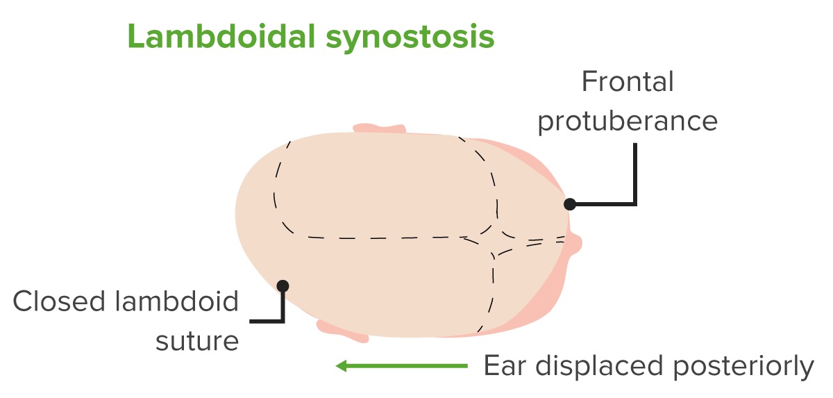
  • Lambdoid/ occipital Occipital Part of the back and base of the cranium that encloses the foramen magnum. Skull: Anatomy plagiocephaly
  • Right side affected in 70% of cases
Unilateral:
  • Flattening of occiput
  • Indentation along synostotic suture
  • Bulging of ipsilateral forehead Forehead The part of the face above the eyes. Melasma leading to rhomboid skull Skull The skull (cranium) is the skeletal structure of the head supporting the face and forming a protective cavity for the brain. The skull consists of 22 bones divided into the viscerocranium (facial skeleton) and the neurocranium. Skull: Anatomy
  • Ipsilateral ear is anterior and inferior
Bilateral:
  • Brachycephaly with bilateral anteriorly and inferiorly displaced ears
Metopic
  • Association with 19p chromosome Chromosome In a prokaryotic cell or in the nucleus of a eukaryotic cell, a structure consisting of or containing DNA which carries the genetic information essential to the cell. Basic Terms of Genetics abnormality
  • Trigonocephaly
  • Pointed forehead Forehead The part of the face above the eyes. Melasma and midline ridge
  • Hypotelorism
Multiple
  • Oxycephaly
  • Cloverleaf skull Skull The skull (cranium) is the skeletal structure of the head supporting the face and forming a protective cavity for the brain. The skull consists of 22 bones divided into the viscerocranium (facial skeleton) and the neurocranium. Skull: Anatomy (Kleeblattschädel)
  • Tower skull Skull The skull (cranium) is the skeletal structure of the head supporting the face and forming a protective cavity for the brain. The skull consists of 22 bones divided into the viscerocranium (facial skeleton) and the neurocranium. Skull: Anatomy with undeveloped sinuses and shallow orbits
  • Elevated intracranial pressure Intracranial Pressure Idiopathic Intracranial Hypertension

Diagnosis and Management

Diagnosis

Diagnosis relies on clinical observation, but imaging may be used to further characterize anatomy for classification or surgery
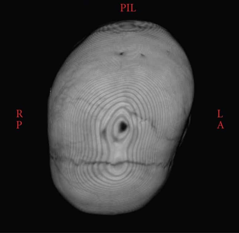
  • Ultrasound of the head is preferred due to lack of radiation Radiation Emission or propagation of acoustic waves (sound), electromagnetic energy waves (such as light; radio waves; gamma rays; or x-rays), or a stream of subatomic particles (such as electrons; neutrons; protons; or alpha particles). Osteosarcoma: can give a prenatal diagnosis
  • CT scan of the head
  • X-rays X-rays X-rays are high-energy particles of electromagnetic radiation used in the medical field for the generation of anatomical images. X-rays are projected through the body of a patient and onto a film, and this technique is called conventional or projectional radiography. X-rays of skull Skull The skull (cranium) is the skeletal structure of the head supporting the face and forming a protective cavity for the brain. The skull consists of 22 bones divided into the viscerocranium (facial skeleton) and the neurocranium. Skull: Anatomy
  • DNA DNA A deoxyribonucleotide polymer that is the primary genetic material of all cells. Eukaryotic and prokaryotic organisms normally contain DNA in a double-stranded state, yet several important biological processes transiently involve single-stranded regions. DNA, which consists of a polysugar-phosphate backbone possessing projections of purines (adenine and guanine) and pyrimidines (thymine and cytosine), forms a double helix that is held together by hydrogen bonds between these purines and pyrimidines (adenine to thymine and guanine to cytosine). DNA Types and Structure testing to look for underlying genetic disorders
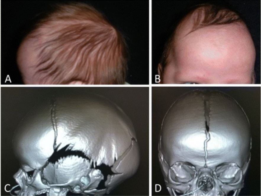
Synostotic sagittal suture with a posteriorly twisted skull

3D CT showing the synostotic sagittal suture with a posteriorly twisted skull

Image: “Atypical craniosynostosis with torticollis and neurological symptoms” by Koljonen V, Leikola J, Valanne L, Hukki J. License: CC BY 3.0

Management

Molded helmet:

  • For mild cases
  • Reshapes head

Surgical intervention: 

  • Goals:
    • Give brain Brain The part of central nervous system that is contained within the skull (cranium). Arising from the neural tube, the embryonic brain is comprised of three major parts including prosencephalon (the forebrain); mesencephalon (the midbrain); and rhombencephalon (the hindbrain). The developed brain consists of cerebrum; cerebellum; and other structures in the brain stem. Nervous System: Anatomy, Structure, and Classification room to grow
    • Release Release Release of a virus from the host cell following virus assembly and maturation. Egress can occur by host cell lysis, exocytosis, or budding through the plasma membrane. Virology developing intracranial pressure Intracranial Pressure Idiopathic Intracranial Hypertension ( pseudotumor cerebri Pseudotumor cerebri Idiopathic intracranial hypertension (IIH), also known as pseudotumor cerebri, is a clinical disorder that presents with symptoms due to increased intracranial pressure (ICP; ≥ 20 mm hg) or CSF pressure (> 250 mm H2O), with no structural changes or other attributable causes. Idiopathic Intracranial Hypertension)
    • Correction of appearance
  • Needed in:
  • Options available include:
    • Shunting
    • Ventriculoperitoneal shunting 
    • Ventriculopleural shunting

Prognosis Prognosis A prediction of the probable outcome of a disease based on a individual’s condition and the usual course of the disease as seen in similar situations. Non-Hodgkin Lymphomas

  • Simple cases: 5% risk of recurrence
  • Complex cases: up to 50% risk of recurrence
  • Recurrence is ↑ in presence of genetic syndromes

Complications

  • Permanent facial deformity Deformity Examination of the Upper Limbs
  • Poor self-esteem
  • intracranial pressure Intracranial Pressure Idiopathic Intracranial Hypertension can lead to:
    • Developmental delay
    • Cognitive impairment
    • Lethargy Lethargy A general state of sluggishness, listless, or uninterested, with being tired, and having difficulty concentrating and doing simple tasks. It may be related to depression or drug addiction. Hyponatremia
    • Seizures Seizures A seizure is abnormal electrical activity of the neurons in the cerebral cortex that can manifest in numerous ways depending on the region of the brain affected. Seizures consist of a sudden imbalance that occurs between the excitatory and inhibitory signals in cortical neurons, creating a net excitation. The 2 major classes of seizures are focal and generalized. Seizures
    • Death

Differential Diagnosis

  • Positional plagiocephaly: a common occurrence in infants that presents as a flat spot on a baby’s head that is caused by the baby spending too much time lying on their back. Positional plagiocephaly does not affect brain Brain The part of central nervous system that is contained within the skull (cranium). Arising from the neural tube, the embryonic brain is comprised of three major parts including prosencephalon (the forebrain); mesencephalon (the midbrain); and rhombencephalon (the hindbrain). The developed brain consists of cerebrum; cerebellum; and other structures in the brain stem. Nervous System: Anatomy, Structure, and Classification development or create increased intracranial pressure Intracranial Pressure Idiopathic Intracranial Hypertension. The condition is treated by changing the position of the head and/or using a helmet in severe cases. 
  • Infant torticollis Torticollis A symptom, not a disease, of a twisted neck. In most instances, the head is tipped toward one side and the chin rotated toward the other. The involuntary muscle contractions in the neck region of patients with torticollis can be due to congenital defects, trauma, inflammation, tumors, and neurological or other factors. Cranial Nerve Palsies: contracture of the sternocleidomastoid Sternocleidomastoid Muscles of the Neck: Anatomy muscle due to abnormal fetal positioning or birth trauma. Typically noted by one month of age when the head is tilted to one side. The condition can be associated with facial asymmetry Asymmetry Examination of the Upper Limbs. Treatment is physical therapy Physical Therapy Becker Muscular Dystrophy and reassuring parents as the condition typically resolves by 6 months of age.

References

  1. Johnson D, Wilkie AO. (2011). Craniosynostosis. European journal of human genetics. EJHG, 19(4), 369–376. https://doi.org/10.1038/ejhg.2010.235
  2. Okamoto T, Nuri T, Harada A, Kyutoku S, Ueda K. (2019). Cranial Suture Measurement by 2-point Method in Ultrasound Screening of Craniosynostosis. Plastic and reconstructive surgery. Global open, 7(5), e2225. https://doi.org/10.1097/GOX.0000000000002225

Create your free account or log in to continue reading!

Sign up now and get free access to Lecturio with concept pages, medical videos, and questions for your medical education.

User Reviews

Details