Advertisement

Advertisement

Advertisement

Advertisement

Congenital Duodenal Obstruction

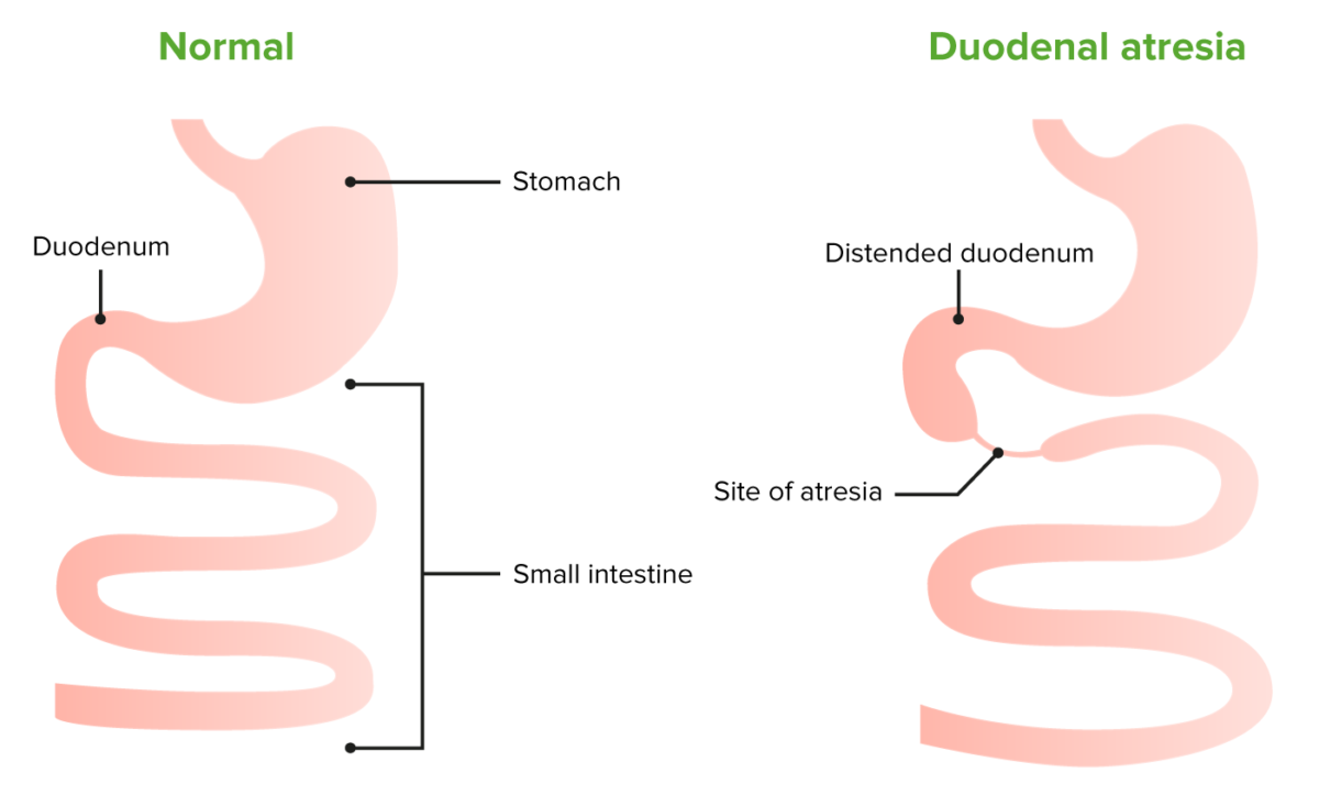
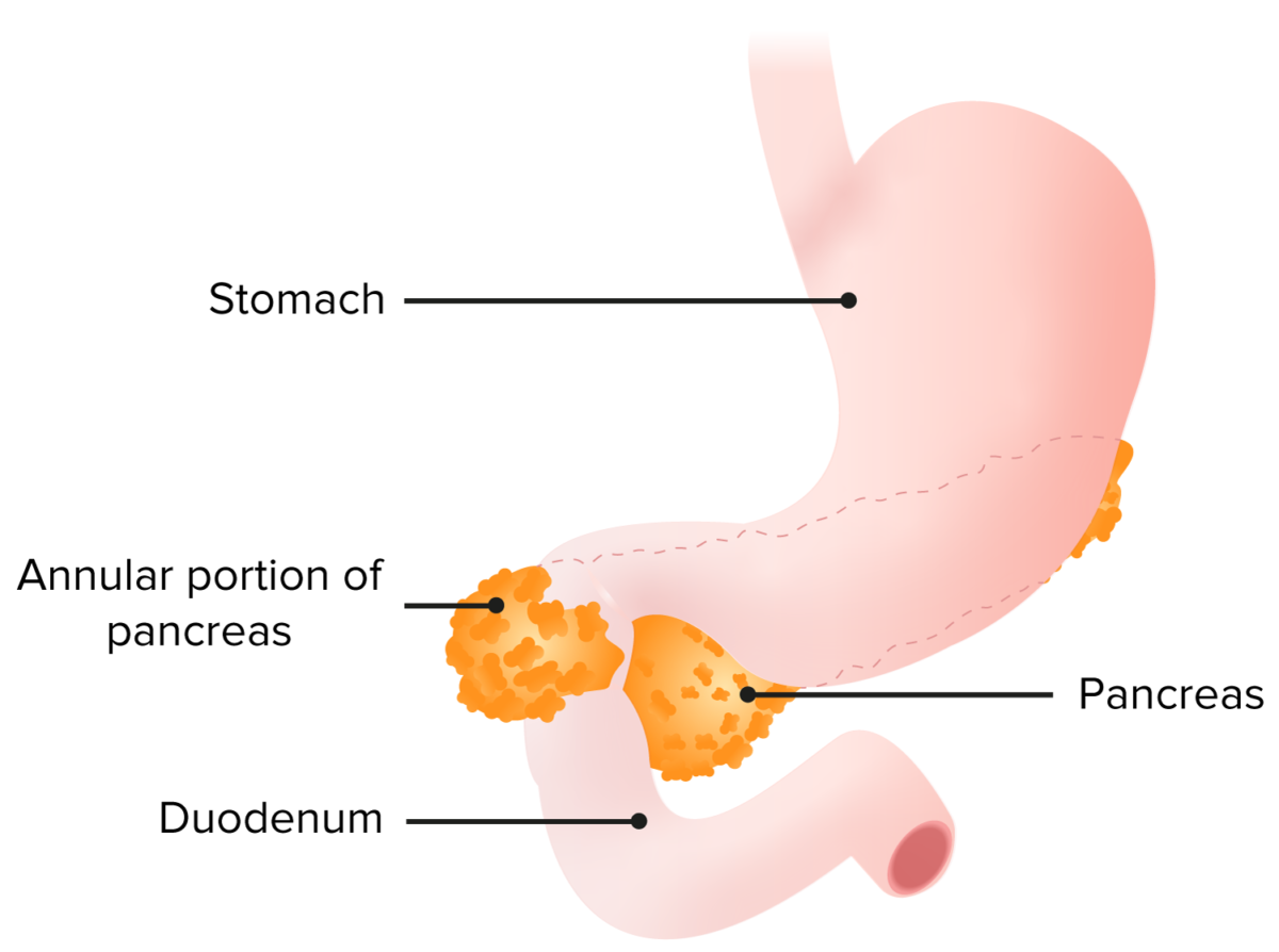
Congenital Congenital Chorioretinitis duodenal obstructions are a group of inborn conditions that cause obstruction of the lumen of the duodenal segment of the small intestine Small intestine The small intestine is the longest part of the GI tract, extending from the pyloric orifice of the stomach to the ileocecal junction. The small intestine is the major organ responsible for chemical digestion and absorption of nutrients. It is divided into 3 segments: the duodenum, the jejunum, and the ileum. Small Intestine: Anatomy, impeding the passage of food and gastric juices. The condition may have intrinsic etiologies, such as duodenal atresia Atresia Hypoplastic Left Heart Syndrome (HLHS) (most common) and/or extrinsic etiologies, such as an annular Annular Dermatologic Examination pancreas Pancreas The pancreas lies mostly posterior to the stomach and extends across the posterior abdominal wall from the duodenum on the right to the spleen on the left. This organ has both exocrine and endocrine tissue. Pancreas: Anatomy. The hallmark of duodenal obstruction is bilious vomiting Vomiting The forcible expulsion of the contents of the stomach through the mouth. Hypokalemia. The condition can be diagnosed through prenatal or postnatal ultrasound. Associated anomalies, including congenital Congenital Chorioretinitis heart disease, are common. Treatment includes fluid resuscitation Resuscitation The restoration to life or consciousness of one apparently dead. . Neonatal Respiratory Distress Syndrome and surgical duodenoduodenostomy.

Last updated: Jan 18, 2024
Editorial responsibility: Stanley Oiseth, Lindsay Jones, Evelin Maza

Advertisement

Advertisement

Advertisement

Advertisement

Advertisement

Advertisement

Epidemiology and Etiology

Epidemiology

  • Overall incidence Incidence The number of new cases of a given disease during a given period in a specified population. It also is used for the rate at which new events occur in a defined population. It is differentiated from prevalence, which refers to all cases in the population at a given time. Measures of Disease Frequency: 2.5–10 per 100,000 live births
  • Duodenal atresia Atresia Hypoplastic Left Heart Syndrome (HLHS)
    • Incidence Incidence The number of new cases of a given disease during a given period in a specified population. It also is used for the rate at which new events occur in a defined population. It is differentiated from prevalence, which refers to all cases in the population at a given time. Measures of Disease Frequency: 1 in 10,000 live births
    • 50% of infants with duodenal atresia Atresia Hypoplastic Left Heart Syndrome (HLHS) are premature Premature Childbirth before 37 weeks of pregnancy (259 days from the first day of the mother’s last menstrual period, or 245 days after fertilization). Necrotizing Enterocolitis.
    • Common concomitant anomalies:
      • Down’s syndrome (25%–40%)
      • Congenital Congenital Chorioretinitis heart disease (30%): associated with increased mortality Mortality All deaths reported in a given population. Measures of Health Status
      • Annular Annular Dermatologic Examination pancreas Pancreas The pancreas lies mostly posterior to the stomach and extends across the posterior abdominal wall from the duodenum on the right to the spleen on the left. This organ has both exocrine and endocrine tissue. Pancreas: Anatomy (30%): associated with late pancreatitis Pancreatitis Inflammation of the pancreas. Pancreatitis is classified as acute unless there are computed tomographic or endoscopic retrograde cholangiopancreatographic findings of chronic pancreatitis. The two most common forms of acute pancreatitis are alcoholic pancreatitis and gallstone pancreatitis. Acute Pancreatitis, gastroesophageal reflux disease Gastroesophageal Reflux Disease Gastroesophageal reflux disease (GERD) occurs when the stomach acid frequently flows back into the esophagus. This backwash (acid reflux) can irritate the lining of the esophagus, causing symptoms such as retrosternal burning pain (heartburn). Gastroesophageal Reflux Disease (GERD), peptic ulcer Peptic ulcer Peptic ulcer disease (PUD) refers to the full-thickness ulcerations of duodenal or gastric mucosa. The ulcerations form when exposure to acid and digestive enzymes overcomes mucosal defense mechanisms. The most common etiologies include Helicobacter pylori (H. pylori) infection and prolonged use of non-steroidal anti-inflammatory drugs (NSAIDs). Peptic Ulcer Disease disease, and gastric cancer Gastric cancer Gastric cancer is the 3rd-most common cause of cancer-related deaths worldwide. The majority of cases are from adenocarcinoma. The modifiable risk factors include Helicobacter pylori infection, smoking, and nitrate-rich diets. Gastric Cancer
      • Malrotation Malrotation Pediatric Gastrointestinal Abnormalities (20%–30%)
      • Renal anomalies (5%–15%)
      • Esophageal atresia Atresia Hypoplastic Left Heart Syndrome (HLHS) (5%–10%)
      • Skeletal malformations (5%)
      • Anorectal anomalies (5%)
  • Annular Annular Dermatologic Examination pancreas Pancreas The pancreas lies mostly posterior to the stomach and extends across the posterior abdominal wall from the duodenum on the right to the spleen on the left. This organ has both exocrine and endocrine tissue. Pancreas: Anatomy:
    • Incidence Incidence The number of new cases of a given disease during a given period in a specified population. It also is used for the rate at which new events occur in a defined population. It is differentiated from prevalence, which refers to all cases in the population at a given time. Measures of Disease Frequency: 5–15 per 100,000 adults
    • Common concomitant abnormalities:
  • Duplication cysts Cysts Any fluid-filled closed cavity or sac that is lined by an epithelium. Cysts can be of normal, abnormal, non-neoplastic, or neoplastic tissues. Fibrocystic Change:
    • Incidence Incidence The number of new cases of a given disease during a given period in a specified population. It also is used for the rate at which new events occur in a defined population. It is differentiated from prevalence, which refers to all cases in the population at a given time. Measures of Disease Frequency: 1 in 45,000 births
    • Duodenal duplication cysts Cysts Any fluid-filled closed cavity or sac that is lined by an epithelium. Cysts can be of normal, abnormal, non-neoplastic, or neoplastic tissues. Fibrocystic Change make up only 5% of reported cases.
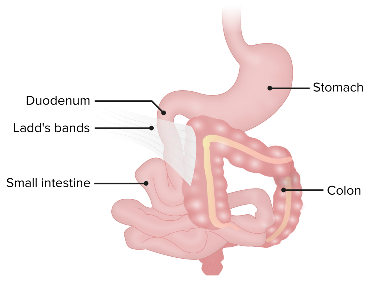
  • Congenital Congenital Chorioretinitis bands: Incidence Incidence The number of new cases of a given disease during a given period in a specified population. It also is used for the rate at which new events occur in a defined population. It is differentiated from prevalence, which refers to all cases in the population at a given time. Measures of Disease Frequency of duodenal obstruction due to Ladd’s bands is not reported. 

Etiology

  • Intrinsic (most common):
  • Extrinsic:
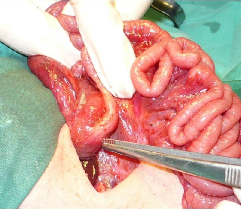
    • Annular Annular Dermatologic Examination pancreas Pancreas The pancreas lies mostly posterior to the stomach and extends across the posterior abdominal wall from the duodenum on the right to the spleen on the left. This organ has both exocrine and endocrine tissue. Pancreas: Anatomy: band of pancreatic tissue surrounding the duodenum Duodenum The shortest and widest portion of the small intestine adjacent to the pylorus of the stomach. It is named for having the length equal to about the width of 12 fingers. Small Intestine: Anatomy arising from incorrect rotation Rotation Motion of an object in which either one or more points on a line are fixed. It is also the motion of a particle about a fixed point. X-rays of the pancreatic bud during embryonal development 
    • Congenital Congenital Chorioretinitis bands (Ladd’s bands): fibrous Fibrous Fibrocystic Change bands of peritoneum Peritoneum The peritoneum is a serous membrane lining the abdominopelvic cavity. This lining is formed by connective tissue and originates from the mesoderm. The membrane lines both the abdominal walls (as parietal peritoneum) and all of the visceral organs (as visceral peritoneum). Peritoneum: Anatomy that persist due to malrotation Malrotation Pediatric Gastrointestinal Abnormalities of the small intestines
    • Duplication cyst: cystic Cystic Fibrocystic Change malformations of the small intestines that can deform the lumen of the duodenum Duodenum The shortest and widest portion of the small intestine adjacent to the pylorus of the stomach. It is named for having the length equal to about the width of 12 fingers. Small Intestine: Anatomy
  • Intrinsic and extrinsic causes may occur concomitantly.

Clinical Presentation

Signs and symptoms of intestinal obstruction Intestinal obstruction Any impairment, arrest, or reversal of the normal flow of intestinal contents toward the anal canal. Ascaris/Ascariasis:

  • Polyhydramnios Polyhydramnios Polyhydramnios is a pathological excess of amniotic fluid. Common causes of polyhydramnios include fetal anomalies, gestational diabetes, multiple gestations, and congenital infections. Patients are often asymptomatic but may present with dyspnea, extremity swelling, or abdominal distention. Polyhydramnios and bile-stained amniotic fluid Amniotic fluid A clear, yellowish liquid that envelopes the fetus inside the sac of amnion. In the first trimester, it is likely a transudate of maternal or fetal plasma. In the second trimester, amniotic fluid derives primarily from fetal lung and kidney. Cells or substances in this fluid can be removed for prenatal diagnostic tests (amniocentesis). Placenta, Umbilical Cord, and Amniotic Cavity may be present at birth.
  • Bilious vomiting Vomiting The forcible expulsion of the contents of the stomach through the mouth. Hypokalemia without abdominal distension on 1st day of life (hallmark)
  • Delayed symptoms or prolonged vomiting Vomiting The forcible expulsion of the contents of the stomach through the mouth. Hypokalemia and failure to thrive Failure to Thrive Failure to thrive (FTT), or faltering growth, describes suboptimal weight gain and growth in children. The majority of cases are due to inadequate caloric intake; however, genetic, infectious, and oncological etiologies are also common. Failure to Thrive based on severity of occlusion
  • Abdominal peristaltic waves may be present, especially early in the course.

Associated abnormalities:

Diagnosis and Management

Diagnosis

History

  • Prenatal:
    • Duodenal obstruction in other pregnancy Pregnancy The status during which female mammals carry their developing young (embryos or fetuses) in utero before birth, beginning from fertilization to birth. Pregnancy: Diagnosis, Physiology, and Care
    • Bowel dilatation or polyhydramnios Polyhydramnios Polyhydramnios is a pathological excess of amniotic fluid. Common causes of polyhydramnios include fetal anomalies, gestational diabetes, multiple gestations, and congenital infections. Patients are often asymptomatic but may present with dyspnea, extremity swelling, or abdominal distention. Polyhydramnios on prenatal screening Screening Preoperative Care
    • Maternal diabetes Diabetes Diabetes mellitus (DM) is a metabolic disease characterized by hyperglycemia and dysfunction of the regulation of glucose metabolism by insulin. Type 1 DM is diagnosed mostly in children and young adults as the result of autoimmune destruction of β cells in the pancreas and the resulting lack of insulin. Type 2 DM has a significant association with obesity and is characterized by insulin resistance. Diabetes Mellitus mellitus
  • Perinatal:
    • Gestational age Gestational age The age of the conceptus, beginning from the time of fertilization. In clinical obstetrics, the gestational age is often estimated as the time from the last day of the last menstruation which is about 2 weeks before ovulation and fertilization. Pregnancy: Diagnosis, Physiology, and Care
    • Hours from birth to onset of vomiting Vomiting The forcible expulsion of the contents of the stomach through the mouth. Hypokalemia
    • Timing of meconium Meconium The thick green-to-black mucilaginous material found in the intestines of a full-term fetus. It consists of secretions of the intestinal glands; bile pigments; fatty acids; amniotic fluid; and intrauterine debris. It constitutes the first stools passed by a newborn. Prenatal and Postnatal Physiology of the Neonate passing
  • Family history Family History Adult Health Maintenance:

Physical exam

  • General:
  • Skin Skin The skin, also referred to as the integumentary system, is the largest organ of the body. The skin is primarily composed of the epidermis (outer layer) and dermis (deep layer). The epidermis is primarily composed of keratinocytes that undergo rapid turnover, while the dermis contains dense layers of connective tissue. Skin: Structure and Functions: jaundice Jaundice Jaundice is the abnormal yellowing of the skin and/or sclera caused by the accumulation of bilirubin. Hyperbilirubinemia is caused by either an increase in bilirubin production or a decrease in the hepatic uptake, conjugation, or excretion of bilirubin. Jaundice
  • Abdomen: Distension suggests that obstruction is not duodenal but more distal.
  • Rectal exam: anal atresia Atresia Hypoplastic Left Heart Syndrome (HLHS) associated with duodenal atresia Atresia Hypoplastic Left Heart Syndrome (HLHS)

Imaging

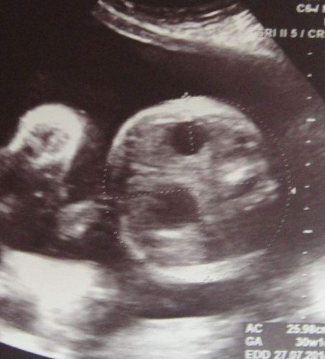
  • Prenatal ultrasound:
    • Shows “double bubble” sign (air-filled stomach Stomach The stomach is a muscular sac in the upper left portion of the abdomen that plays a critical role in digestion. The stomach develops from the foregut and connects the esophagus with the duodenum. Structurally, the stomach is C-shaped and forms a greater and lesser curvature and is divided grossly into regions: the cardia, fundus, body, and pylorus. Stomach: Anatomy and duodenum Duodenum The shortest and widest portion of the small intestine adjacent to the pylorus of the stomach. It is named for having the length equal to about the width of 12 fingers. Small Intestine: Anatomy
    • May reveal polyhydramnios Polyhydramnios Polyhydramnios is a pathological excess of amniotic fluid. Common causes of polyhydramnios include fetal anomalies, gestational diabetes, multiple gestations, and congenital infections. Patients are often asymptomatic but may present with dyspnea, extremity swelling, or abdominal distention. Polyhydramnios 
  • Radiography:
    • Double bubble sign confirms diagnosis of duodenal obstruction. 
    • Contrast studies may occasionally be needed to rule out malrotation Malrotation Pediatric Gastrointestinal Abnormalities and volvulus Volvulus A volvulus is the twisting or axial rotation of a portion of the bowel around its mesentery. The most common site of volvulus in adults is the colon; most frequently the sigmoid volvulus. Patients typically present with symptoms of bowel obstruction such as abdominal pain, distension, vomiting, and constipation/obstipation. Volvulus.
  • Computed tomography (CT) or magnetic resonance imaging (MRI) may be used for better visualization.

Management

Medical therapy

Surgical management

References

  1. Kliegman RB, ST Geme JW, Blum MJ, Shah SS, Tasker RC, Wilson KM, Behrman RE. (2016). In Nelson’s Textbook of Pediatrics. (20th ed.) Elsevier.
  2. Ravitch MM. (1975). The pancreas in infants and children. Surg Clin North Am, 55(2):377–85. https://doi.org/10.1016/s0039-6109(16)40587-6
  3. Sangüesa Nebot C, Llorens Salvador R, Carazo Palacios E, Picó Aliaga S, Ibañez Pradas V. (2018). Enteric duplication cysts in children: varied presentations, varied imaging findings. Insights Imaging, 9(6):1097–1106. https://doi.org/10.1007/s13244-018-0660-z
  4. Sadler TW, Langman J. (2012). Langman’s medical embryology (12th ed.) Wolters Kluwer Health/Lippincott Williams & Wilkins.

Create your free account or log in to continue reading!

Sign up now and get free access to Lecturio with concept pages, medical videos, and questions for your medical education.

User Reviews

Details