Advertisement

Advertisement

Advertisement

Advertisement

Colorectal Cancer Screening

Colorectal cancer (CRC) is the 2nd-leading cause of cancer-related death in the United States. Almost all cases of CRC are adenocarcinoma and the majority of lesions come from the malignant transformation of an adenomatous polyp. As most CRCs are asymptomatic, screening is essential in detecting early disease. Screening is recommended to start at the age of 45 years, utilizing various screening tools available with colonoscopy, flexible sigmoidoscopy, and fecal tests among them. For high-risk individuals, earlier and more frequent screening Screening Preoperative Care is recommended. Other stool-based strategies and visualization tests are also available for CRC screening Screening Preoperative Care.

Last updated: May 17, 2024

Editorial responsibility: Stanley Oiseth, Lindsay Jones, Evelin Maza

Advertisement

Advertisement

Advertisement

Advertisement

Advertisement

Advertisement

Overview

Colorectal cancer Colorectal cancer Colorectal cancer (CRC) is the 2nd leading cause of cancer-related deaths in the United States. Colorectal cancer is a heterogeneous disease that arises from genetic and epigenetic abnormalities, with influence from environmental factors. Colorectal Cancer (CRC)

  • The majority of CRCs are adenocarcinomas.
  • Most CRCs arise from polyps (adenomatous or serrated).
  • Importance of screening Screening Preoperative Care for CRC:
    • 3rd-most common cancer (among both men and women)
    • 2nd-most common cause of death due to cancer in the United States
    • 94% of new cases of colorectal cancer Colorectal cancer Colorectal cancer (CRC) is the 2nd leading cause of cancer-related deaths in the United States. Colorectal cancer is a heterogeneous disease that arises from genetic and epigenetic abnormalities, with influence from environmental factors. Colorectal Cancer occur in adults aged 45 years and older.
  • In 2016, about 25% of eligible adults had not been screened for CRC.

Risk factors

  • Age: 94% of new cases of CRC occur in adults aged 45 years and older.
  • Hereditary syndromes:
    • Hereditary non-polyposis colorectal cancer Hereditary non-polyposis colorectal cancer Lynch syndrome, also called hereditary non-polyposis colorectal cancer (HNPCC), is the most common inherited colon cancer syndrome, and carries a significantly increased risk for endometrial cancer and other malignancies. Lynch syndrome has an autosomal dominant inheritance pattern involving pathogenic variants in one of the mismatch repair (MMR) genes or epithelial cell adhesion molecule (EpCAM). Lynch syndrome ( HNPCC HNPCC Lynch syndrome, also called hereditary non-polyposis colorectal cancer (HNPCC), is the most common inherited colon cancer syndrome, and carries a significantly increased risk for endometrial cancer and other malignancies. Lynch syndrome has an autosomal dominant inheritance pattern involving pathogenic variants in one of the mismatch repair (MMR) genes or epithelial cell adhesion molecule (EpCAM). Lynch syndrome) or Lynch syndrome Lynch syndrome Lynch syndrome, also called hereditary non-polyposis colorectal cancer (HNPCC), is the most common inherited colon cancer syndrome, and carries a significantly increased risk for endometrial cancer and other malignancies. Lynch syndrome has an autosomal dominant inheritance pattern involving pathogenic variants in one of the mismatch repair (MMR) genes or epithelial cell adhesion molecule (EpCAM). Lynch syndrome
    • Familial adenomatous polyposis Familial Adenomatous Polyposis Familial adenomatous polyposis (FAP) is an autosomal dominant inherited genetic disorder that presents with numerous adenomatous polyps in the colon. Familial adenomatous polyposis is the most common of the polyposis syndromes, which is a group of inherited or acquired conditions characterized by the growth of polyps in the GI tract, associated with other extracolonic features. Familial Adenomatous Polyposis ( FAP FAP Familial adenomatous polyposis (FAP) is an autosomal dominant inherited genetic disorder that presents with numerous adenomatous polyps in the colon. Familial adenomatous polyposis is the most common of the polyposis syndromes, which is a group of inherited or acquired conditions characterized by the growth of polyps in the GI tract, associated with other extracolonic features. Familial Adenomatous Polyposis)
    • Peutz-Jeghers syndrome Peutz-Jeghers Syndrome Peutz-Jeghers syndrome (PJS) is an autosomal-dominant inherited disorder characterized by GI polyps and mucocutaneous-pigmented macules. Peutz-Jeghers syndrome is 1 of the polyposis syndromes, a group of inherited or acquired conditions characterized by the growth of polyps in the GI tract and associated with other extracolonic features. Peutz-Jeghers Syndrome ( PJS PJS Peutz-Jeghers syndrome (PJS) is an autosomal-dominant inherited disorder characterized by GI polyps and mucocutaneous-pigmented macules. Peutz-Jeghers syndrome is 1 of the polyposis syndromes, a group of inherited or acquired conditions characterized by the growth of polyps in the GI tract and associated with other extracolonic features. Peutz-Jeghers Syndrome)
    • MUTYH-associated polyposis (MAP): a mutation Mutation Genetic mutations are errors in DNA that can cause protein misfolding and dysfunction. There are various types of mutations, including chromosomal, point, frameshift, and expansion mutations. Types of Mutations in the base excision repair Base excision repair DNA Repair Mechanisms gene Gene A category of nucleic acid sequences that function as units of heredity and which code for the basic instructions for the development, reproduction, and maintenance of organisms. Basic Terms of Genetics mutY homolog
    • Family history Family History Adult Health Maintenance of CRC
  • Colon Colon The large intestines constitute the last portion of the digestive system. The large intestine consists of the cecum, appendix, colon (with ascending, transverse, descending, and sigmoid segments), rectum, and anal canal. The primary function of the colon is to remove water and compact the stool prior to expulsion from the body via the rectum and anal canal. Colon, Cecum, and Appendix: Anatomy pathologies:
    • Inflammatory bowel disease (IBD)
    • Large adenomatous polyps Adenomatous polyps Benign neoplasms derived from glandular epithelium. Colorectal Cancer or previous diagnosis of CRC
  • Other medical conditions:
    • Cystic Cystic Fibrocystic Change fibrosis Fibrosis Any pathological condition where fibrous connective tissue invades any organ, usually as a consequence of inflammation or other injury. Bronchiolitis Obliterans
    • Abdominopelvic radiation Radiation Emission or propagation of acoustic waves (sound), electromagnetic energy waves (such as light; radio waves; gamma rays; or x-rays), or a stream of subatomic particles (such as electrons; neutrons; protons; or alpha particles). Osteosarcoma
    • Conditions with increased insulin Insulin Insulin is a peptide hormone that is produced by the beta cells of the pancreas. Insulin plays a role in metabolic functions such as glucose uptake, glycolysis, glycogenesis, lipogenesis, and protein synthesis. Exogenous insulin may be needed for individuals with diabetes mellitus, in whom there is a deficiency in endogenous insulin or increased insulin resistance. Insulin or insulin-like growth factor (e.g., diabetes Diabetes Diabetes mellitus (DM) is a metabolic disease characterized by hyperglycemia and dysfunction of the regulation of glucose metabolism by insulin. Type 1 DM is diagnosed mostly in children and young adults as the result of autoimmune destruction of β cells in the pancreas and the resulting lack of insulin. Type 2 DM has a significant association with obesity and is characterized by insulin resistance. Diabetes Mellitus, acromegaly Acromegaly A condition caused by prolonged exposure to excessive human growth hormone in adults. It is characterized by bony enlargement of the face; lower jaw (prognathism); hands; feet; head; and thorax. The most common etiology is a growth hormone-secreting pituitary adenoma. Acromegaly and Gigantism, obesity Obesity Obesity is a condition associated with excess body weight, specifically with the deposition of excessive adipose tissue. Obesity is considered a global epidemic. Major influences come from the western diet and sedentary lifestyles, but the exact mechanisms likely include a mixture of genetic and environmental factors. Obesity)
    • Streptococcus Streptococcus Streptococcus is one of the two medically important genera of gram-positive cocci, the other being Staphylococcus. Streptococci are identified as different species on blood agar on the basis of their hemolytic pattern and sensitivity to optochin and bacitracin. There are many pathogenic species of streptococci, including S. pyogenes, S. agalactiae, S. pneumoniae, and the viridans streptococci. Streptococcus bovis bacteremia Bacteremia The presence of viable bacteria circulating in the blood. Fever, chills, tachycardia, and tachypnea are common acute manifestations of bacteremia. The majority of cases are seen in already hospitalized patients, most of whom have underlying diseases or procedures which render their bloodstreams susceptible to invasion. Glycopeptides
    • Renal transplantation
  • Lifestyle and social factors:
    • Smoking Smoking Willful or deliberate act of inhaling and exhaling smoke from burning substances or agents held by hand. Interstitial Lung Diseases and alcohol consumption (> 4 drinks/day)
    • Diet (increased fat, red and processed meat, low fiber)
    • Low socioeconomic status

Screening Rationale

Colorectal polyps

  • Abnormal tissue outgrowths arising from the colonic mucosa and extending into the lumen
  • Most common lesions from which CRC arises
  • Most common progression: small to large polyps → dysplasia → carcinoma
  • Development of CRC takes an average of 10 years.
  • 30% of the population has polyps by the age of 50.
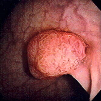
Colon polyp

Polyp of sigmoid colon revealed by colonoscopy: The polyp is pedunculated (with a short stalk).

Image: “Colon polyp” by Dr. F.C. Turner. License: CC BY 2.5

Benefits of screening Screening Preoperative Care

Colorectal cancer Colorectal cancer Colorectal cancer (CRC) is the 2nd leading cause of cancer-related deaths in the United States. Colorectal cancer is a heterogeneous disease that arises from genetic and epigenetic abnormalities, with influence from environmental factors. Colorectal Cancer is generally a preventable cancer when proper screening Screening Preoperative Care is performed. Screening Screening Preoperative Care:
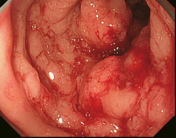
  • Prevents CRC by detection and removal of polyps
  • Identifies early-stage CRC, thereby improving prognosis Prognosis A prediction of the probable outcome of a disease based on a individual’s condition and the usual course of the disease as seen in similar situations. Non-Hodgkin Lymphomas and decreasing mortality Mortality All deaths reported in a given population. Measures of Health Status rate

Strategies for Screening

Stool-based tests

  • High-sensitivity guaiac-based fecal occult blood test Fecal occult blood test Colorectal Cancer (HSgFOBT):
    • Identifies hemoglobin in stool
    • Positive test: Guaiac reagent turns paper blue (peroxidase reaction).
    • Restrictions:
      • Eliminate red meat (3 days before test).
      • Limit Limit A value (e.g., pressure or time) that should not be exceeded and which is specified by the operator to protect the lung Invasive Mechanical Ventilation vitamin C Vitamin C A six carbon compound related to glucose. It is found naturally in citrus fruits and many vegetables. Ascorbic acid is an essential nutrient in human diets, and necessary to maintain connective tissue and bone. Its biologically active form, vitamin C, functions as a reducing agent and coenzyme in several metabolic pathways. Vitamin C is considered an antioxidant. Water-soluble Vitamins and their Deficiencies to < 250 mg a day (3 days before test).
      • Nonsteroidal anti-inflammatory drug ( NSAID NSAID Nonsteroidal antiinflammatory drugs (NSAIDs) are a class of medications consisting of aspirin, reversible NSAIDs, and selective NSAIDs. NSAIDs are used as antiplatelet, analgesic, antipyretic, and antiinflammatory agents. Nonsteroidal Antiinflammatory Drugs (NSAIDs)) intake may affect the test.
    • 3 consecutive stool samples needed 
  • Fecal immunochemical test (FIT):
    • Measures hemoglobin in the stool
    • No medication or diet restrictions prior to the test
    • Only 1 sample needed
    • Should be submitted within 24 hours of collection
  • Stool DNA DNA A deoxyribonucleotide polymer that is the primary genetic material of all cells. Eukaryotic and prokaryotic organisms normally contain DNA in a double-stranded state, yet several important biological processes transiently involve single-stranded regions. DNA, which consists of a polysugar-phosphate backbone possessing projections of purines (adenine and guanine) and pyrimidines (thymine and cytosine), forms a double helix that is held together by hydrogen bonds between these purines and pyrimidines (adenine to thymine and guanine to cytosine). DNA Types and Structure test:
    • Also called multitarget stool DNA DNA A deoxyribonucleotide polymer that is the primary genetic material of all cells. Eukaryotic and prokaryotic organisms normally contain DNA in a double-stranded state, yet several important biological processes transiently involve single-stranded regions. DNA, which consists of a polysugar-phosphate backbone possessing projections of purines (adenine and guanine) and pyrimidines (thymine and cytosine), forms a double helix that is held together by hydrogen bonds between these purines and pyrimidines (adenine to thymine and guanine to cytosine). DNA Types and Structure testing (MT-sDNA)
    • Composite of tests:
      • Molecular assays testing for DNA DNA A deoxyribonucleotide polymer that is the primary genetic material of all cells. Eukaryotic and prokaryotic organisms normally contain DNA in a double-stranded state, yet several important biological processes transiently involve single-stranded regions. DNA, which consists of a polysugar-phosphate backbone possessing projections of purines (adenine and guanine) and pyrimidines (thymine and cytosine), forms a double helix that is held together by hydrogen bonds between these purines and pyrimidines (adenine to thymine and guanine to cytosine). DNA Types and Structure (KRAS) mutations
      • Gene Gene A category of nucleic acid sequences that function as units of heredity and which code for the basic instructions for the development, reproduction, and maintenance of organisms. Basic Terms of Genetics amplification technique testing for methylation Methylation Addition of methyl groups. In histo-chemistry methylation is used to esterify carboxyl groups and remove sulfate groups by treating tissue sections with hot methanol in the presence of hydrochloric acid. . Glucocorticoids biomarkers
      • FIT
    • Test available by prescription
    • No dietary or medication restrictions
    • A full stool sample is required.

Direct visualization tests

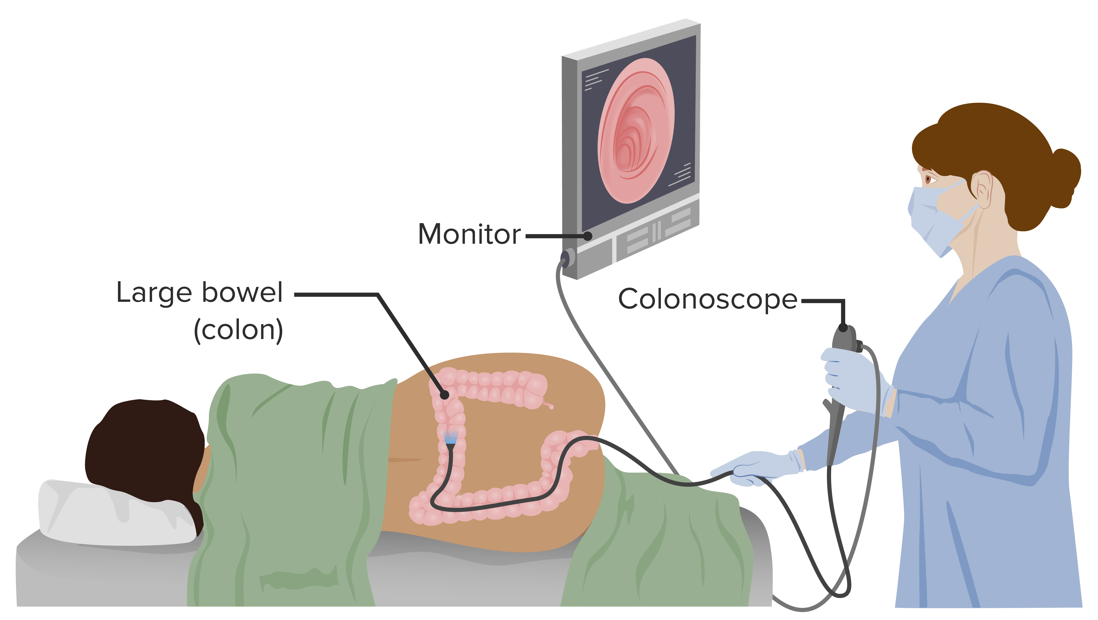
  • Colonoscopy:
    • Gold standard for detecting precancerous Precancerous Pathological conditions that tend eventually to become malignant. Barrett Esophagus adenomas and CRC
    • Visualizes rectum Rectum The rectum and anal canal are the most terminal parts of the lower GI tract/large intestine that form a functional unit and control defecation. Fecal continence is maintained by several important anatomic structures including rectal folds, anal valves, the sling-like puborectalis muscle, and internal and external anal sphincters. Rectum and Anal Canal: Anatomy, colon Colon The large intestines constitute the last portion of the digestive system. The large intestine consists of the cecum, appendix, colon (with ascending, transverse, descending, and sigmoid segments), rectum, and anal canal. The primary function of the colon is to remove water and compact the stool prior to expulsion from the body via the rectum and anal canal. Colon, Cecum, and Appendix: Anatomy, and part of the terminal ileum Ileum The distal and narrowest portion of the small intestine, between the jejunum and the ileocecal valve of the large intestine. Small Intestine: Anatomy
    • Both diagnostic and therapeutic (lesions can be biopsied and removed)
    • Requires medication adjustment ( NSAIDs NSAIDS Primary vs Secondary Headaches, aspirin Aspirin The prototypical analgesic used in the treatment of mild to moderate pain. It has anti-inflammatory and antipyretic properties and acts as an inhibitor of cyclooxygenase which results in the inhibition of the biosynthesis of prostaglandins. Aspirin also inhibits platelet aggregation and is used in the prevention of arterial and venous thrombosis. Nonsteroidal Antiinflammatory Drugs (NSAIDs), anticoagulant)
    • Requires bowel preparation
    • Sedation generally used
    • Procedure-related risks:
      • Perforation Perforation A pathological hole in an organ, blood vessel or other soft part of the body, occurring in the absence of external force. Esophagitis
      • Bleeding
      • Infection
  • Sigmoidoscopy:
    • Endoscope reaches up to the splenic flexure Splenic flexure Small Intestine: Anatomy.
    • Allows visualization but only in the left colon Colon The large intestines constitute the last portion of the digestive system. The large intestine consists of the cecum, appendix, colon (with ascending, transverse, descending, and sigmoid segments), rectum, and anal canal. The primary function of the colon is to remove water and compact the stool prior to expulsion from the body via the rectum and anal canal. Colon, Cecum, and Appendix: Anatomy (so right-sided lesions are missed)
    • Allows removal of lesions, but lesions > 1 cm usually done in subsequent colonoscopy
    • Requires medication adjustment ( NSAIDs NSAIDS Primary vs Secondary Headaches, aspirin Aspirin The prototypical analgesic used in the treatment of mild to moderate pain. It has anti-inflammatory and antipyretic properties and acts as an inhibitor of cyclooxygenase which results in the inhibition of the biosynthesis of prostaglandins. Aspirin also inhibits platelet aggregation and is used in the prevention of arterial and venous thrombosis. Nonsteroidal Antiinflammatory Drugs (NSAIDs), anticoagulant)
    • Performed in-office without sedation
    • Procedure-related risks:
      • Perforation Perforation A pathological hole in an organ, blood vessel or other soft part of the body, occurring in the absence of external force. Esophagitis (most common)
      • Bleeding
      • Infection
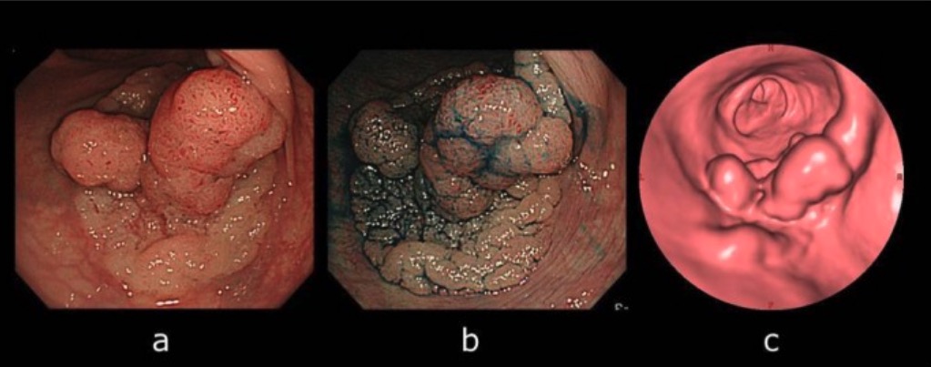
  • Computed tomography (CT) colonography:
    • Use of CT scanning to reconstruct bowel mucosa images in 2 and 3 dimensions
    • Needs bowel preparation, but sedation not required
    • The bowel is distended by introducing air or carbon dioxide via the rectum Rectum The rectum and anal canal are the most terminal parts of the lower GI tract/large intestine that form a functional unit and control defecation. Fecal continence is maintained by several important anatomic structures including rectal folds, anal valves, the sling-like puborectalis muscle, and internal and external anal sphincters. Rectum and Anal Canal: Anatomy
    • Limitation(s): biopsy Biopsy Removal and pathologic examination of specimens from the living body. Ewing Sarcoma or lesion removal not possible
  • Colon Colon The large intestines constitute the last portion of the digestive system. The large intestine consists of the cecum, appendix, colon (with ascending, transverse, descending, and sigmoid segments), rectum, and anal canal. The primary function of the colon is to remove water and compact the stool prior to expulsion from the body via the rectum and anal canal. Colon, Cecum, and Appendix: Anatomy capsule Capsule An envelope of loose gel surrounding a bacterial cell which is associated with the virulence of pathogenic bacteria. Some capsules have a well-defined border, whereas others form a slime layer that trails off into the medium. Most capsules consist of relatively simple polysaccharides but there are some bacteria whose capsules are made of polypeptides. Bacteroides endoscopy Endoscopy Procedures of applying endoscopes for disease diagnosis and treatment. Endoscopy involves passing an optical instrument through a small incision in the skin i.e., percutaneous; or through a natural orifice and along natural body pathways such as the digestive tract; and/or through an incision in the wall of a tubular structure or organ, i.e. Transluminal, to examine or perform surgery on the interior parts of the body. Gastroesophageal Reflux Disease (GERD) (PillCam Colon Colon The large intestines constitute the last portion of the digestive system. The large intestine consists of the cecum, appendix, colon (with ascending, transverse, descending, and sigmoid segments), rectum, and anal canal. The primary function of the colon is to remove water and compact the stool prior to expulsion from the body via the rectum and anal canal. Colon, Cecum, and Appendix: Anatomy 2):
    • The patient swallows a capsule Capsule An envelope of loose gel surrounding a bacterial cell which is associated with the virulence of pathogenic bacteria. Some capsules have a well-defined border, whereas others form a slime layer that trails off into the medium. Most capsules consist of relatively simple polysaccharides but there are some bacteria whose capsules are made of polypeptides. Bacteroides with wireless video cameras. 
    • Images are taken as the capsule Capsule An envelope of loose gel surrounding a bacterial cell which is associated with the virulence of pathogenic bacteria. Some capsules have a well-defined border, whereas others form a slime layer that trails off into the medium. Most capsules consist of relatively simple polysaccharides but there are some bacteria whose capsules are made of polypeptides. Bacteroides goes through the intestine.
    • Requires bowel preparation
    • Not indicated for screening Screening Preoperative Care, but to be used in case of incomplete colonoscopy
    • Limitation: biopsy Biopsy Removal and pathologic examination of specimens from the living body. Ewing Sarcoma or lesion removal not possible

Colorectal Cancer Screening for Average-Risk Individuals

For an average-risk individual, screening Screening Preoperative Care is initiated at 45 years of age.

  • Characteristics of an average-risk individual:
  • Options for screening Screening Preoperative Care:
    • Annual HSgFOBT
    • Annual FIT
    • sDNA-FIT test every 3 years
    • Flexible sigmoidoscopy every 5 years (limited to distal part of the colon Colon The large intestines constitute the last portion of the digestive system. The large intestine consists of the cecum, appendix, colon (with ascending, transverse, descending, and sigmoid segments), rectum, and anal canal. The primary function of the colon is to remove water and compact the stool prior to expulsion from the body via the rectum and anal canal. Colon, Cecum, and Appendix: Anatomy)
    • Flexible sigmoidoscopy every 10 years plus annual FIT
    • Colonoscopy every 10 years
    • CT colonography every 5 years
  • A positive screening Screening Preoperative Care stool-based test, CT colonography, or sigmoidoscopy warrants a colonoscopy.
  • Selective screening Screening Preoperative Care for adults aged 76–85 years, which may be based on:
    • Patient preferences
    • Prior screening Screening Preoperative Care results
    • Overall health status/ comorbidities Comorbidities The presence of co-existing or additional diseases with reference to an initial diagnosis or with reference to the index condition that is the subject of study. Comorbidity may affect the ability of affected individuals to function and also their survival; it may be used as a prognostic indicator for length of hospital stay, cost factors, and outcome or survival. St. Louis Encephalitis Virus

Colorectal Cancer Screening for High-Risk Individuals

  • Individuals with a family history Family History Adult Health Maintenance of CRC (including advanced adenoma or advanced serrated lesion):
    • Colonoscopy at age 40 or 10 years earlier than the age of diagnosis of the 1st-degree relative (whichever comes first)
    • Repeat every 5 years.
    • If the patient declines colonoscopy, annual FIT testing is recommended.
  • Individuals with inflammatory bowel disease:
    • Begin 8 years post-diagnosis.
    • Colonoscopy with biopsies every 1–2 years
  • Individuals with classic familial adenomatous polyposis Familial Adenomatous Polyposis Familial adenomatous polyposis (FAP) is an autosomal dominant inherited genetic disorder that presents with numerous adenomatous polyps in the colon. Familial adenomatous polyposis is the most common of the polyposis syndromes, which is a group of inherited or acquired conditions characterized by the growth of polyps in the GI tract, associated with other extracolonic features. Familial Adenomatous Polyposis ( FAP FAP Familial adenomatous polyposis (FAP) is an autosomal dominant inherited genetic disorder that presents with numerous adenomatous polyps in the colon. Familial adenomatous polyposis is the most common of the polyposis syndromes, which is a group of inherited or acquired conditions characterized by the growth of polyps in the GI tract, associated with other extracolonic features. Familial Adenomatous Polyposis):
    • Associated with 100 or more adenomatous colorectal polyps
    • 100% risk of CRC
    • Screening Screening Preoperative Care:
      • Begin at 10–15 years of age.
      • Annual colonoscopy while awaiting colectomy
  • Individuals with hereditary nonpolyposis colorectal cancer Colorectal cancer Colorectal cancer (CRC) is the 2nd leading cause of cancer-related deaths in the United States. Colorectal cancer is a heterogeneous disease that arises from genetic and epigenetic abnormalities, with influence from environmental factors. Colorectal Cancer ( HNPCC HNPCC Lynch syndrome, also called hereditary non-polyposis colorectal cancer (HNPCC), is the most common inherited colon cancer syndrome, and carries a significantly increased risk for endometrial cancer and other malignancies. Lynch syndrome has an autosomal dominant inheritance pattern involving pathogenic variants in one of the mismatch repair (MMR) genes or epithelial cell adhesion molecule (EpCAM). Lynch syndrome) or Lynch syndrome Lynch syndrome Lynch syndrome, also called hereditary non-polyposis colorectal cancer (HNPCC), is the most common inherited colon cancer syndrome, and carries a significantly increased risk for endometrial cancer and other malignancies. Lynch syndrome has an autosomal dominant inheritance pattern involving pathogenic variants in one of the mismatch repair (MMR) genes or epithelial cell adhesion molecule (EpCAM). Lynch syndrome:
    • Begin at age 20–25 years or 2–5 years prior to the earliest age of CRC diagnosis in the family (whichever comes first).
    • Colonoscopy every 1–2 years
  • Individuals with Peutz-Jeghers Syndrome Peutz-Jeghers Syndrome Peutz-Jeghers syndrome (PJS) is an autosomal-dominant inherited disorder characterized by GI polyps and mucocutaneous-pigmented macules. Peutz-Jeghers syndrome is 1 of the polyposis syndromes, a group of inherited or acquired conditions characterized by the growth of polyps in the GI tract and associated with other extracolonic features. Peutz-Jeghers Syndrome:
  • Individuals with juvenile polyposis:
    • Begin screening Screening Preoperative Care at 12 years of age (earlier if with symptoms).
    • If polyps are found, repeat colonoscopy yearly.
    • If no polyps are found, repeat colonoscopy every 1–3 years.

References

  1. Colorectal screening. United States Preventive Services Task Force. Retrieved December 9, 2020, from https://uspreventiveservicestaskforce.org/uspstf/draft-recommendation/colorectal-cancer-screening3#tab2
  2. Doubeni, C., Lamont, J., Elmore, J., Givens., J. (2020). Tests for screening for colorectal cancer. UpToDate. Retrieved December 9, 2020, from https://www.uptodate.com/contents/tests-for-screening-for-colorectal-cancer?search=colorectal%20screening&source=search_result&selectedTitle=1~150&usage_type=default&display_rank=1
  3. Macrae, F., Goldberg, R. & Seres, D., Savarese, D. (Eds.) (2020) Colorectal cancer: Epidemiology, risk factors and protective factors. UpToDate. Retrieved December 3, 2020, from https://www.uptodate.com/contents/colorectal-cancer-epidemiology-risk-factors-and-protective-factors?search=colorectal%20cancer%20epidemiology&source=search_result&selectedTitle=1~150&usage_type=default&display_rank=1
  4. Macrae, F., Lamont, T. & Grover, MD, S. (2020) Overview of colon polyps. UpToDate. Retrieved December 3, 2020, from https://www.uptodate.com/contents/overview-of-colon-polyps?search=overview%20of%20managememnt%20of%20colorectal%20cancer&source=search_result&selectedTitle=15~150&usage_type=default&display_rank=15
  5. Ursem C, & McQuaid K.R. (2021). Colorectal cancer. Papadakis M.A., & McPhee S.J., & Rabow M.W. (Eds.), Current Medical Diagnosis & Treatment 2021. McGraw-Hill.

Create your free account or log in to continue reading!

Sign up now and get free access to Lecturio with concept pages, medical videos, and questions for your medical education.

User Reviews

Details