Advertisement

Advertisement

Advertisement

Advertisement

Class 2 Antiarrhythmic Drugs (Beta Blockers)

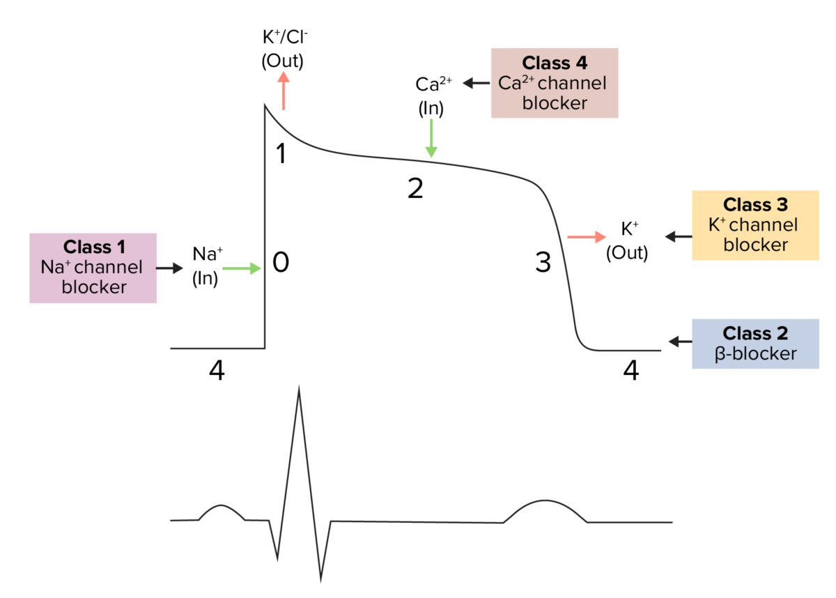
Class 2 antiarrhythmics include beta-blockers, which exert their therapeutic effects by blocking epinephrine Epinephrine The active sympathomimetic hormone from the adrenal medulla. It stimulates both the alpha- and beta- adrenergic systems, causes systemic vasoconstriction and gastrointestinal relaxation, stimulates the heart, and dilates bronchi and cerebral vessels. Sympathomimetic Drugs and norepinephrine Norepinephrine Precursor of epinephrine that is secreted by the adrenal medulla and is a widespread central and autonomic neurotransmitter. Norepinephrine is the principal transmitter of most postganglionic sympathetic fibers, and of the diffuse projection system in the brain that arises from the locus ceruleus. Receptors and Neurotransmitters of the CNS from binding to the beta-adrenergic receptors Receptors Receptors are proteins located either on the surface of or within a cell that can bind to signaling molecules known as ligands (e.g., hormones) and cause some type of response within the cell. Receptors in cardiac tissue. The outcome is an antiarrhythmic effect, which results from decreased sinoatrial node Sinoatrial node The small mass of modified cardiac muscle fibers located at the junction of the superior vena cava and right atrium. Contraction impulses probably start in this node, spread over the atrium (heart atrium) and are then transmitted by the atrioventricular bundle (bundle of His) to the ventricle (heart ventricle). Heart: Anatomy activity and increased atrioventricular conduction time and refractory period. Additional effects include decreased cardiac contractility, afterload Afterload Afterload is the resistance in the aorta that prevents blood from leaving the heart. Afterload represents the pressure the LV needs to overcome to eject blood into the aorta. Cardiac Mechanics, and blood pressure. Class 2 antiarrhythmics are used in the management of atrial fibrillation Atrial fibrillation Atrial fibrillation (AF or Afib) is a supraventricular tachyarrhythmia and the most common kind of arrhythmia. It is caused by rapid, uncontrolled atrial contractions and uncoordinated ventricular responses. Atrial Fibrillation, atrial flutter Atrial flutter Atrial flutter is a regular supraventricular tachycardia characterized by an atrial heart rate between 240/min and 340/min (typically 300/min), atrioventricular (AV) node conduction block, and a "sawtooth" pattern on an electrocardiogram (ECG). Atrial Flutter, supraventricular tachycardia Tachycardia Abnormally rapid heartbeat, usually with a heart rate above 100 beats per minute for adults. Tachycardia accompanied by disturbance in the cardiac depolarization (cardiac arrhythmia) is called tachyarrhythmia. Sepsis in Children, and ventricular arrhythmias. Adverse effects include bradycardia Bradycardia Bradyarrhythmia is a rhythm in which the heart rate is less than 60/min. Bradyarrhythmia can be physiologic, without symptoms or hemodynamic change. Pathologic bradyarrhythmia results in reduced cardiac output and hemodynamic instability causing syncope, dizziness, or dyspnea. Bradyarrhythmias, hypotension Hypotension Hypotension is defined as low blood pressure, specifically < 90/60 mm Hg, and is most commonly a physiologic response. Hypotension may be mild, serious, or life threatening, depending on the cause. Hypotension, bronchospasm Bronchospasm Asthma Drugs, fluid retention, and fatigue Fatigue The state of weariness following a period of exertion, mental or physical, characterized by a decreased capacity for work and reduced efficiency to respond to stimuli. Fibromyalgia. Beta-blockers should not be used in individuals with decompensated heart failure Heart Failure A heterogeneous condition in which the heart is unable to pump out sufficient blood to meet the metabolic need of the body. Heart failure can be caused by structural defects, functional abnormalities (ventricular dysfunction), or a sudden overload beyond its capacity. Chronic heart failure is more common than acute heart failure which results from sudden insult to cardiac function, such as myocardial infarction. Total Anomalous Pulmonary Venous Return (TAPVR), shock Shock Shock is a life-threatening condition associated with impaired circulation that results in tissue hypoxia. The different types of shock are based on the underlying cause: distributive (↑ cardiac output (CO), ↓ systemic vascular resistance (SVR)), cardiogenic (↓ CO, ↑ SVR), hypovolemic (↓ CO, ↑ SVR), obstructive (↓ CO), and mixed. Types of Shock, and severe bradycardia Bradycardia Bradyarrhythmia is a rhythm in which the heart rate is less than 60/min. Bradyarrhythmia can be physiologic, without symptoms or hemodynamic change. Pathologic bradyarrhythmia results in reduced cardiac output and hemodynamic instability causing syncope, dizziness, or dyspnea. Bradyarrhythmias.

Last updated: Jul 21, 2023

Editorial responsibility: Stanley Oiseth, Lindsay Jones, Evelin Maza

Advertisement

Advertisement

Advertisement

Advertisement

Advertisement

Advertisement

Chemistry and Pharmacodynamics

Definition

Class 2 antiarrhythmics include beta-blockers, which competitively inhibit epinephrine Epinephrine The active sympathomimetic hormone from the adrenal medulla. It stimulates both the alpha- and beta- adrenergic systems, causes systemic vasoconstriction and gastrointestinal relaxation, stimulates the heart, and dilates bronchi and cerebral vessels. Sympathomimetic Drugs and norepinephrine Norepinephrine Precursor of epinephrine that is secreted by the adrenal medulla and is a widespread central and autonomic neurotransmitter. Norepinephrine is the principal transmitter of most postganglionic sympathetic fibers, and of the diffuse projection system in the brain that arises from the locus ceruleus. Receptors and Neurotransmitters of the CNS from binding to the beta-adrenergic receptors Receptors Receptors are proteins located either on the surface of or within a cell that can bind to signaling molecules known as ligands (e.g., hormones) and cause some type of response within the cell. Receptors on vascular and cardiac cells.
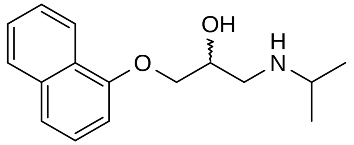
Chemical structure

  • Contain an aromatic or heteroaromatic ring with an amine group
  • Propranolol Propranolol A widely used non-cardioselective beta-adrenergic antagonist. Propranolol has been used for myocardial infarction; arrhythmia; angina pectoris; hypertension; hyperthyroidism; migraine; pheochromocytoma; and anxiety but adverse effects instigate replacement by newer drugs. Antiadrenergic Drugs is the prototype drug.
  • Most clinically used beta-blockers are delivered as racemates, except timolol.
Chemical structure of propranolol

Chemical structure of propranolol

Image: “Chemical structure of propranolol” by catclock. License: Public Domain
Mechanism of action

  • Beta-blockers competitively inhibit the beta-adrenergic receptors Receptors Receptors are proteins located either on the surface of or within a cell that can bind to signaling molecules known as ligands (e.g., hormones) and cause some type of response within the cell. Receptors:
    • Nonselective agents that bind BIND Hyperbilirubinemia of the Newborn to and induce antagonistic effects at both beta-1 (B1) and beta-2 (B2) receptors Receptors Receptors are proteins located either on the surface of or within a cell that can bind to signaling molecules known as ligands (e.g., hormones) and cause some type of response within the cell. Receptors
    • Cardioselective agents only bind BIND Hyperbilirubinemia of the Newborn to B1 receptors Receptors Receptors are proteins located either on the surface of or within a cell that can bind to signaling molecules known as ligands (e.g., hormones) and cause some type of response within the cell. Receptors.
  • Receptor Receptor Receptors are proteins located either on the surface of or within a cell that can bind to signaling molecules known as ligands (e.g., hormones) and cause some type of response within the cell. Receptors locations:
    • B1:
      • Sinoatrial (SA) node
      • Atrioventricular (AV) node
      • Atrial and ventricular muscle
      • Kidneys Kidneys The kidneys are a pair of bean-shaped organs located retroperitoneally against the posterior wall of the abdomen on either side of the spine. As part of the urinary tract, the kidneys are responsible for blood filtration and excretion of water-soluble waste in the urine. Kidneys: Anatomy
    • B2:
      • Blood vessels
      • Bronchi Bronchi The larger air passages of the lungs arising from the terminal bifurcation of the trachea. They include the largest two primary bronchi which branch out into secondary bronchi, and tertiary bronchi which extend into bronchioles and pulmonary alveoli. Bronchial Tree: Anatomy
      • GI tract
      • Uterus Uterus The uterus, cervix, and fallopian tubes are part of the internal female reproductive system. The uterus has a thick wall made of smooth muscle (the myometrium) and an inner mucosal layer (the endometrium). The most inferior portion of the uterus is the cervix, which connects the uterine cavity to the vagina. Uterus, Cervix, and Fallopian Tubes: Anatomy
      • Liver Liver The liver is the largest gland in the human body. The liver is found in the superior right quadrant of the abdomen and weighs approximately 1.5 kilograms. Its main functions are detoxification, metabolism, nutrient storage (e.g., iron and vitamins), synthesis of coagulation factors, formation of bile, filtration, and storage of blood. Liver: Anatomy
      • Urinary tract Urinary tract The urinary tract is located in the abdomen and pelvis and consists of the kidneys, ureters, urinary bladder, and urethra. The structures permit the excretion of urine from the body. Urine flows from the kidneys through the ureters to the urinary bladder and out through the urethra. Urinary Tract: Anatomy
  • Action on cardiac cells:
    • Block the binding of catecholamines Catecholamines A general class of ortho-dihydroxyphenylalkylamines derived from tyrosine. Adrenal Hormones ( epinephrine Epinephrine The active sympathomimetic hormone from the adrenal medulla. It stimulates both the alpha- and beta- adrenergic systems, causes systemic vasoconstriction and gastrointestinal relaxation, stimulates the heart, and dilates bronchi and cerebral vessels. Sympathomimetic Drugs) to G-protein-coupled receptors Receptors Receptors are proteins located either on the surface of or within a cell that can bind to signaling molecules known as ligands (e.g., hormones) and cause some type of response within the cell. Receptors (B1 receptor Receptor Receptors are proteins located either on the surface of or within a cell that can bind to signaling molecules known as ligands (e.g., hormones) and cause some type of response within the cell. Receptors) → inhibit activation of adenylyl cyclase
    • ↓ Conversion of ATP to cAMP cAMP An adenine nucleotide containing one phosphate group which is esterified to both the 3′- and 5′-positions of the sugar moiety. It is a second messenger and a key intracellular regulator, functioning as a mediator of activity for a number of hormones, including epinephrine, glucagon, and acth. Phosphodiesterase Inhibitors → ↓ protein kinase Protein kinase A family of enzymes that catalyze the conversion of ATP and a protein to adp and a phosphoprotein. Interferons A activation
    • Calcium Calcium A basic element found in nearly all tissues. It is a member of the alkaline earth family of metals with the atomic symbol ca, atomic number 20, and atomic weight 40. Calcium is the most abundant mineral in the body and combines with phosphorus to form calcium phosphate in the bones and teeth. It is essential for the normal functioning of nerves and muscles and plays a role in blood coagulation (as factor IV) and in many enzymatic processes. Electrolytes ( Ca CA Condylomata acuminata are a clinical manifestation of genital HPV infection. Condylomata acuminata are described as raised, pearly, flesh-colored, papular, cauliflower-like lesions seen in the anogenital region that may cause itching, pain, or bleeding. Condylomata Acuminata (Genital Warts)) channels Channels The Cell: Cell Membrane are not phosphorylated → do not open → ↓ Ca CA Condylomata acuminata are a clinical manifestation of genital HPV infection. Condylomata acuminata are described as raised, pearly, flesh-colored, papular, cauliflower-like lesions seen in the anogenital region that may cause itching, pain, or bleeding. Condylomata Acuminata (Genital Warts) influx into the myocardial cells
    • Nodal effect:
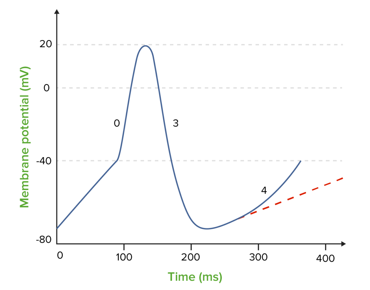
      • ↓ SA node activity
      • ↑ AV node conduction time and refractory period
      • ↓ Slope and prolonged phase 4

Physiologic effects

  • Antiarrhythmic effects:
    • Negative chronotropic effects (HR)
    • Negative dromotropic effects:
      • Delays speed and ability to instigate an action potential Action Potential Abrupt changes in the membrane potential that sweep along the cell membrane of excitable cells in response to excitation stimuli. Membrane Potential
      • ↓ Conduction velocity
    • Prolonged refractory period
    • Membrane-stabilizing activity ( MSA MSA A syndrome complex composed of three conditions which represent clinical variants of the same disease process: striatonigral degeneration; shy-drager syndrome; and the sporadic form of olivopontocerebellar atrophies. Clinical features include autonomic, cerebellar, and basal ganglia dysfunction. Pathologic examination reveals atrophy of the basal ganglia, cerebellum, pons, and medulla, with prominent loss of autonomic neurons in the brain stem and spinal cord. Atypical Parkinsonian Syndromes) and intrinsic sympathomimetic Sympathomimetic Sympathomimetic drugs, also known as adrenergic agonists, mimic the action of the stimulators (α, β, or dopamine receptors) of the sympathetic autonomic nervous system. Sympathomimetic drugs are classified based on the type of receptors the drugs act on (some agents act on several receptors but 1 is predominate). Sympathomimetic Drugs activity (ISA) do not have a major impact on antiarrhythmic potency.
  • B1 blockade:
    • ↓ Oxygen demand:
      • ↓ HR 
      • ↓ Contractility 
      • Afterload Afterload Afterload is the resistance in the aorta that prevents blood from leaving the heart. Afterload represents the pressure the LV needs to overcome to eject blood into the aorta. Cardiac Mechanics
      • ↓ Blood pressure
    • ↑ Oxygen supply (indirectly):
      • Prolongs diastole Diastole Post-systolic relaxation of the heart, especially the heart ventricles. Cardiac Cycle
      • Improves perfusion
  • B2 blockade:
  • Alpha-1 (A1) blockade:
    • Seen with carvedilol
    • Further ↓ afterload Afterload Afterload is the resistance in the aorta that prevents blood from leaving the heart. Afterload represents the pressure the LV needs to overcome to eject blood into the aorta. Cardiac Mechanics due to ↓ systemic vascular resistance Resistance Physiologically, the opposition to flow of air caused by the forces of friction. As a part of pulmonary function testing, it is the ratio of driving pressure to the rate of air flow. Ventilation: Mechanics of Breathing (SVR)
  • ECG ECG An electrocardiogram (ECG) is a graphic representation of the electrical activity of the heart plotted against time. Adhesive electrodes are affixed to the skin surface allowing measurement of cardiac impulses from many angles. The ECG provides 3-dimensional information about the conduction system of the heart, the myocardium, and other cardiac structures. Electrocardiogram (ECG) effects: PR prolongation

Adrenergic receptor Receptor Receptors are proteins located either on the surface of or within a cell that can bind to signaling molecules known as ligands (e.g., hormones) and cause some type of response within the cell. Receptors types and their properties

Table: Adrenergic receptor Receptor Receptors are proteins located either on the surface of or within a cell that can bind to signaling molecules known as ligands (e.g., hormones) and cause some type of response within the cell. Receptors types and their properties
Receptor Receptor Receptors are proteins located either on the surface of or within a cell that can bind to signaling molecules known as ligands (e.g., hormones) and cause some type of response within the cell. Receptors location Response to stimulus Response to blockade
B1 receptors Receptors Receptors are proteins located either on the surface of or within a cell that can bind to signaling molecules known as ligands (e.g., hormones) and cause some type of response within the cell. Receptors
Heart SA node ↑ HR ↓ HR
Atria ↑ Contractility and conduction speed ↓ Contractility and conduction speed
AV node and His-Purkinje fibers ↑ Automaticity and conduction speed ↓ Automaticity and conduction speed
Ventricles ↑ Contractility, automaticity, conduction speed ↓ Contractility, automaticity, conduction speed
Kidney JG cells Renin Renin A highly specific (leu-leu) endopeptidase that generates angiotensin I from its precursor angiotensinogen, leading to a cascade of reactions which elevate blood pressure and increase sodium retention by the kidney in the renin-angiotensin system. Renal Sodium and Water Regulation release Renin Renin A highly specific (leu-leu) endopeptidase that generates angiotensin I from its precursor angiotensinogen, leading to a cascade of reactions which elevate blood pressure and increase sodium retention by the kidney in the renin-angiotensin system. Renal Sodium and Water Regulation release
B2 receptors Receptors Receptors are proteins located either on the surface of or within a cell that can bind to signaling molecules known as ligands (e.g., hormones) and cause some type of response within the cell. Receptors
Arteries Arteries Arteries are tubular collections of cells that transport oxygenated blood and nutrients from the heart to the tissues of the body. The blood passes through the arteries in order of decreasing luminal diameter, starting in the largest artery (the aorta) and ending in the small arterioles. Arteries are classified into 3 types: large elastic arteries, medium muscular arteries, and small arteries and arterioles. Arteries: Histology
  • Carotid
  • Coronary
  • Peripheral
Vasodilation Vasodilation The physiological widening of blood vessels by relaxing the underlying vascular smooth muscle. Pulmonary Hypertension Drugs Vasoconstriction Vasoconstriction The physiological narrowing of blood vessels by contraction of the vascular smooth muscle. Vascular Resistance, Flow, and Mean Arterial Pressure
Lungs Lungs Lungs are the main organs of the respiratory system. Lungs are paired viscera located in the thoracic cavity and are composed of spongy tissue. The primary function of the lungs is to oxygenate blood and eliminate CO2. Lungs: Anatomy Bronchiolar smooth muscle Bronchodilation Bronchoconstriction
Bladder Bladder A musculomembranous sac along the urinary tract. Urine flows from the kidneys into the bladder via the ureters, and is held there until urination. Pyelonephritis and Perinephric Abscess Wall Relaxation Contraction
Liver Liver The liver is the largest gland in the human body. The liver is found in the superior right quadrant of the abdomen and weighs approximately 1.5 kilograms. Its main functions are detoxification, metabolism, nutrient storage (e.g., iron and vitamins), synthesis of coagulation factors, formation of bile, filtration, and storage of blood. Liver: Anatomy Hepatic tissue Stimulates glycogenolysis Glycogenolysis The release of glucose from glycogen by glycogen phosphorylase (phosphorolysis). The released glucose-1-phosphate is then converted to glucose-6-phosphate by phosphoglucomutase before entering glycolysis. Glycogenolysis is stimulated by glucagon or epinephrine via the activation of phosphorylase kinase. Glycogen Metabolism
  • Insulin Insulin Insulin is a peptide hormone that is produced by the beta cells of the pancreas. Insulin plays a role in metabolic functions such as glucose uptake, glycolysis, glycogenesis, lipogenesis, and protein synthesis. Exogenous insulin may be needed for individuals with diabetes mellitus, in whom there is a deficiency in endogenous insulin or increased insulin resistance. Insulin secretion Secretion Coagulation Studies
  • Glycogenolysis Glycogenolysis The release of glucose from glycogen by glycogen phosphorylase (phosphorolysis). The released glucose-1-phosphate is then converted to glucose-6-phosphate by phosphoglucomutase before entering glycolysis. Glycogenolysis is stimulated by glucagon or epinephrine via the activation of phosphorylase kinase. Glycogen Metabolism
A1 receptors Receptors Receptors are proteins located either on the surface of or within a cell that can bind to signaling molecules known as ligands (e.g., hormones) and cause some type of response within the cell. Receptors
Arteries Arteries Arteries are tubular collections of cells that transport oxygenated blood and nutrients from the heart to the tissues of the body. The blood passes through the arteries in order of decreasing luminal diameter, starting in the largest artery (the aorta) and ending in the small arterioles. Arteries are classified into 3 types: large elastic arteries, medium muscular arteries, and small arteries and arterioles. Arteries: Histology Peripheral Vasoconstriction Vasoconstriction The physiological narrowing of blood vessels by contraction of the vascular smooth muscle. Vascular Resistance, Flow, and Mean Arterial Pressure Vasodilation Vasodilation The physiological widening of blood vessels by relaxing the underlying vascular smooth muscle. Pulmonary Hypertension Drugs
Bladder Bladder A musculomembranous sac along the urinary tract. Urine flows from the kidneys into the bladder via the ureters, and is held there until urination. Pyelonephritis and Perinephric Abscess Sphincter Contraction Relaxation
AV: atrioventricular
SA: sinoatrial
JG: juxtaglomerular

Additional properties of beta-blockers

Some beta-blockers can exhibit the following properties and effects:

Table: Properties of beta-blockers
Beta-blocker property Effect
ISA Partial stimulation of B receptors Receptors Receptors are proteins located either on the surface of or within a cell that can bind to signaling molecules known as ligands (e.g., hormones) and cause some type of response within the cell. Receptors → suboptimal lowering of HR (avoid post-MI)
MSA MSA A syndrome complex composed of three conditions which represent clinical variants of the same disease process: striatonigral degeneration; shy-drager syndrome; and the sporadic form of olivopontocerebellar atrophies. Clinical features include autonomic, cerebellar, and basal ganglia dysfunction. Pathologic examination reveals atrophy of the basal ganglia, cerebellum, pons, and medulla, with prominent loss of autonomic neurons in the brain stem and spinal cord. Atypical Parkinsonian Syndromes Drug can ↓ cardiac conduction velocity by blocking myocyte Na+ channels Channels The Cell: Cell Membrane
NO Drug stimulates NO production → peripheral vasodilation Vasodilation The physiological widening of blood vessels by relaxing the underlying vascular smooth muscle. Pulmonary Hypertension Drugs
ISA: intrinsic sympathomimetic activity
MSA: membrane-stabilizing activity

Classification

Class 2 antiarrhythmics can be classified based on their beta-receptor selectivity:

  • Cardioselective beta-blockers (B1 receptors Receptors Receptors are proteins located either on the surface of or within a cell that can bind to signaling molecules known as ligands (e.g., hormones) and cause some type of response within the cell. Receptors):
  • Noncardioselective beta-blockers (B1 and B2 receptors Receptors Receptors are proteins located either on the surface of or within a cell that can bind to signaling molecules known as ligands (e.g., hormones) and cause some type of response within the cell. Receptors):
    • Nadolol
    • Propranolol Propranolol A widely used non-cardioselective beta-adrenergic antagonist. Propranolol has been used for myocardial infarction; arrhythmia; angina pectoris; hypertension; hyperthyroidism; migraine; pheochromocytoma; and anxiety but adverse effects instigate replacement by newer drugs. Antiadrenergic Drugs
    • Timolol
    • Carvedilol (also has A1 receptor-blocking activity)

Pharmacokinetics

Absorption Absorption Absorption involves the uptake of nutrient molecules and their transfer from the lumen of the GI tract across the enterocytes and into the interstitial space, where they can be taken up in the venous or lymphatic circulation. Digestion and Absorption

  • Most drugs are rapidly and completely absorbed from the GI tract.
  • ↓ Rate of absorption Absorption Absorption involves the uptake of nutrient molecules and their transfer from the lumen of the GI tract across the enterocytes and into the interstitial space, where they can be taken up in the venous or lymphatic circulation. Digestion and Absorption:
    • Elderly
    • Individuals with renal failure Renal failure Conditions in which the kidneys perform below the normal level in the ability to remove wastes, concentrate urine, and maintain electrolyte balance; blood pressure; and calcium metabolism. Renal insufficiency can be classified by the degree of kidney damage (as measured by the level of proteinuria) and reduction in glomerular filtration rate. Crush Syndrome

Distribution

  • Rapid distribution within the body
  • Protein binding is variable Variable Variables represent information about something that can change. The design of the measurement scales, or of the methods for obtaining information, will determine the data gathered and the characteristics of that data. As a result, a variable can be qualitative or quantitative, and may be further classified into subgroups. Types of Variables.
  • Lipophilic agents can cross the blood-brain barrier Blood-brain barrier Specialized non-fenestrated tightly-joined endothelial cells with tight junctions that form a transport barrier for certain substances between the cerebral capillaries and the brain tissue. Systemic and Special Circulations.

Metabolism

  • Most lipophilic agents undergo significant 1st-pass hepatic metabolism:
    • Carvedilol
    • Metoprolol Metoprolol A selective adrenergic beta-1 blocking agent that is commonly used to treat angina pectoris; hypertension; and cardiac arrhythmias. Antiadrenergic Drugs
    • Propranolol Propranolol A widely used non-cardioselective beta-adrenergic antagonist. Propranolol has been used for myocardial infarction; arrhythmia; angina pectoris; hypertension; hyperthyroidism; migraine; pheochromocytoma; and anxiety but adverse effects instigate replacement by newer drugs. Antiadrenergic Drugs
  • Some drugs are converted to their active metabolites.
  • Hydrophobic compounds tend to undergo limited hepatic metabolism.

Excretion

  • Hepatic metabolism (lipophilic agents) → bile Bile An emulsifying agent produced in the liver and secreted into the duodenum. Its composition includes bile acids and salts; cholesterol; and electrolytes. It aids digestion of fats in the duodenum. Gallbladder and Biliary Tract: Anatomy
  • Hydrophilic Hydrophilic Aminoglycosides agents → renal excretion
  • Impaired liver Liver The liver is the largest gland in the human body. The liver is found in the superior right quadrant of the abdomen and weighs approximately 1.5 kilograms. Its main functions are detoxification, metabolism, nutrient storage (e.g., iron and vitamins), synthesis of coagulation factors, formation of bile, filtration, and storage of blood. Liver: Anatomy and kidney function can ↓ rate of elimination Elimination The initial damage and destruction of tumor cells by innate and adaptive immunity. Completion of the phase means no cancer growth. Cancer Immunotherapy

Indications

Arrhythmia

Class 2 antiarrhythmics are used in the management of:

  • Atrial fibrillation Atrial fibrillation Atrial fibrillation (AF or Afib) is a supraventricular tachyarrhythmia and the most common kind of arrhythmia. It is caused by rapid, uncontrolled atrial contractions and uncoordinated ventricular responses. Atrial Fibrillation
  • Atrial flutter Atrial flutter Atrial flutter is a regular supraventricular tachycardia characterized by an atrial heart rate between 240/min and 340/min (typically 300/min), atrioventricular (AV) node conduction block, and a “sawtooth” pattern on an electrocardiogram (ECG). Atrial Flutter
  • Supraventricular tachycardias
  • Ventricular arrhythmias

Additional uses

Beta-blockers, as a class, encompass FDA approvals for treating:

  • Coronary heart disease Coronary heart disease Coronary heart disease (CHD), or ischemic heart disease, describes a situation in which an inadequate supply of blood to the myocardium exists due to a stenosis of the coronary arteries, typically from atherosclerosis. Coronary Heart Disease:
    • Angina
    • Post-MI
  • Essential tremors
  • Glaucoma Glaucoma Glaucoma is an optic neuropathy characterized by typical visual field defects and optic nerve atrophy seen as optic disc cupping on examination. The acute form of glaucoma is a medical emergency. Glaucoma is often, but not always, caused by increased intraocular pressure (IOP). Glaucoma (timolol)
  • Heart failure Heart Failure A heterogeneous condition in which the heart is unable to pump out sufficient blood to meet the metabolic need of the body. Heart failure can be caused by structural defects, functional abnormalities (ventricular dysfunction), or a sudden overload beyond its capacity. Chronic heart failure is more common than acute heart failure which results from sudden insult to cardiac function, such as myocardial infarction. Total Anomalous Pulmonary Venous Return (TAPVR) with reduced ejection fraction Ejection fraction Cardiac Cycle
  • Hypertension Hypertension Hypertension, or high blood pressure, is a common disease that manifests as elevated systemic arterial pressures. Hypertension is most often asymptomatic and is found incidentally as part of a routine physical examination or during triage for an unrelated medical encounter. Hypertension
  • Hypertrophic subaortic stenosis Stenosis Hypoplastic Left Heart Syndrome (HLHS)
  • Migraine Migraine Migraine headache is a primary headache disorder and is among the most prevalent disorders in the world. Migraine is characterized by episodic, moderate to severe headaches that may be associated with increased sensitivity to light and sound, as well as nausea and/or vomiting. Migraine Headache prophylaxis Prophylaxis Cephalosporins
  • Pheochromocytoma Pheochromocytoma Pheochromocytoma is a catecholamine-secreting tumor derived from chromaffin cells. The majority of tumors originate in the adrenal medulla, but they may also arise from sympathetic ganglia (also referred to as paraganglioma). Symptoms are associated with excessive catecholamine production and commonly include hypertension, tachycardia, headache, and sweating. Pheochromocytoma; adjunct
  • Variceal hemorrhage prophylaxis Prophylaxis Cephalosporins

Indications for class 2 antiarrhythmic medications

Table: Properties of beta-blockers
Medication Arrhythmia Angina MI MI MI is ischemia and death of an area of myocardial tissue due to insufficient blood flow and oxygenation, usually from thrombus formation on a ruptured atherosclerotic plaque in the epicardial arteries. Clinical presentation is most commonly with chest pain, but women and patients with diabetes may have atypical symptoms. Myocardial Infarction HF Hypertension Hypertension Hypertension, or high blood pressure, is a common disease that manifests as elevated systemic arterial pressures. Hypertension is most often asymptomatic and is found incidentally as part of a routine physical examination or during triage for an unrelated medical encounter. Hypertension MSA MSA A syndrome complex composed of three conditions which represent clinical variants of the same disease process: striatonigral degeneration; shy-drager syndrome; and the sporadic form of olivopontocerebellar atrophies. Clinical features include autonomic, cerebellar, and basal ganglia dysfunction. Pathologic examination reveals atrophy of the basal ganglia, cerebellum, pons, and medulla, with prominent loss of autonomic neurons in the brain stem and spinal cord. Atypical Parkinsonian Syndromes
Atenolol*
  • Afib Afib Atrial fibrillation (AF or Afib) is a supraventricular tachyarrhythmia and the most common kind of arrhythmia. It is caused by rapid, uncontrolled atrial contractions and uncoordinated ventricular responses. Atrial Fibrillation/flutter
  • SVT SVT Supraventricular tachycardias are related disorders in which the elevation in heart rate is driven by pathophysiology in the atria. This group falls under the larger umbrella of tachyarrhythmias and includes paroxysmal supraventricular tachycardias (PSVTs), ventricular pre-excitation syndromes (i.e. Wolff-Parkinson-White syndrome), atrial flutter, multifocal atrial tachycardia, and atrial fibrillation. Supraventricular Tachycardias
  • V
X X X
Betaxolol* Afib Afib Atrial fibrillation (AF or Afib) is a supraventricular tachyarrhythmia and the most common kind of arrhythmia. It is caused by rapid, uncontrolled atrial contractions and uncoordinated ventricular responses. Atrial Fibrillation X X
Bisoprolol*
  • Afib Afib Atrial fibrillation (AF or Afib) is a supraventricular tachyarrhythmia and the most common kind of arrhythmia. It is caused by rapid, uncontrolled atrial contractions and uncoordinated ventricular responses. Atrial Fibrillation/flutter
  • V
X X X
Metoprolol Metoprolol A selective adrenergic beta-1 blocking agent that is commonly used to treat angina pectoris; hypertension; and cardiac arrhythmias. Antiadrenergic Drugs*
  • Afib Afib Atrial fibrillation (AF or Afib) is a supraventricular tachyarrhythmia and the most common kind of arrhythmia. It is caused by rapid, uncontrolled atrial contractions and uncoordinated ventricular responses. Atrial Fibrillation/flutter
  • SVT SVT Supraventricular tachycardias are related disorders in which the elevation in heart rate is driven by pathophysiology in the atria. This group falls under the larger umbrella of tachyarrhythmias and includes paroxysmal supraventricular tachycardias (PSVTs), ventricular pre-excitation syndromes (i.e. Wolff-Parkinson-White syndrome), atrial flutter, multifocal atrial tachycardia, and atrial fibrillation. Supraventricular Tachycardias
  • V
X X X X X
Esmolol Esmolol Antiadrenergic Drugs*
  • Afib Afib Atrial fibrillation (AF or Afib) is a supraventricular tachyarrhythmia and the most common kind of arrhythmia. It is caused by rapid, uncontrolled atrial contractions and uncoordinated ventricular responses. Atrial Fibrillation/flutter
  • SVT SVT Supraventricular tachycardias are related disorders in which the elevation in heart rate is driven by pathophysiology in the atria. This group falls under the larger umbrella of tachyarrhythmias and includes paroxysmal supraventricular tachycardias (PSVTs), ventricular pre-excitation syndromes (i.e. Wolff-Parkinson-White syndrome), atrial flutter, multifocal atrial tachycardia, and atrial fibrillation. Supraventricular Tachycardias
  • V
X
Acebutolol* V X X X X
Nadolol
  • Afib Afib Atrial fibrillation (AF or Afib) is a supraventricular tachyarrhythmia and the most common kind of arrhythmia. It is caused by rapid, uncontrolled atrial contractions and uncoordinated ventricular responses. Atrial Fibrillation
  • SVT SVT Supraventricular tachycardias are related disorders in which the elevation in heart rate is driven by pathophysiology in the atria. This group falls under the larger umbrella of tachyarrhythmias and includes paroxysmal supraventricular tachycardias (PSVTs), ventricular pre-excitation syndromes (i.e. Wolff-Parkinson-White syndrome), atrial flutter, multifocal atrial tachycardia, and atrial fibrillation. Supraventricular Tachycardias
  • V
X X X
Propranolol Propranolol A widely used non-cardioselective beta-adrenergic antagonist. Propranolol has been used for myocardial infarction; arrhythmia; angina pectoris; hypertension; hyperthyroidism; migraine; pheochromocytoma; and anxiety but adverse effects instigate replacement by newer drugs. Antiadrenergic Drugs
  • Afib Afib Atrial fibrillation (AF or Afib) is a supraventricular tachyarrhythmia and the most common kind of arrhythmia. It is caused by rapid, uncontrolled atrial contractions and uncoordinated ventricular responses. Atrial Fibrillation/flutter
  • SVT SVT Supraventricular tachycardias are related disorders in which the elevation in heart rate is driven by pathophysiology in the atria. This group falls under the larger umbrella of tachyarrhythmias and includes paroxysmal supraventricular tachycardias (PSVTs), ventricular pre-excitation syndromes (i.e. Wolff-Parkinson-White syndrome), atrial flutter, multifocal atrial tachycardia, and atrial fibrillation. Supraventricular Tachycardias
  • V
X X X X
Timolol Afib Afib Atrial fibrillation (AF or Afib) is a supraventricular tachyarrhythmia and the most common kind of arrhythmia. It is caused by rapid, uncontrolled atrial contractions and uncoordinated ventricular responses. Atrial Fibrillation X X X
Carvedilol Afib Afib Atrial fibrillation (AF or Afib) is a supraventricular tachyarrhythmia and the most common kind of arrhythmia. It is caused by rapid, uncontrolled atrial contractions and uncoordinated ventricular responses. Atrial Fibrillation/flutter X X X X X
* denotes cardioselective beta-blockers
MSA: membrane-stabilizing activity
Afib: atrial fibrillation
SVT: supraventricular tachycardia
V: ventricular arrhythmias
HF: heart failure

Adverse Effects and Contraindications

Adverse effects

  • Cardiovascular:
    • Bradycardia Bradycardia Bradyarrhythmia is a rhythm in which the heart rate is less than 60/min. Bradyarrhythmia can be physiologic, without symptoms or hemodynamic change. Pathologic bradyarrhythmia results in reduced cardiac output and hemodynamic instability causing syncope, dizziness, or dyspnea. Bradyarrhythmias
    • Hypotension Hypotension Hypotension is defined as low blood pressure, specifically < 90/60 mm Hg, and is most commonly a physiologic response. Hypotension may be mild, serious, or life threatening, depending on the cause. Hypotension
    • Heart failure Heart Failure A heterogeneous condition in which the heart is unable to pump out sufficient blood to meet the metabolic need of the body. Heart failure can be caused by structural defects, functional abnormalities (ventricular dysfunction), or a sudden overload beyond its capacity. Chronic heart failure is more common than acute heart failure which results from sudden insult to cardiac function, such as myocardial infarction. Total Anomalous Pulmonary Venous Return (TAPVR) exacerbation (with initial therapy)
    • Withdrawal with discontinuation that can precipitate (from ↑ sympathetic activity):
      • MI MI MI is ischemia and death of an area of myocardial tissue due to insufficient blood flow and oxygenation, usually from thrombus formation on a ruptured atherosclerotic plaque in the epicardial arteries. Clinical presentation is most commonly with chest pain, but women and patients with diabetes may have atypical symptoms. Myocardial Infarction (in individuals with coronary artery Coronary Artery Truncus Arteriosus disease)
      • Ventricular tachyarrhythmias
      • Sudden cardiac death Sudden cardiac death Cardiac arrest is the sudden, complete cessation of cardiac output with hemodynamic collapse. Patients present as pulseless, unresponsive, and apneic. Rhythms associated with cardiac arrest are ventricular fibrillation/tachycardia, asystole, or pulseless electrical activity. Cardiac Arrest
  • CNS:
    • Fatigue Fatigue The state of weariness following a period of exertion, mental or physical, characterized by a decreased capacity for work and reduced efficiency to respond to stimuli. Fibromyalgia
    • Depression
    • Vivid dreams
    • Sexual dysfunction Sexual dysfunction Physiological disturbances in normal sexual performance in either the male or the female. Sexual Physiology
  • Respiratory: bronchoconstriction
  • Endocrine:
    • Hypoglycemia Hypoglycemia Hypoglycemia is an emergency condition defined as a serum glucose level ≤ 70 mg/dL (≤ 3.9 mmol/L) in diabetic patients. In nondiabetic patients, there is no specific or defined limit for normal serum glucose levels, and hypoglycemia is defined mainly by its clinical features. Hypoglycemia
    • Hyperglycemia Hyperglycemia Abnormally high blood glucose level. Diabetes Mellitus (usually in individuals with noninsulin-dependent diabetes Diabetes Diabetes mellitus (DM) is a metabolic disease characterized by hyperglycemia and dysfunction of the regulation of glucose metabolism by insulin. Type 1 DM is diagnosed mostly in children and young adults as the result of autoimmune destruction of β cells in the pancreas and the resulting lack of insulin. Type 2 DM has a significant association with obesity and is characterized by insulin resistance. Diabetes Mellitus mellitus)
    • Dyslipidemia
    • Weight gain
    • Fluid retention
  • Dermatologic: potential exacerbation of psoriasis Psoriasis Psoriasis is a common T-cell-mediated inflammatory skin condition. The etiology is unknown, but is thought to be due to genetic inheritance and environmental triggers. There are 4 major subtypes, with the most common form being chronic plaque psoriasis. Psoriasis

Contraindications Contraindications A condition or factor associated with a recipient that makes the use of a drug, procedure, or physical agent improper or inadvisable. Contraindications may be absolute (life threatening) or relative (higher risk of complications in which benefits may outweigh risks). Noninvasive Ventilation

  • Decompensated heart failure Heart Failure A heterogeneous condition in which the heart is unable to pump out sufficient blood to meet the metabolic need of the body. Heart failure can be caused by structural defects, functional abnormalities (ventricular dysfunction), or a sudden overload beyond its capacity. Chronic heart failure is more common than acute heart failure which results from sudden insult to cardiac function, such as myocardial infarction. Total Anomalous Pulmonary Venous Return (TAPVR)
  • Cardiogenic shock Cardiogenic shock Shock resulting from diminution of cardiac output in heart disease. Types of Shock
  • Hypotension Hypotension Hypotension is defined as low blood pressure, specifically < 90/60 mm Hg, and is most commonly a physiologic response. Hypotension may be mild, serious, or life threatening, depending on the cause. Hypotension
  • Severe bradycardia Bradycardia Bradyarrhythmia is a rhythm in which the heart rate is less than 60/min. Bradyarrhythmia can be physiologic, without symptoms or hemodynamic change. Pathologic bradyarrhythmia results in reduced cardiac output and hemodynamic instability causing syncope, dizziness, or dyspnea. Bradyarrhythmias
  • 2nd- or 3rd-degree AV block AV block Atrioventricular (AV) block is a bradyarrhythmia caused by delay, or interruption, in the electrical conduction between the atria and the ventricles. Atrioventricular block occurs due to either anatomic or functional impairment, and is classified into 3 types. Atrioventricular block (AV block)
  • Sick-sinus syndrome (unless a pacemaker Pacemaker A device designed to stimulate, by electric impulses, contraction of the heart muscles. It may be temporary (external) or permanent (internal or internal-external). Bradyarrhythmias is present)
  • Severe reactive airway Airway ABCDE Assessment disease
  • Pulmonary hypertension Pulmonary Hypertension Pulmonary hypertension (PH) or pulmonary arterial hypertension (PAH) is characterized by elevated pulmonary arterial pressure, which can lead to chronic progressive right heart failure. Pulmonary hypertension is grouped into 5 categories based on etiology, which include primary PAH, and PH due to cardiac disease, lung or hypoxic disease, chronic thromboembolic disease, and multifactorial or unclear etiologies. Pulmonary Hypertension ( esmolol Esmolol Antiadrenergic Drugs)

Drug interactions

  • Enhance bradycardia Bradycardia Bradyarrhythmia is a rhythm in which the heart rate is less than 60/min. Bradyarrhythmia can be physiologic, without symptoms or hemodynamic change. Pathologic bradyarrhythmia results in reduced cardiac output and hemodynamic instability causing syncope, dizziness, or dyspnea. Bradyarrhythmias:
    • Ca-channel blockers
    • Amiodarone Amiodarone An antianginal and class III antiarrhythmic drug. It increases the duration of ventricular and atrial muscle action by inhibiting potassium channels and voltage-gated sodium channels. There is a resulting decrease in heart rate and in vascular resistance. Pulmonary Fibrosis
    • A2 agonists
    • Digoxin Digoxin A cardiotonic glycoside obtained mainly from digitalis lanata; it consists of three sugars and the aglycone digoxigenin. Digoxin has positive inotropic and negative chronotropic activity. It is used to control ventricular rate in atrial fibrillation and in the management of congestive heart failure with atrial fibrillation. Its use in congestive heart failure and sinus rhythm is less certain. The margin between toxic and therapeutic doses is small. Cardiac Glycosides
  • Enhanced hypoglycemia Hypoglycemia Hypoglycemia is an emergency condition defined as a serum glucose level ≤ 70 mg/dL (≤ 3.9 mmol/L) in diabetic patients. In nondiabetic patients, there is no specific or defined limit for normal serum glucose levels, and hypoglycemia is defined mainly by its clinical features. Hypoglycemia:
    • Sulfonylureas Sulfonylureas Sulfonamides and Trimethoprim
    • Insulin Insulin Insulin is a peptide hormone that is produced by the beta cells of the pancreas. Insulin plays a role in metabolic functions such as glucose uptake, glycolysis, glycogenesis, lipogenesis, and protein synthesis. Exogenous insulin may be needed for individuals with diabetes mellitus, in whom there is a deficiency in endogenous insulin or increased insulin resistance. Insulin
  • Excessive vasoconstriction Vasoconstriction The physiological narrowing of blood vessels by contraction of the vascular smooth muscle. Vascular Resistance, Flow, and Mean Arterial Pressure: ergot alkaloids Ergot alkaloids Alkaloids originally isolated from the ergot fungus claviceps purpurea (hypocreaceae). They include compounds that are structurally related to ergoline (ergolines) and ergotamine (ergotamines). Many of the ergot alkaloids act as alpha-adrenergic antagonists. Triptans and Ergot Alkaloids

Comparison of Antiarrhythmic Drug Classes

The following table compares antiarrhythmic classes 1‒4. Class 5 is not included due to the varied mechanisms of action and effects.

Table: Comparison of antiarrhythmic drug classes 1‒4
Class Mechanism of action Effects Arrhythmia indications
1 1A
  • ↓ Phase 0 slope
  • ↓ Conduction velocity in nonnodal tissue
  • Atrial and ventricular
  • WPW
1B Ventricular
1C Mostly atrial
2
  • Block beta receptors Receptors Receptors are proteins located either on the surface of or within a cell that can bind to signaling molecules known as ligands (e.g., hormones) and cause some type of response within the cell. Receptors
  • Ca CA Condylomata acuminata are a clinical manifestation of genital HPV infection. Condylomata acuminata are described as raised, pearly, flesh-colored, papular, cauliflower-like lesions seen in the anogenital region that may cause itching, pain, or bleeding. Condylomata Acuminata (Genital Warts) influx into myocardial cells
  • Affects refractory period
  • ↓ Phase 4 slope
  • ↑ Phase 4 duration
  • ↓ Conduction velocity in nodal and nonnodal tissue
Atrial and ventricular
3 Atrial and ventricular
4
  • Block Ca CA Condylomata acuminata are a clinical manifestation of genital HPV infection. Condylomata acuminata are described as raised, pearly, flesh-colored, papular, cauliflower-like lesions seen in the anogenital region that may cause itching, pain, or bleeding. Condylomata Acuminata (Genital Warts) channels Channels The Cell: Cell Membrane
  • Ca CA Condylomata acuminata are a clinical manifestation of genital HPV infection. Condylomata acuminata are described as raised, pearly, flesh-colored, papular, cauliflower-like lesions seen in the anogenital region that may cause itching, pain, or bleeding. Condylomata Acuminata (Genital Warts) influx into myocardial cells
Atrial
AV: atrioventricular
Ca: calcium
SA: sinoatrial
WPW: Wolff-Parkinson-White syndrome

References

  1. Hall, J.E., Guyton, A.C. (2016). The Autonomic Nervous System and the Adrenal Medulla. Guyton and Hall Textbook of Medical Physiology, 13e. Philadelphia, PA: Elsevier.
  2. Opie, L.H. (2013). β-blocking agents. Drugs for the Heart, 8e. Philadelphia, PA: Elsevier Saunders.
  3. Pencina, M.J., Navar, A.M., Wojdyla, D., et al. (2019). Quantifying Importance of Major Risk Factors for Coronary Heart Disease. Circulation; 139, 1603–1611. https://doi.org/10.1161/CIRCULATIONAHA.117.031855.
  4. Benjamin, E.J., Muntner, P., Alonso, A., Bittencourt, M.S., et al. (2019). Heart disease and stroke statistics—2019 update: A report from the American Heart Association. Circulation; 139:e56–e528. https://doi.org/10.1161/CIR.0000000000000659.
  5. Lipton, R.B., Stewart, W.F., Diamond, S., et al. (2001). Prevalence and burden of migraine in the United States: Data from the American Migraine Study II. Headache; 41, 646–657. https://doi.org/10.1046/j.1526-4610.2001.041007646.x.
  6. Jackson, J.L., Kuriyama, A., Kuwatsuka, Y., et al. (2019). Beta-blockers for the prevention of headache in adults, a systematic review and meta-analysis. PLoS One; 14, e0212785. https://doi.org/10.1371/journal.pone.0212785.
  7. De Leo, S., Lee, S.Y., Braverman, L.E. (2016). Hyperthyroidism. The Lancet; 388, 906–918. http://dx.doi.org/10.1016/S0140-6736(16)00278-6.
  8. Podrid, P. (2020). Major side effects of beta blockers. In Levy, S. (Ed.) UpToDate. Retrieved June 13, 2021, from https://www.uptodate.com/contents/major-side-effects-of-beta-blockers.
  9. Kannam, J., Gersh, B. (2021). Beta blockers in the management of chronic coronary syndrome. Verheugt, F. UpToDate. Retrieved June 13, 2021, from https://www.uptodate.com/contents/beta-blockers-in-the-management-of-chronic-coronary-syndrome.

Create your free account or log in to continue reading!

Sign up now and get free access to Lecturio with concept pages, medical videos, and questions for your medical education.

User Reviews

Details