Chronic Shoulder Pain

Shoulder pain Pain An unpleasant sensation induced by noxious stimuli which are detected by nerve endings of nociceptive neurons. Pain: Types and Pathways is considered chronic when present for > 6 months. Common conditions resulting in chronic shoulder pain Pain An unpleasant sensation induced by noxious stimuli which are detected by nerve endings of nociceptive neurons. Pain: Types and Pathways include rotator cuff disorders, adhesive capsulitis, shoulder instability, and joint arthritis Arthritis Acute or chronic inflammation of joints. Osteoarthritis. Shoulder pain Pain An unpleasant sensation induced by noxious stimuli which are detected by nerve endings of nociceptive neurons. Pain: Types and Pathways can be intrinsic or extrinsic to the joint. The potential of serious causes of referred pain Referred Pain Spinal Disk Herniation to the shoulder such as cardiovascular and abdominal etiologies should be considered. A focused history and physical examination is essential and should include inspection Inspection Dermatologic Examination, palpation Palpation Application of fingers with light pressure to the surface of the body to determine consistency of parts beneath in physical diagnosis; includes palpation for determining the outlines of organs. Dermatologic Examination, range of motion Range of motion The distance and direction to which a bone joint can be extended. Range of motion is a function of the condition of the joints, muscles, and connective tissues involved. Joint flexibility can be improved through appropriate muscle strength exercises. Examination of the Upper Limbs (ROM), and provocative testing. Imaging often includes plain radiographs occasionally supplemented with MRI. Management varies related to specific disorders and frequently requires physical therapy Physical Therapy Becker Muscular Dystrophy.

Last updated: May 16, 2024

Editorial responsibility: Stanley Oiseth, Lindsay Jones, Evelin Maza

Overview

Epidemiology

  • Shoulder pain Pain An unpleasant sensation induced by noxious stimuli which are detected by nerve endings of nociceptive neurons. Pain: Types and Pathways is responsible for approximately 10%–20% of all musculoskeletal complaints.
  • Annual incidence Incidence The number of new cases of a given disease during a given period in a specified population. It also is used for the rate at which new events occur in a defined population. It is differentiated from prevalence, which refers to all cases in the population at a given time. Measures of Disease Frequency: 15 per 1,000 individuals in the primary care setting
  • In 2000, shoulder complaints in the United States totaled $7,000,000,000.

Etiology

  • Risk factors:
    • Age: 
      • < 40: overuse injuries, instability, or labral tear
      • > 40: rotator cuff tear, adhesive capsulitis, osteoarthritis Osteoarthritis Osteoarthritis (OA) is the most common form of arthritis, and is due to cartilage destruction and changes of the subchondral bone. The risk of developing this disorder increases with age, obesity, and repetitive joint use or trauma. Patients develop gradual joint pain, stiffness lasting < 30 minutes, and decreased range of motion. Osteoarthritis ( OA OA Osteoarthritis (OA) is the most common form of arthritis, and is due to cartilage destruction and changes of the subchondral bone. The risk of developing this disorder increases with age, obesity, and repetitive joint use or trauma. Patients develop gradual joint pain, stiffness lasting < 30 minutes, and decreased range of motion. Osteoarthritis), or bicep disorders
    • Vocation or recreational activities
    • History of previous trauma
  • Intrinsic causes: pain Pain An unpleasant sensation induced by noxious stimuli which are detected by nerve endings of nociceptive neurons. Pain: Types and Pathways originates from the shoulder complex Shoulder complex The shoulder complex comprises the glenohumeral joint, sternoclavicular joint, acromioclavicular joint, and the scapulothoracic articulation, and connects the upper limb to the trunk. This group of joints consists of the clavicle, scapula, and humerus bones, multiple muscles and supporting ligaments, cartilage, and bursae. Shoulder Joint: Anatomy:
    • Most common: rotator cuff related disorders (e.g., rotator cuff tears, tendinopathy, bursitis, impingement)
    • Common:
      • Glenohumeral joint disorders (e.g., glenohumeral OA OA Osteoarthritis (OA) is the most common form of arthritis, and is due to cartilage destruction and changes of the subchondral bone. The risk of developing this disorder increases with age, obesity, and repetitive joint use or trauma. Patients develop gradual joint pain, stiffness lasting < 30 minutes, and decreased range of motion. Osteoarthritis, adhesive capsulitis)
      • Biceps Biceps Arm: Anatomy tendinopathy or rupture
      • Acromioclavicular joint Acromioclavicular joint The gliding joint formed by the outer extremity of the clavicle and the inner margin of the acromion process of the scapula. Examination of the Upper Limbs OA OA Osteoarthritis (OA) is the most common form of arthritis, and is due to cartilage destruction and changes of the subchondral bone. The risk of developing this disorder increases with age, obesity, and repetitive joint use or trauma. Patients develop gradual joint pain, stiffness lasting < 30 minutes, and decreased range of motion. Osteoarthritis
      • Labral tears Labral Tears Legg-Calvé-Perthes Disease
    • Less common: inflammatory disorders (e.g., rheumatoid arthritis Arthritis Acute or chronic inflammation of joints. Osteoarthritis, gout Gout Gout is a heterogeneous metabolic disease associated with elevated serum uric acid levels (> 6.8 mg/dL) and abnormal deposits of monosodium urate in tissues. The condition is often familial and is initially characterized by painful, recurring, and usually monoarticular acute arthritis, or “gout flare,” followed later by chronic deforming arthritis. Gout, pseudogout, septic arthritis Arthritis Acute or chronic inflammation of joints. Osteoarthritis, polymyalgia rheumatica Polymyalgia rheumatica A syndrome in the elderly characterized by proximal joint and muscle pain, high erythrocyte sedimentation rate, and a self-limiting course. Pain is usually accompanied by evidence of an inflammatory reaction. Women are affected twice as commonly as men and caucasians more frequently than other groups. The condition is frequently associated with giant cell arteritis and some theories pose the possibility that the two diseases arise from a single etiology or even that they are the same entity. Giant Cell Arteritis)
  • Extrinsic causes:
    • Neurologic:
      • Cervical radiculopathy Cervical Radiculopathy Spinal Disk Herniation
      • Brachial plexopathy Plexopathy Neuropathy is a nerve pathology presenting with sensory, motor, or autonomic impairment secondary to dysfunction of the affected nerve. The peripheral nerves are derived from several plexuses, with the brachial and lumbosacral plexuses supplying the major innervation to the extremities. Mononeuropathy (affecting a single nerve) and plexopathy (affecting the plexus) can occur from trauma, compression, and systemic diseases. Mononeuropathy and Plexopathy
      • Herpes zoster Herpes Zoster Varicella-zoster virus (VZV) is a linear, double-stranded DNA virus in the Herpesviridae family. Shingles (also known as herpes zoster) is more common in adults and occurs due to the reactivation of VZV. Varicella-Zoster Virus/Chickenpox
      • Thoracic outlet Thoracic Outlet Thoracic Outlet Syndrome syndrome ( TOS TOS Thoracic outlet syndrome (TOS) is a broad term used for a spectrum of syndromes related to the general region of the thoracic outlet, which involves the compression or irritation of elements of the brachial plexus, subclavian artery, or subclavian vein. Thoracic Outlet Syndrome)
    • Cardiovascular/pulmonary:
      • Myocardial infarction Myocardial infarction MI is ischemia and death of an area of myocardial tissue due to insufficient blood flow and oxygenation, usually from thrombus formation on a ruptured atherosclerotic plaque in the epicardial arteries. Clinical presentation is most commonly with chest pain, but women and patients with diabetes may have atypical symptoms. Myocardial Infarction
      • Angina pectoris Angina pectoris The symptom of paroxysmal pain consequent to myocardial ischemia usually of distinctive character, location and radiation. It is thought to be provoked by a transient stressful situation during which the oxygen requirements of the myocardium exceed that supplied by the coronary circulation. Stable and Unstable Angina
      • Aortic dissection Aortic dissection Aortic dissection occurs due to shearing stress from pulsatile pressure causing a tear in the tunica intima of the aortic wall. This tear allows blood to flow into the media, creating a “false lumen.” Aortic dissection is most commonly caused by uncontrolled hypertension. Aortic Dissection
      • Pulmonary embolism Pulmonary Embolism Pulmonary embolism (PE) is a potentially fatal condition that occurs as a result of intraluminal obstruction of the main pulmonary artery or its branches. The causative factors include thrombi, air, amniotic fluid, and fat. In PE, gas exchange is impaired due to the decreased return of deoxygenated blood to the lungs. Pulmonary Embolism (PE)
      • Pancoast tumor Pancoast Tumor Thoracic Outlet Syndrome (apical lung tumor Tumor Inflammation)
      • Other malignancies
      • Vascular thrombosis Thrombosis Formation and development of a thrombus or blood clot in the blood vessel. Epidemic Typhus
    • Abdominal:
      • Radiates to left shoulder: splenic rupture Splenic rupture Splenic rupture is a medical emergency that carries a significant risk of hypovolemic shock and death. Injury to the spleen accounts for nearly half of all injuries to intra-abdominal organs. The most common reason for a rupture of the spleen is blunt abdominal trauma, specifically, motor vehicle accidents. Rupture of the Spleen (Kehr sign)
      • Radiates to right shoulder: gallbladder Gallbladder The gallbladder is a pear-shaped sac, located directly beneath the liver, that sits on top of the superior part of the duodenum. The primary functions of the gallbladder include concentrating and storing up to 50 mL of bile. Gallbladder and Biliary Tract: Anatomy or hepatic disorders (e.g., cholecystitis Cholecystitis Cholecystitis is the inflammation of the gallbladder (GB) usually caused by the obstruction of the cystic duct (acute cholecystitis). Mechanical irritation by gallstones can also produce chronic GB inflammation. Cholecystitis is one of the most common complications of cholelithiasis but inflammation without gallstones can occur in a minority of patients. Cholecystitis, abscess Abscess Accumulation of purulent material in tissues, organs, or circumscribed spaces, usually associated with signs of infection. Chronic Granulomatous Disease)
      • Radiates to either shoulder: abdominal (e.g., abscess Abscess Accumulation of purulent material in tissues, organs, or circumscribed spaces, usually associated with signs of infection. Chronic Granulomatous Disease, hemorrhage, ruptured viscus, pancreatitis Pancreatitis Inflammation of the pancreas. Pancreatitis is classified as acute unless there are computed tomographic or endoscopic retrograde cholangiopancreatographic findings of chronic pancreatitis. The two most common forms of acute pancreatitis are alcoholic pancreatitis and gallstone pancreatitis. Acute Pancreatitis) or esophageal disorders

Anatomy of the shoulder joint Shoulder joint The articulation between the head of the humerus and the glenoid cavity of the scapula. Examination of the Upper Limbs

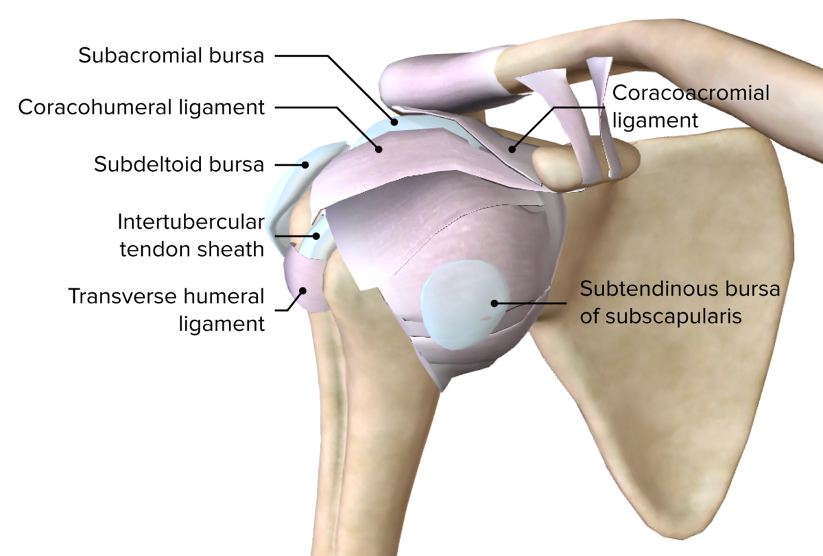
  • The shoulder complex Shoulder complex The shoulder complex comprises the glenohumeral joint, sternoclavicular joint, acromioclavicular joint, and the scapulothoracic articulation, and connects the upper limb to the trunk. This group of joints consists of the clavicle, scapula, and humerus bones, multiple muscles and supporting ligaments, cartilage, and bursae. Shoulder Joint: Anatomy is made up of multiple joints, bones, ligaments, and tendons.
  • The glenohumeral joint has the most mobility of any joint in the body.
  • Glenohumeral joint:
    • Passive restraints:
      • Joint capsule Capsule An envelope of loose gel surrounding a bacterial cell which is associated with the virulence of pathogenic bacteria. Some capsules have a well-defined border, whereas others form a slime layer that trails off into the medium. Most capsules consist of relatively simple polysaccharides but there are some bacteria whose capsules are made of polypeptides. Bacteroides
      • Glenohumeral ligaments
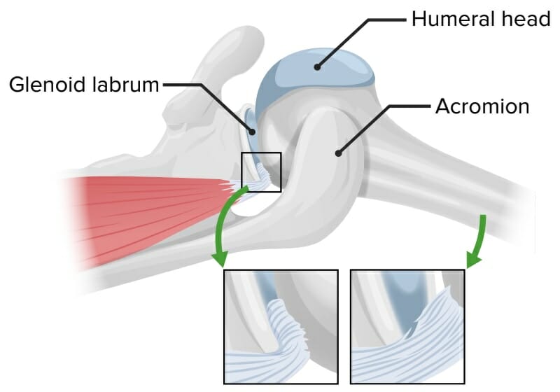
      • Glenoid labrum
    • Active stabilizers:
      • Rotator cuff muscles
      • Periscapular muscles
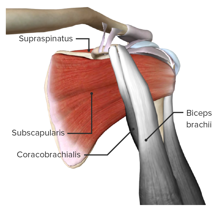
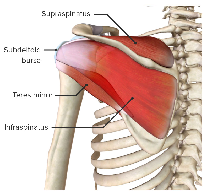
  • Rotator cuff muscles (active stabilizers):
    • Active stabilizers:
      • Supraspinatus
      • Infraspinatus
      • Teres minor
      • Subscapularis
    • Form a cuff around the head of the humerus Head of The Humerus The upper rounded extremity of the humerus fitting into the glenoid cavity of the scapula. Arm: Anatomy
    • Stabilize the joint while allowing motion
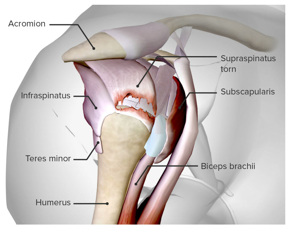
    • Supraspinatus is the most commonly torn rotator cuff tendon.
  • Shoulder complex Shoulder complex The shoulder complex comprises the glenohumeral joint, sternoclavicular joint, acromioclavicular joint, and the scapulothoracic articulation, and connects the upper limb to the trunk. This group of joints consists of the clavicle, scapula, and humerus bones, multiple muscles and supporting ligaments, cartilage, and bursae. Shoulder Joint: Anatomy also includes:
  • Cervical spine Spine The human spine, or vertebral column, is the most important anatomical and functional axis of the human body. It consists of 7 cervical vertebrae, 12 thoracic vertebrae, and 5 lumbar vertebrae and is limited cranially by the skull and caudally by the sacrum. Vertebral Column: Anatomy evaluation is included in the assessment of shoulder pain Pain An unpleasant sensation induced by noxious stimuli which are detected by nerve endings of nociceptive neurons. Pain: Types and Pathways.
  • A thorough neurovascular examination is essential after trauma to the upper extremity.

Mnemonic:

SITS: Supraspinatus, Infraspinatus, Teres minor, Subscapularis
Evaluation of Chronic Shoulder Pain

History

  • Pain Pain An unpleasant sensation induced by noxious stimuli which are detected by nerve endings of nociceptive neurons. Pain: Types and Pathways > 6 months
  • Previous shoulder injury, trauma, or surgery
  • History and pain Pain An unpleasant sensation induced by noxious stimuli which are detected by nerve endings of nociceptive neurons. Pain: Types and Pathways patterns are important in the diagnosis of shoulder pathology.
  • Location of pain Pain An unpleasant sensation induced by noxious stimuli which are detected by nerve endings of nociceptive neurons. Pain: Types and Pathways:
  • Pain Pain An unpleasant sensation induced by noxious stimuli which are detected by nerve endings of nociceptive neurons. Pain: Types and Pathways at night: rotator cuff disease
  • Cervical spine Spine The human spine, or vertebral column, is the most important anatomical and functional axis of the human body. It consists of 7 cervical vertebrae, 12 thoracic vertebrae, and 5 lumbar vertebrae and is limited cranially by the skull and caudally by the sacrum. Vertebral Column: Anatomy etiology:
    • Periscapular pain Pain An unpleasant sensation induced by noxious stimuli which are detected by nerve endings of nociceptive neurons. Pain: Types and Pathways
    • Pain Pain An unpleasant sensation induced by noxious stimuli which are detected by nerve endings of nociceptive neurons. Pain: Types and Pathways or numbness below the elbow

Physical exam

  • Exposure of shoulder
  • Compare to unaffected side
  • Inspection Inspection Dermatologic Examination/observation:
  • Cervical spine Spine The human spine, or vertebral column, is the most important anatomical and functional axis of the human body. It consists of 7 cervical vertebrae, 12 thoracic vertebrae, and 5 lumbar vertebrae and is limited cranially by the skull and caudally by the sacrum. Vertebral Column: Anatomy exam: Always consider associated injury.
  • Active and passive range of motion Range of motion The distance and direction to which a bone joint can be extended. Range of motion is a function of the condition of the joints, muscles, and connective tissues involved. Joint flexibility can be improved through appropriate muscle strength exercises. Examination of the Upper Limbs (ROM):
    • ROM can be evaluated either sitting or supine.
    • Lack of passive ROM: adhesive capsulitis versus OA OA Osteoarthritis (OA) is the most common form of arthritis, and is due to cartilage destruction and changes of the subchondral bone. The risk of developing this disorder increases with age, obesity, and repetitive joint use or trauma. Patients develop gradual joint pain, stiffness lasting < 30 minutes, and decreased range of motion. Osteoarthritis
    • Assessment of scapular motion: Look for winging and scapular dyskinesis ( asymmetry Asymmetry Examination of the Upper Limbs of motion).
  • Palpation Palpation Application of fingers with light pressure to the surface of the body to determine consistency of parts beneath in physical diagnosis; includes palpation for determining the outlines of organs. Dermatologic Examination:
  • Neurologic and vascular exam:
    • Evaluation of brachial plexus Brachial Plexus The large network of nerve fibers which distributes the innervation of the upper extremity. The brachial plexus extends from the neck into the axilla. In humans, the nerves of the plexus usually originate from the lower cervical and the first thoracic spinal cord segments (c5-c8 and T1), but variations are not uncommon. Peripheral Nerve Injuries in the Cervicothoracic Region
    • Testing for TOS TOS Thoracic outlet syndrome (TOS) is a broad term used for a spectrum of syndromes related to the general region of the thoracic outlet, which involves the compression or irritation of elements of the brachial plexus, subclavian artery, or subclavian vein. Thoracic Outlet Syndrome
    • Evaluation for vascular effort thrombosis Thrombosis Formation and development of a thrombus or blood clot in the blood vessel. Epidemic Typhus (i.e., overhead athletes/workers)

Imaging

Imaging studies might be helpful to support or confirm the diagnosis:
  • Plain radiographs:
    • AP shoulder
    • AP glenoid
    • Scapular Y view
    • Axillary view
  • MRI or ultrasound for more difficult cases

Important Causes and Management of Chronic or Subacute Shoulder Pain

Rotator cuff pathology

  • Rotator cuff tendinopathy or tendinitis Tendinitis Ankylosing Spondylitis:
    • One of the most common causes of shoulder pain Pain An unpleasant sensation induced by noxious stimuli which are detected by nerve endings of nociceptive neurons. Pain: Types and Pathways
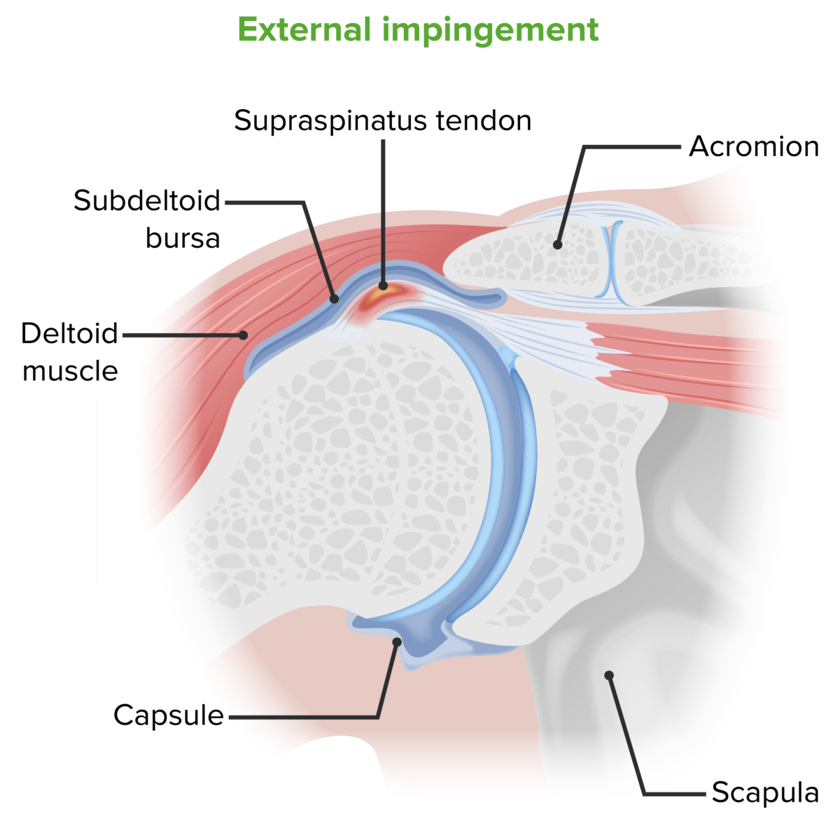
    • The rotator cuff tendons (especially the supraspinatus due to the anatomical position) undergo compression Compression Blunt Chest Trauma in the subacromial space with abduction Abduction Examination of the Upper Limbs of the shoulder.
    • Features similar to impingement syndrome:
    • Diagnosis is clinically based.
    • X-rays X-rays X-rays are high-energy particles of electromagnetic radiation used in the medical field for the generation of anatomical images. X-rays are projected through the body of a patient and onto a film, and this technique is called conventional or projectional radiography. X-rays are often normal and may include calcification of the tendon.
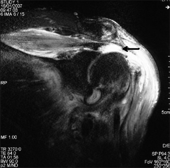
    • MRI may be indicated to exclude significant rotator cuff tear.
    • Management:
      • Modification of activities: PT for rotator cuff strengthening, ROM, and balance of the shoulder
      • Consider steroid injections for pain Pain An unpleasant sensation induced by noxious stimuli which are detected by nerve endings of nociceptive neurons. Pain: Types and Pathways management.
      • Surgical evaluation for recalcitrant cases
  • Shoulder impingement syndrome:
    • Etiology:
      • Repetitive overhead motions
      • Calcification of the coracoacromial ligament
      • Subacromial bursitis
      • Supraspinatus tendonitis Tendonitis Reactive Arthritis or tendinosis
      • Any condition further narrowing the subacromial space
    • Features similar to rotator cuff tendinopathy:
    • Diagnosis is clinically based.
    • X-ray X-ray Penetrating electromagnetic radiation emitted when the inner orbital electrons of an atom are excited and release radiant energy. X-ray wavelengths range from 1 pm to 10 nm. Hard x-rays are the higher energy, shorter wavelength x-rays. Soft x-rays or grenz rays are less energetic and longer in wavelength. The short wavelength end of the x-ray spectrum overlaps the gamma rays wavelength range. The distinction between gamma rays and x-rays is based on their radiation source. Pulmonary Function Tests is often normal and may include calcification of the tendon.
    • MRI is generally not indicated.
    • Management:
      • Modification of activities: PT for rotator cuff strengthening and ROM
      • Consider steroid injection for pain Pain An unpleasant sensation induced by noxious stimuli which are detected by nerve endings of nociceptive neurons. Pain: Types and Pathways management.
      • Surgical evaluation for recalcitrant cases
  • Rotator cuff tendon tears:
    • Generally, age is helpful in classifying the tear:
      • Younger individuals commonly have acute tears.
      • Older individuals often have degenerative tears.
      • Important: A significant number of individuals > 60 years of age have asymptomatic degenerative rotator cuff tears on MRI studies.
    • Supraspinatus is the most commonly torn rotator cuff.
    • Features:
      • Shoulder weakness with similar pain Pain An unpleasant sensation induced by noxious stimuli which are detected by nerve endings of nociceptive neurons. Pain: Types and Pathways when reaching overhead or behind
      • Often bothersome at night
    • Perform physical examination and muscle strength Muscle strength The amount of force generated by muscle contraction. Muscle strength can be measured during isometric, isotonic, or isokinetic contraction, either manually or using a device such as a muscle strength dynamometer. Neurological Examination testing specific to the location of the rotator cuff tendon tear.
    • Initial diagnosis is based on clinical examination.
    • MRI is used to confirm the diagnosis.
    • Management:
      • Based on multiple factors: age, duration of symptoms, partial versus full tear, and comorbidities Comorbidities The presence of co-existing or additional diseases with reference to an initial diagnosis or with reference to the index condition that is the subject of study. Comorbidity may affect the ability of affected individuals to function and also their survival; it may be used as a prognostic indicator for length of hospital stay, cost factors, and outcome or survival. St. Louis Encephalitis Virus
      • A full thickness tear generally requires surgical repair.
      • A partial thickness tear may be treated with PT.
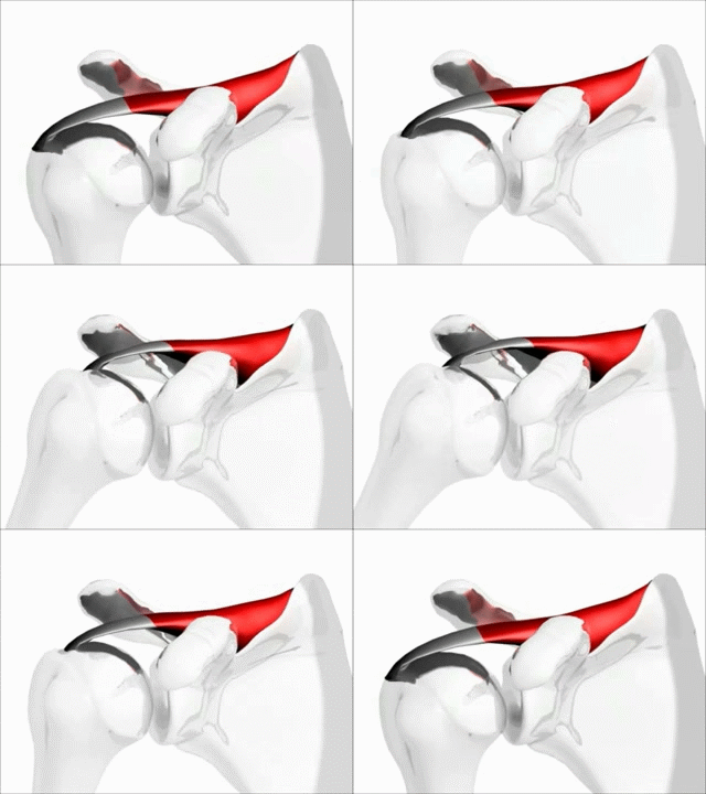
Shoulder motion with rotator cuff

Function of the supraspinatus muscle

Image: “Shoulder motion with rotator cuff” by Young Lae, Moon M.D. Chair of 3D Based Medical Application Working group. Chairman and Professor of Orthopaedics, Chosun University Hospital, Korea. License: CC BY 3.0
Other shoulder pathology

Biceps Biceps Arm: Anatomy tendinopathy or rupture:
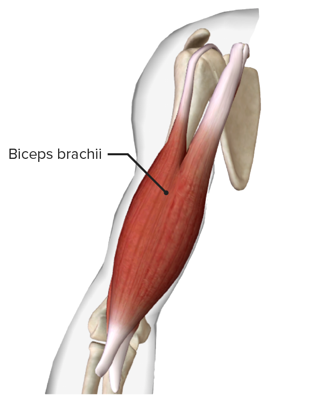
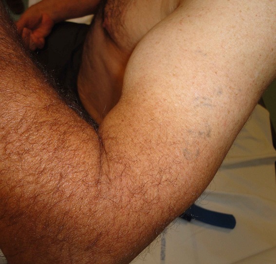
  • A degenerative condition affecting the long head of the biceps Biceps Arm: Anatomy tendon
  • Repetitive lifting and overhead reaching → irritation, microtears, tendinopathy → damage to bicep tendons
  • The individual complains of anterior shoulder pain Pain An unpleasant sensation induced by noxious stimuli which are detected by nerve endings of nociceptive neurons. Pain: Types and Pathways and weakness.
  • Aggravated by lifting and carrying
  • Resultant swelling Swelling Inflammation appears like a “Popeye” muscle on exam.
  • Management:
    • The majority are nonsurgical.
    • Surgical repair may be done in younger individuals or for cosmetic reasons.

Acromioclavicular joint Acromioclavicular joint The gliding joint formed by the outer extremity of the clavicle and the inner margin of the acromion process of the scapula. Examination of the Upper Limbs pain Pain An unpleasant sensation induced by noxious stimuli which are detected by nerve endings of nociceptive neurons. Pain: Types and Pathways:

  • The acromioclavicular joint Acromioclavicular joint The gliding joint formed by the outer extremity of the clavicle and the inner margin of the acromion process of the scapula. Examination of the Upper Limbs is a common site of degenerative joint disease (DJD) with ↑ age or previous trauma.
  • Examination: pain Pain An unpleasant sensation induced by noxious stimuli which are detected by nerve endings of nociceptive neurons. Pain: Types and Pathways to palpation Palpation Application of fingers with light pressure to the surface of the body to determine consistency of parts beneath in physical diagnosis; includes palpation for determining the outlines of organs. Dermatologic Examination of joint, positive cross-arm test 
  • Plain X-ray X-ray Penetrating electromagnetic radiation emitted when the inner orbital electrons of an atom are excited and release radiant energy. X-ray wavelengths range from 1 pm to 10 nm. Hard x-rays are the higher energy, shorter wavelength x-rays. Soft x-rays or grenz rays are less energetic and longer in wavelength. The short wavelength end of the x-ray spectrum overlaps the gamma rays wavelength range. The distinction between gamma rays and x-rays is based on their radiation source. Pulmonary Function Tests: DJD of the acromioclavicular joint Acromioclavicular joint The gliding joint formed by the outer extremity of the clavicle and the inner margin of the acromion process of the scapula. Examination of the Upper Limbs
  • Management: steroid joint injections and surgical evaluation

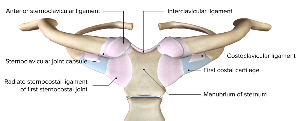
Sternoclavicular joint Sternoclavicular Joint Examination of the Upper Limbs pathology:

Adhesive capsulitis (frozen shoulder):

  • Painful shoulder with often disabling loss of ROM
  • Self-limited, but can be prolonged (2–3 years)
  • Most common in the 5th and 6th decade of life; women > men
  • Exact pathophysiology is not definitively understood.
  • Associated with diabetes Diabetes Diabetes mellitus (DM) is a metabolic disease characterized by hyperglycemia and dysfunction of the regulation of glucose metabolism by insulin. Type 1 DM is diagnosed mostly in children and young adults as the result of autoimmune destruction of β cells in the pancreas and the resulting lack of insulin. Type 2 DM has a significant association with obesity and is characterized by insulin resistance. Diabetes Mellitus, hypothyroidism Hypothyroidism Hypothyroidism is a condition characterized by a deficiency of thyroid hormones. Iodine deficiency is the most common cause worldwide, but Hashimoto’s disease (autoimmune thyroiditis) is the leading cause in non-iodine-deficient regions. Hypothyroidism, or immobilization Immobilization Delirium
  • Painful restriction of both active and passive movement of the shoulder 
  • Diagnosis is clinical.
  • 3 phases:
    • Initial phase Initial Phase Sepsis in Children: pain Pain An unpleasant sensation induced by noxious stimuli which are detected by nerve endings of nociceptive neurons. Pain: Types and Pathways and loss of motion
    • Intermediate phase: stiffness, severe loss of ROM, ↓ in pain Pain An unpleasant sensation induced by noxious stimuli which are detected by nerve endings of nociceptive neurons. Pain: Types and Pathways
    • Recovery phase: motion gradually improves
  • Examination:
  • Radiology:
    • X-ray X-ray Penetrating electromagnetic radiation emitted when the inner orbital electrons of an atom are excited and release radiant energy. X-ray wavelengths range from 1 pm to 10 nm. Hard x-rays are the higher energy, shorter wavelength x-rays. Soft x-rays or grenz rays are less energetic and longer in wavelength. The short wavelength end of the x-ray spectrum overlaps the gamma rays wavelength range. The distinction between gamma rays and x-rays is based on their radiation source. Pulmonary Function Tests (usually normal)
    • MRI (usually not necessary) may show minimal signs of edema Edema Edema is a condition in which excess serous fluid accumulates in the body cavity or interstitial space of connective tissues. Edema is a symptom observed in several medical conditions. It can be categorized into 2 types, namely, peripheral (in the extremities) and internal (in an organ or body cavity). Edema and ligament thickening.
  • Management:
    • Lack of consensus on the optimal management
    • PT, glucocorticoids Glucocorticoids Glucocorticoids are a class within the corticosteroid family. Glucocorticoids are chemically and functionally similar to endogenous cortisol. There are a wide array of indications, which primarily benefit from the antiinflammatory and immunosuppressive effects of this class of drugs. Glucocorticoids, injections, and surgical management are all used with varying effect.

Glenohumeral OA OA Osteoarthritis (OA) is the most common form of arthritis, and is due to cartilage destruction and changes of the subchondral bone. The risk of developing this disorder increases with age, obesity, and repetitive joint use or trauma. Patients develop gradual joint pain, stiffness lasting < 30 minutes, and decreased range of motion. Osteoarthritis:

  • Uncommon site for primary OA Primary OA Osteoarthritis
  • Often related to distant trauma or large rotator cuff tears: In the absence of any previous trauma, consider metabolic abnormalities.
  • Gradual onset of anterior or deep shoulder pain Pain An unpleasant sensation induced by noxious stimuli which are detected by nerve endings of nociceptive neurons. Pain: Types and Pathways and ↓ ROM
  • Examination:
    • ↓ ROM often with crepitus Crepitus Osteoarthritis
    • Shoulder atrophy Atrophy Decrease in the size of a cell, tissue, organ, or multiple organs, associated with a variety of pathological conditions such as abnormal cellular changes, ischemia, malnutrition, or hormonal changes. Cellular Adaptation often develops
  • Diagnosis:
    • Plain X-ray X-ray Penetrating electromagnetic radiation emitted when the inner orbital electrons of an atom are excited and release radiant energy. X-ray wavelengths range from 1 pm to 10 nm. Hard x-rays are the higher energy, shorter wavelength x-rays. Soft x-rays or grenz rays are less energetic and longer in wavelength. The short wavelength end of the x-ray spectrum overlaps the gamma rays wavelength range. The distinction between gamma rays and x-rays is based on their radiation source. Pulmonary Function Tests is used to confirm the diagnosis.
    • Laboratory evaluation may be needed to exclude other diagnoses.
  • Management:

Chronic instability of the shoulder:

  • Can be secondary to previous traumatic dislocation/subluxations
  • Frequently present with symptoms of instability
  • Episodes of “dead arm Arm The arm, or “upper arm” in common usage, is the region of the upper limb that extends from the shoulder to the elbow joint and connects inferiorly to the forearm through the cubital fossa. It is divided into 2 fascial compartments (anterior and posterior). Arm: Anatomy
  • Diagnosis:
    • Signs of generalized hyperlaxity
    • Positive sulcus sign
    • Signs of excessive mobility
  • Management:
    • PT is the cornerstone of therapy.
    • Surgery evaluation in recalcitrant cases

Superior Labral tear from Anterior to Posterior (SLAP) lesion:

  • Injury to the glenoid labrum (SLAP)
  • Etiology: most commonly in overhead athletes or trauma
  • Clinical manifestations: dull pain Pain An unpleasant sensation induced by noxious stimuli which are detected by nerve endings of nociceptive neurons. Pain: Types and Pathways or discomfort (especially with overhead activities or a change in velocity) in overhead athletes
  • Imaging:
    • Plain X-rays X-rays X-rays are high-energy particles of electromagnetic radiation used in the medical field for the generation of anatomical images. X-rays are projected through the body of a patient and onto a film, and this technique is called conventional or projectional radiography. X-rays to rule out other causes
    • MRI to confirm diagnosis
  • Classification:
  • Management:
    • The majority of type I injuries are nonoperative (i.e., NSAIDs NSAIDS Primary vs Secondary Headaches and PT).
    • Consider surgical evaluation with persistent pain Pain An unpleasant sensation induced by noxious stimuli which are detected by nerve endings of nociceptive neurons. Pain: Types and Pathways.
    • Additional factors to consider:
      • Age of the individual
      • Overhead athlete
      • Functional requirements

Clinical Relevance

  • Thoracic outlet Thoracic Outlet Thoracic Outlet Syndrome syndrome ( TOS TOS Thoracic outlet syndrome (TOS) is a broad term used for a spectrum of syndromes related to the general region of the thoracic outlet, which involves the compression or irritation of elements of the brachial plexus, subclavian artery, or subclavian vein. Thoracic Outlet Syndrome): a broad term used for a spectrum of syndromes related to the general region of the thoracic outlet Thoracic Outlet Thoracic Outlet Syndrome involving the compression Compression Blunt Chest Trauma or irritation of elements of the brachial plexus Brachial Plexus The large network of nerve fibers which distributes the innervation of the upper extremity. The brachial plexus extends from the neck into the axilla. In humans, the nerves of the plexus usually originate from the lower cervical and the first thoracic spinal cord segments (c5-c8 and T1), but variations are not uncommon. Peripheral Nerve Injuries in the Cervicothoracic Region, the subclavian artery, or the subclavian vein. The diagnosis is clinically based and supported by radiography and a number of provocation maneuvers. Management can be medical and surgical.
  • Acute shoulder pain Pain An unpleasant sensation induced by noxious stimuli which are detected by nerve endings of nociceptive neurons. Pain: Types and Pathways: commonly caused by acute shoulder injuries, including acromioclavicular joint injuries Acromioclavicular Joint Injuries Acute Shoulder Pain, clavicle Clavicle A bone on the ventral side of the shoulder girdle, which in humans is commonly called the collar bone. Clavicle Fracture fractures, glenohumeral dislocations, proximal humerus Humerus Bone in humans and primates extending from the shoulder joint to the elbow joint. Arm: Anatomy fractures, and rotator cuff tears. Management includes pain Pain An unpleasant sensation induced by noxious stimuli which are detected by nerve endings of nociceptive neurons. Pain: Types and Pathways control and varies based on the diagnosis.
  • Hypothyroidism Hypothyroidism Hypothyroidism is a condition characterized by a deficiency of thyroid hormones. Iodine deficiency is the most common cause worldwide, but Hashimoto’s disease (autoimmune thyroiditis) is the leading cause in non-iodine-deficient regions. Hypothyroidism: a condition characterized by the deficiency of thyroid Thyroid The thyroid gland is one of the largest endocrine glands in the human body. The thyroid gland is a highly vascular, brownish-red gland located in the visceral compartment of the anterior region of the neck. Thyroid Gland: Anatomy hormones Hormones Hormones are messenger molecules that are synthesized in one part of the body and move through the bloodstream to exert specific regulatory effects on another part of the body. Hormones play critical roles in coordinating cellular activities throughout the body in response to the constant changes in both the internal and external environments. Hormones: Overview and Types. Thyroid Thyroid The thyroid gland is one of the largest endocrine glands in the human body. The thyroid gland is a highly vascular, brownish-red gland located in the visceral compartment of the anterior region of the neck. Thyroid Gland: Anatomy hormones Hormones Hormones are messenger molecules that are synthesized in one part of the body and move through the bloodstream to exert specific regulatory effects on another part of the body. Hormones play critical roles in coordinating cellular activities throughout the body in response to the constant changes in both the internal and external environments. Hormones: Overview and Types are integral in metabolic processes and the development of the brain Brain The part of central nervous system that is contained within the skull (cranium). Arising from the neural tube, the embryonic brain is comprised of three major parts including prosencephalon (the forebrain); mesencephalon (the midbrain); and rhombencephalon (the hindbrain). The developed brain consists of cerebrum; cerebellum; and other structures in the brain stem. Nervous System: Anatomy, Structure, and Classification and other organs. Diagnosis is by obtaining thyroid function tests Thyroid Function Tests Blood tests used to evaluate the functioning of the thyroid gland. Ion Channel Myopathy. Elevated thyroid Thyroid The thyroid gland is one of the largest endocrine glands in the human body. The thyroid gland is a highly vascular, brownish-red gland located in the visceral compartment of the anterior region of the neck. Thyroid Gland: Anatomy stimulating hormone and a low free thyroxine Thyroxine The major hormone derived from the thyroid gland. Thyroxine is synthesized via the iodination of tyrosines (monoiodotyrosine) and the coupling of iodotyrosines (diiodotyrosine) in the thyroglobulin. Thyroxine is released from thyroglobulin by proteolysis and secreted into the blood. Thyroid Hormones ( T4 T4 The major hormone derived from the thyroid gland. Thyroxine is synthesized via the iodination of tyrosines (monoiodotyrosine) and the coupling of iodotyrosines (diiodotyrosine) in the thyroglobulin. Thyroxine is released from thyroglobulin by proteolysis and secreted into the blood. Thyroxine is peripherally deiodinated to form triiodothyronine which exerts a broad spectrum of stimulatory effects on cell metabolism. Thyroid Hormones) are noted. Management is with synthetic T4 T4 The major hormone derived from the thyroid gland. Thyroxine is synthesized via the iodination of tyrosines (monoiodotyrosine) and the coupling of iodotyrosines (diiodotyrosine) in the thyroglobulin. Thyroxine is released from thyroglobulin by proteolysis and secreted into the blood. Thyroxine is peripherally deiodinated to form triiodothyronine which exerts a broad spectrum of stimulatory effects on cell metabolism. Thyroid Hormones. Hypothyroidism Hypothyroidism Hypothyroidism is a condition characterized by a deficiency of thyroid hormones. Iodine deficiency is the most common cause worldwide, but Hashimoto’s disease (autoimmune thyroiditis) is the leading cause in non-iodine-deficient regions. Hypothyroidism is also associated with adhesive capsulitis.
  • Ehlers-Danlos syndrome Ehlers-Danlos syndrome Ehlers-Danlos syndrome (EDS) is a heterogeneous group of inherited connective tissue disorders that are characterized by hyperextensible skin, hypermobile joints, and fragility of the skin and connective tissue. Ehlers-Danlos Syndrome: a heterogenous group of inherited connective tissue disorders Inherited Connective Tissue Disorders Ehlers-Danlos Syndrome characterized by hyperextensible skin Skin The skin, also referred to as the integumentary system, is the largest organ of the body. The skin is primarily composed of the epidermis (outer layer) and dermis (deep layer). The epidermis is primarily composed of keratinocytes that undergo rapid turnover, while the dermis contains dense layers of connective tissue. Skin: Structure and Functions, hypermobile joints, and fragility of the skin Skin The skin, also referred to as the integumentary system, is the largest organ of the body. The skin is primarily composed of the epidermis (outer layer) and dermis (deep layer). The epidermis is primarily composed of keratinocytes that undergo rapid turnover, while the dermis contains dense layers of connective tissue. Skin: Structure and Functions and connective tissue Connective tissue Connective tissues originate from embryonic mesenchyme and are present throughout the body except inside the brain and spinal cord. The main function of connective tissues is to provide structural support to organs. Connective tissues consist of cells and an extracellular matrix. Connective Tissue: Histology. The syndrome is due to genetic defects affecting collagen Collagen A polypeptide substance comprising about one third of the total protein in mammalian organisms. It is the main constituent of skin; connective tissue; and the organic substance of bones (bone and bones) and teeth (tooth). Connective Tissue: Histology processing and synthesis Synthesis Polymerase Chain Reaction (PCR). The diagnosis is clinical but confirmed via genetic testing Genetic Testing Detection of a mutation; genotype; karyotype; or specific alleles associated with genetic traits, heritable diseases, or predisposition to a disease, or that may lead to the disease in descendants. It includes prenatal genetic testing. Myotonic Dystrophies. No curative treatment exists.
  • Osteoarthritis Osteoarthritis Osteoarthritis (OA) is the most common form of arthritis, and is due to cartilage destruction and changes of the subchondral bone. The risk of developing this disorder increases with age, obesity, and repetitive joint use or trauma. Patients develop gradual joint pain, stiffness lasting < 30 minutes, and decreased range of motion. Osteoarthritis: the most common form of arthritis Arthritis Acute or chronic inflammation of joints. Osteoarthritis due to cartilage Cartilage Cartilage is a type of connective tissue derived from embryonic mesenchyme that is responsible for structural support, resilience, and the smoothness of physical actions. Perichondrium (connective tissue membrane surrounding cartilage) compensates for the absence of vasculature in cartilage by providing nutrition and support. Cartilage: Histology destruction and changes of the subchondral bone Subchondral Bone Osteochondritis Dissecans. Individuals develop gradual joint pain Pain An unpleasant sensation induced by noxious stimuli which are detected by nerve endings of nociceptive neurons. Pain: Types and Pathways, stiffness, and decreased ROM. The diagnosis is clinical and supported with radiographic joint findings. Management includes conservative measures, analgesic medications, glucocorticoid intra-articular injections, and surgery for advanced disease. 

References

  1. Burbank, K.M., Stevenson, J.H., Czarnecki, G.R., Dorfman, J. (2008). Chronic shoulder pain: part I. Evaluation and diagnosis. Am Fam Physician. 77(4), 453–60. https://pubmed.ncbi.nlm.nih.gov/18326164/
  2. Meislin, R.J., Sperling, J.W., Stitik, T.P. (2005). Persistent shoulder pain: epidemiology, pathophysiology, and diagnosis. Am J Orthop (Belle Mead NJ). 34(12 Suppl), 5–9. https://pubmed.ncbi.nlm.nih.gov/16450690/
  3. Van der Windt, D.A., Koes, B.W., de Jong, B.A., Bouter, L.M. (1995). Shoulder disorders in general practice: incidence, patient characteristics, and management. Ann Rheum Dis. 54(12), 959–964. https://pubmed.ncbi.nlm.nih.gov/8546527/
  4. Iannotti, J.P., Kwon, Y.W. (2005). Management of persistent shoulder pain. a treatment algorithm. Am J Orthop. 34(12 suppl), 16–23. https://pubmed.ncbi.nlm.nih.gov/16450692/
  5. Woodward, T.W., Best, T.M. (2000). The painful shoulder: part I. Clinical evaluation. Am Fam Physician. 61(10), 3079–3088. https://pubmed.ncbi.nlm.nih.gov/10839557/
  6. Woodward, T.W., Best, T.M. (2000). The painful shoulder: part II. Acute and chronic disorders. Am Fam Physician. 61(11), 3291–3300. https://pubmed.ncbi.nlm.nih.gov/10865925/
  7. Ebell, M.H. (2005). Diagnosing rotator cuff tears. Am Fam Physician. 71(8), 1587–1588. https://pubmed.ncbi.nlm.nih.gov/15864899/

Create your free account or log in to continue reading!

Sign up now and get free access to Lecturio with concept pages, medical videos, and questions for your medical education.

User Reviews

Details