Chronic Myeloid Leukemia

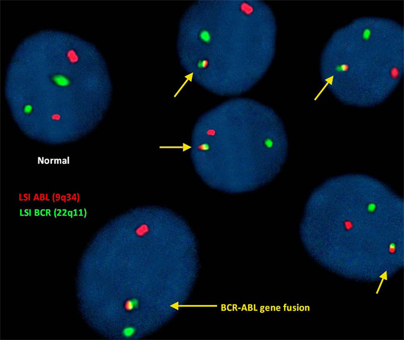
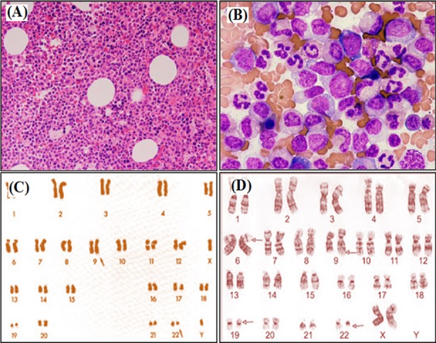
Chronic myeloid leukemia is a malignant proliferation of the granulocytic cell line characterized by a fairly normal differentiation. The underlying genetic abnormality is the Philadelphia chromosome Philadelphia chromosome An aberrant form of human chromosome 22 characterized by translocation of the distal end of chromosome 9 from 9q34, to the long arm of chromosome 22 at 22q11. It is present in the bone marrow cells of 80 to 90 percent of patients with chronic myelocytic leukemia. Acute Lymphoblastic Leukemia, an abbreviated chromosome Chromosome In a prokaryotic cell or in the nucleus of a eukaryotic cell, a structure consisting of or containing DNA which carries the genetic information essential to the cell. Basic Terms of Genetics 22, resulting from reciprocal (9;22)(q34;q11) translocation. The chromosome Chromosome In a prokaryotic cell or in the nucleus of a eukaryotic cell, a structure consisting of or containing DNA which carries the genetic information essential to the cell. Basic Terms of Genetics contains the BCR-ABL1 fusion gene Gene A category of nucleic acid sequences that function as units of heredity and which code for the basic instructions for the development, reproduction, and maintenance of organisms. Basic Terms of Genetics (from ABL1 on chromosome 9 Chromosome 9 Friedreich Ataxia and BCR BCR Lymphocytes: Histology on chromosome Chromosome In a prokaryotic cell or in the nucleus of a eukaryotic cell, a structure consisting of or containing DNA which carries the genetic information essential to the cell. Basic Terms of Genetics 22), which induces constitutive tyrosine Tyrosine A non-essential amino acid. In animals it is synthesized from phenylalanine. It is also the precursor of epinephrine; thyroid hormones; and melanin. Synthesis of Nonessential Amino Acids kinase activation and consequently, uncontrolled granulocyte production. Patients Patients Individuals participating in the health care system for the purpose of receiving therapeutic, diagnostic, or preventive procedures. Clinician–Patient Relationship with CML can be asymptomatic or have constitutional symptoms Constitutional Symptoms Antineutrophil Cytoplasmic Antibody (ANCA)-Associated Vasculitis, sternal pain Pain An unpleasant sensation induced by noxious stimuli which are detected by nerve endings of nociceptive neurons. Pain: Types and Pathways, and splenomegaly Splenomegaly Splenomegaly is pathologic enlargement of the spleen that is attributable to numerous causes, including infections, hemoglobinopathies, infiltrative processes, and outflow obstruction of the portal vein. Splenomegaly. Laboratory studies show an elevated WBC, and a peripheral blood smear Peripheral Blood Smear Anemia: Overview and Types shows increased numbers of immature cells. Demonstration of Philadelphia translocation using cytogenetic techniques is considered the gold standard diagnostic test. Without treatment, CML generally has a triphasic course progressing from a chronic phase Chronic phase HIV Infection and AIDS to an accelerated phase and leading to a terminal blast crisis. Management includes therapy with tyrosine Tyrosine A non-essential amino acid. In animals it is synthesized from phenylalanine. It is also the precursor of epinephrine; thyroid hormones; and melanin. Synthesis of Nonessential Amino Acids kinase inhibitors and palliative agents, as well as allogeneic hematopoietic cell transplantation.

Last updated: May 17, 2024

Editorial responsibility: Stanley Oiseth, Lindsay Jones, Evelin Maza

Overview

Definition

Chronic myeloid leukemia is a chronic myeloproliferative neoplasm characterized by uncontrolled and dysregulated proliferation of the granulocytic lineage (mature and maturing cells), with a maintained capacity for differentiation. Chronic myeloid leukemia is also known as chronic myelocytic leukemia or chronic myelogenous leukemia.

Epidemiology

  • 20% of all leukemia in adults
  • Incidence Incidence The number of new cases of a given disease during a given period in a specified population. It also is used for the rate at which new events occur in a defined population. It is differentiated from prevalence, which refers to all cases in the population at a given time. Measures of Disease Frequency:
    • In the United States: 1–2 cases per 100,000 inhabitants 
    • Increases with age
  • Median age at presentation: approximately 55 years
  • Slight male predominance
  • No known familial predisposition 
  • Prevalence Prevalence The total number of cases of a given disease in a specified population at a designated time. It is differentiated from incidence, which refers to the number of new cases in the population at a given time. Measures of Disease Frequency of CML is increasing in developed countries due to the effect of ABL1 kinase inhibitors.

Etiology

  • Not known in most cases
  • High-dose ionizing radiation Radiation Emission or propagation of acoustic waves (sound), electromagnetic energy waves (such as light; radio waves; gamma rays; or x-rays), or a stream of subatomic particles (such as electrons; neutrons; protons; or alpha particles). Osteosarcoma is an important risk factor.

Pathophysiology

Hematopoiesis Hematopoiesis The development and formation of various types of blood cells. Hematopoiesis can take place in the bone marrow (medullary) or outside the bone marrow (extramedullary hematopoiesis). Bone Marrow: Composition and Hematopoiesis

Hematopoiesis Hematopoiesis The development and formation of various types of blood cells. Hematopoiesis can take place in the bone marrow (medullary) or outside the bone marrow (extramedullary hematopoiesis). Bone Marrow: Composition and Hematopoiesis starts with a hematopoietic stem cell, which is prompted to divide and differentiate with appropriate chemical stimuli (hemopoietic growth factors).
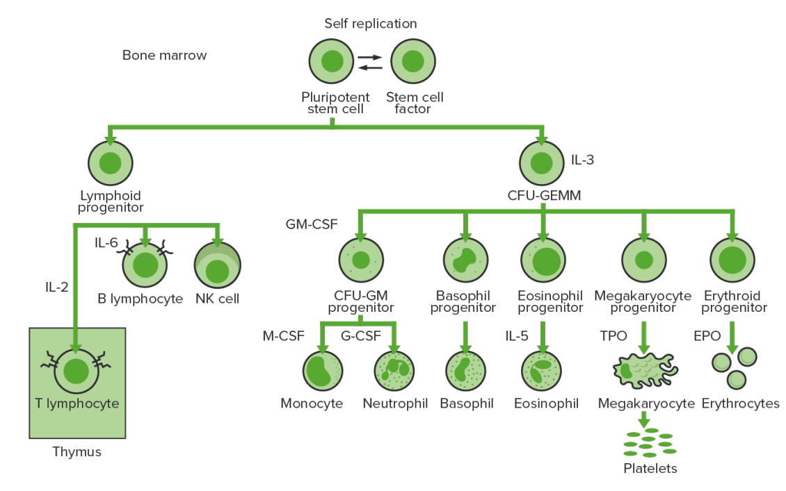
  • Lymphoid stem cells give rise to lymphocytes Lymphocytes Lymphocytes are heterogeneous WBCs involved in immune response. Lymphocytes develop from the bone marrow, starting from hematopoietic stem cells (HSCs) and progressing to common lymphoid progenitors (CLPs). B and T lymphocytes and natural killer (NK) cells arise from the lineage. Lymphocytes: Histology.
  • Myeloid stem cells eventually differentiate into platelets Platelets Platelets are small cell fragments involved in hemostasis. Thrombopoiesis takes place primarily in the bone marrow through a series of cell differentiation and is influenced by several cytokines. Platelets are formed after fragmentation of the megakaryocyte cytoplasm. Platelets: Histology, erythrocytes Erythrocytes Erythrocytes, or red blood cells (RBCs), are the most abundant cells in the blood. While erythrocytes in the fetus are initially produced in the yolk sac then the liver, the bone marrow eventually becomes the main site of production. Erythrocytes: Histology, granulocytes Granulocytes Leukocytes with abundant granules in the cytoplasm. They are divided into three groups according to the staining properties of the granules: neutrophilic, eosinophilic, and basophilic. Mature granulocytes are the neutrophils; eosinophils; and basophils. White Myeloid Cells: Histology ( neutrophils Neutrophils Granular leukocytes having a nucleus with three to five lobes connected by slender threads of chromatin, and cytoplasm containing fine inconspicuous granules and stainable by neutral dyes. Innate Immunity: Phagocytes and Antigen Presentation, basophils Basophils Granular leukocytes characterized by a relatively pale-staining, lobate nucleus and cytoplasm containing coarse dark-staining granules of variable size and stainable by basic dyes. Innate Immunity: Phagocytes and Antigen Presentation, eosinophils Eosinophils Granular leukocytes with a nucleus that usually has two lobes connected by a slender thread of chromatin, and cytoplasm containing coarse, round granules that are uniform in size and stainable by eosin. Innate Immunity: Phagocytes and Antigen Presentation), and monocytes Monocytes Large, phagocytic mononuclear leukocytes produced in the vertebrate bone marrow and released into the blood; contain a large, oval or somewhat indented nucleus surrounded by voluminous cytoplasm and numerous organelles. Innate Immunity: Phagocytes and Antigen Presentation:
    • IL-3 stimulates the differentiation of multipotent hematopoietic stem cells Hematopoietic stem cells Progenitor cells from which all blood cells derived. They are found primarily in the bone marrow and also in small numbers in the peripheral blood. Bone Marrow: Composition and Hematopoiesis into myeloid progenitor cells Myeloid progenitor cells Stem cells derived from hematopoietic stem cells. Derived from these myeloid progenitor cells are the megakaryocytes; erythroid cells; myeloid cells; and some dendritic cells. Acute Myeloid Leukemia.
    • Granulocyte macrophage colony-stimulating factor Macrophage colony-stimulating factor A mononuclear phagocyte colony-stimulating factor (M-CSF) synthesized by mesenchymal cells. The compound stimulates the survival, proliferation, and differentiation of hematopoietic cells of the monocyte-macrophage series. M-CSF is a disulfide-bonded glycoprotein dimer with a mw of 70 kda. It binds to a specific high affinity receptor. White Myeloid Cells: Histology ( GM-CSF GM-CSF An acidic glycoprotein of mw 23 kda with internal disulfide bonds. The protein is produced in response to a number of inflammatory mediators by mesenchymal cells present in the hemopoietic environment and at peripheral sites of inflammation. GM-CSF is able to stimulate the production of neutrophilic granulocytes, macrophages, and mixed granulocyte-macrophage colonies from bone marrow cells and can stimulate the formation of eosinophil colonies from fetal liver progenitor cells. GM-CSF can also stimulate some functional activities in mature granulocytes and macrophages. White Myeloid Cells: Histology) → differentiation from myeloid progenitors to granulocytes Granulocytes Leukocytes with abundant granules in the cytoplasm. They are divided into three groups according to the staining properties of the granules: neutrophilic, eosinophilic, and basophilic. Mature granulocytes are the neutrophils; eosinophils; and basophils. White Myeloid Cells: Histology ( neutrophils Neutrophils Granular leukocytes having a nucleus with three to five lobes connected by slender threads of chromatin, and cytoplasm containing fine inconspicuous granules and stainable by neutral dyes. Innate Immunity: Phagocytes and Antigen Presentation) and monocytes Monocytes Large, phagocytic mononuclear leukocytes produced in the vertebrate bone marrow and released into the blood; contain a large, oval or somewhat indented nucleus surrounded by voluminous cytoplasm and numerous organelles. Innate Immunity: Phagocytes and Antigen Presentation 
    • IL-5 → differentiation to eosinophils Eosinophils Granular leukocytes with a nucleus that usually has two lobes connected by a slender thread of chromatin, and cytoplasm containing coarse, round granules that are uniform in size and stainable by eosin. Innate Immunity: Phagocytes and Antigen Presentation
    • Thrombopoietin Thrombopoietin A humoral factor that stimulates the production of thrombocytes (blood platelets). Thrombopoietin stimulates the proliferation of bone marrow megakaryocytes and their release of blood platelets. The process is called thrombopoiesis. Platelets: Histology (TPO) → differentiation to thrombocytes ( platelets Platelets Platelets are small cell fragments involved in hemostasis. Thrombopoiesis takes place primarily in the bone marrow through a series of cell differentiation and is influenced by several cytokines. Platelets are formed after fragmentation of the megakaryocyte cytoplasm. Platelets: Histology)
    • EPO EPO Glycoprotein hormone, secreted chiefly by the kidney in the adult and the liver in the fetus, that acts on erythroid stem cells of the bone marrow to stimulate proliferation and differentiation. Erythrocytes: Histology → differentiation to erythrocytes Erythrocytes Erythrocytes, or red blood cells (RBCs), are the most abundant cells in the blood. While erythrocytes in the fetus are initially produced in the yolk sac then the liver, the bone marrow eventually becomes the main site of production. Erythrocytes: Histology ( RBCs RBCs Erythrocytes, or red blood cells (RBCs), are the most abundant cells in the blood. While erythrocytes in the fetus are initially produced in the yolk sac then the liver, the bone marrow eventually becomes the main site of production. Erythrocytes: Histology)
Bone marrow hematopoiesis

Bone marrow hematopoiesis:
Proliferation and differentiation of the formed elements of blood:
In CML, there is sustained proliferation of cells in the granulocytic line (myeloblasts → neutrophils, basophils, eosinophils). Both mature and maturing cells are seen; thus, there are cells that are only partially effective.
CFU-GEMM: colony-forming unit–granulocyte, erythrocyte, monocyte, megakaryocyte
GM-CSF: granulocyte macrophage colony-stimulating factor
M-CSF: macrophage colony-stimulating factor
G-CSF: granulocyte colony-stimulating factor
NK: natural killer

Image by Lecturio.

Mutation Mutation Genetic mutations are errors in DNA that can cause protein misfolding and dysfunction. There are various types of mutations, including chromosomal, point, frameshift, and expansion mutations. Types of Mutations

Chronic myeloid leukemia is an acquired abnormality of the hematopoietic stem cells Hematopoietic stem cells Progenitor cells from which all blood cells derived. They are found primarily in the bone marrow and also in small numbers in the peripheral blood. Bone Marrow: Composition and Hematopoiesis in the bone marrow Bone marrow The soft tissue filling the cavities of bones. Bone marrow exists in two types, yellow and red. Yellow marrow is found in the large cavities of large bones and consists mostly of fat cells and a few primitive blood cells. Red marrow is a hematopoietic tissue and is the site of production of erythrocytes and granular leukocytes. Bone marrow is made up of a framework of connective tissue containing branching fibers with the frame being filled with marrow cells. Bone Marrow: Composition and Hematopoiesis.
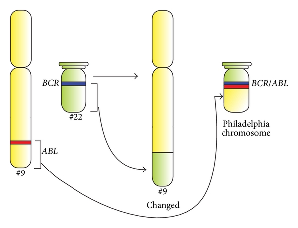
  • Mutation Mutation Genetic mutations are errors in DNA that can cause protein misfolding and dysfunction. There are various types of mutations, including chromosomal, point, frameshift, and expansion mutations. Types of Mutations occurs in the BCR-ABL1 fusion gene Gene A category of nucleic acid sequences that function as units of heredity and which code for the basic instructions for the development, reproduction, and maintenance of organisms. Basic Terms of Genetics:
    • Involves BCR BCR Lymphocytes: Histology on chromosome Chromosome In a prokaryotic cell or in the nucleus of a eukaryotic cell, a structure consisting of or containing DNA which carries the genetic information essential to the cell. Basic Terms of Genetics 22 and ABL1 on chromosome 9 Chromosome 9 Friedreich Ataxia
    • Typically results from a reciprocal (9;22)(q34;q11) translocation giving rise to the Philadelphia ( Ph pH The quantitative measurement of the acidity or basicity of a solution. Acid-Base Balance) chromosome Chromosome In a prokaryotic cell or in the nucleus of a eukaryotic cell, a structure consisting of or containing DNA which carries the genetic information essential to the cell. Basic Terms of Genetics:
      • Abnormal chromosome Chromosome In a prokaryotic cell or in the nucleus of a eukaryotic cell, a structure consisting of or containing DNA which carries the genetic information essential to the cell. Basic Terms of Genetics 22
      • Hallmark genetic aberration of CML
    • BCR-ABL1 fusion gene Gene A category of nucleic acid sequences that function as units of heredity and which code for the basic instructions for the development, reproduction, and maintenance of organisms. Basic Terms of Genetics produces a unique product Product A molecule created by the enzymatic reaction. Basics of Enzymes, the BCR-ABL1 fusion protein Fusion protein Proteins that catalyze membrane fusion. Measles Virus.
  • BCR-ABL1 protein:
    • Oncogenic protein that induces a persistently enhanced tyrosine Tyrosine A non-essential amino acid. In animals it is synthesized from phenylalanine. It is also the precursor of epinephrine; thyroid hormones; and melanin. Synthesis of Nonessential Amino Acids kinase activity
    • Deregulated tyrosine Tyrosine A non-essential amino acid. In animals it is synthesized from phenylalanine. It is also the precursor of epinephrine; thyroid hormones; and melanin. Synthesis of Nonessential Amino Acids kinase activity leads to CML through:
      • Overproliferation of myeloid cells Myeloid Cells The classes of bone marrow-derived blood cells in the monocytic series (monocytes and their precursors) and granulocytic series (granulocytes and their precursors). White Myeloid Cells: Histology in the bone marrow Bone marrow The soft tissue filling the cavities of bones. Bone marrow exists in two types, yellow and red. Yellow marrow is found in the large cavities of large bones and consists mostly of fat cells and a few primitive blood cells. Red marrow is a hematopoietic tissue and is the site of production of erythrocytes and granular leukocytes. Bone marrow is made up of a framework of connective tissue containing branching fibers with the frame being filled with marrow cells. Bone Marrow: Composition and Hematopoiesis
      • Discordant maturation of cells
      • Apoptosis Apoptosis A regulated cell death mechanism characterized by distinctive morphologic changes in the nucleus and cytoplasm, including the endonucleolytic cleavage of genomic DNA, at regularly spaced, internucleosomal sites, I.e., DNA fragmentation. It is genetically-programmed and serves as a balance to mitosis in regulating the size of animal tissues and in mediating pathologic processes associated with tumor growth. Ischemic Cell Damage 
  • The clinical hallmark of CML is the uncontrolled production of mature and immature granulocytes Granulocytes Leukocytes with abundant granules in the cytoplasm. They are divided into three groups according to the staining properties of the granules: neutrophilic, eosinophilic, and basophilic. Mature granulocytes are the neutrophils; eosinophils; and basophils. White Myeloid Cells: Histology:
  • ↑ Division of cells → ↑ expansion of the bone marrow Bone marrow The soft tissue filling the cavities of bones. Bone marrow exists in two types, yellow and red. Yellow marrow is found in the large cavities of large bones and consists mostly of fat cells and a few primitive blood cells. Red marrow is a hematopoietic tissue and is the site of production of erythrocytes and granular leukocytes. Bone marrow is made up of a framework of connective tissue containing branching fibers with the frame being filled with marrow cells. Bone Marrow: Composition and Hematopoiesis 
Schematic diagram of translocation in philadelphia chromosome

Schematic diagram of the translocation that forms the Philadelphia chromosome, and schematic representation of the BCR and ABL genes.
ABL1 and BCR genes reside on the long arms of chromosomes 9 and 22, respectively. The fusion BCR-ABL gene is formed with the translocation of the ABL1 gene (from chromosome 9) to the long arm of chromosome 22, creating a changed chromosome 22 (the so-called Philadelphia chromosome).

Image: “Figure 1” by Ewelina Trela, Sylwester Glowacki, and Janusz Błasiak. License: CC BY 3.0

Clinical Presentation

General clinical features

  • From anemia Anemia Anemia is a condition in which individuals have low Hb levels, which can arise from various causes. Anemia is accompanied by a reduced number of RBCs and may manifest with fatigue, shortness of breath, pallor, and weakness. Subtypes are classified by the size of RBCs, chronicity, and etiology. Anemia: Overview and Types and increased cell turnover:
    • Fever Fever Fever is defined as a measured body temperature of at least 38°C (100.4°F). Fever is caused by circulating endogenous and/or exogenous pyrogens that increase levels of prostaglandin E2 in the hypothalamus. Fever is commonly associated with chills, rigors, sweating, and flushing of the skin. Fever, night sweats Night sweats Tuberculosis
    • Malaise Malaise Tick-borne Encephalitis Virus, fatigue Fatigue The state of weariness following a period of exertion, mental or physical, characterized by a decreased capacity for work and reduced efficiency to respond to stimuli. Fibromyalgia
    • Weight loss Weight loss Decrease in existing body weight. Bariatric Surgery
  • From splenomegaly Splenomegaly Splenomegaly is pathologic enlargement of the spleen that is attributable to numerous causes, including infections, hemoglobinopathies, infiltrative processes, and outflow obstruction of the portal vein. Splenomegaly and splenic infarction Splenic Infarction Insufficiency of arterial or venous blood supply to the spleen due to emboli, thrombi, vascular torsion, or pressure that produces a macroscopic area of necrosis. . Imaging of the Spleen:
  • From platelet dysfunction: 
  • From expanding bone marrow Bone marrow The soft tissue filling the cavities of bones. Bone marrow exists in two types, yellow and red. Yellow marrow is found in the large cavities of large bones and consists mostly of fat cells and a few primitive blood cells. Red marrow is a hematopoietic tissue and is the site of production of erythrocytes and granular leukocytes. Bone marrow is made up of a framework of connective tissue containing branching fibers with the frame being filled with marrow cells. Bone Marrow: Composition and Hematopoiesis
    • Sternal pain Pain An unpleasant sensation induced by noxious stimuli which are detected by nerve endings of nociceptive neurons. Pain: Types and Pathways/tenderness
    • Bone Bone Bone is a compact type of hardened connective tissue composed of bone cells, membranes, an extracellular mineralized matrix, and central bone marrow. The 2 primary types of bone are compact and spongy. Bones: Structure and Types pain Pain An unpleasant sensation induced by noxious stimuli which are detected by nerve endings of nociceptive neurons. Pain: Types and Pathways
  • From ↑ uric acid Uric acid An oxidation product, via xanthine oxidase, of oxypurines such as xanthine and hypoxanthine. It is the final oxidation product of purine catabolism in humans and primates, whereas in most other mammals urate oxidase further oxidizes it to allantoin. Nephrolithiasis production ( uric acid Uric acid An oxidation product, via xanthine oxidase, of oxypurines such as xanthine and hypoxanthine. It is the final oxidation product of purine catabolism in humans and primates, whereas in most other mammals urate oxidase further oxidizes it to allantoin. Nephrolithiasis released as a result of cell degradation): gouty arthritis Arthritis Acute or chronic inflammation of joints. Osteoarthritis
  • From hyperviscosity-related or thrombotic events (due to severe leukocytosis Leukocytosis A transient increase in the number of leukocytes in a body fluid. West Nile Virus and/or thrombocytosis):
    • Confusion
    • Headache Headache The symptom of pain in the cranial region. It may be an isolated benign occurrence or manifestation of a wide variety of headache disorders. Brain Abscess
    • Blurred vision Blurred Vision Retinal Detachment
    • Priapism Priapism A prolonged painful erection that may lasts hours and is not associated with sexual activity. It is seen in patients with sickle cell anemia, advanced malignancy, spinal trauma; and certain drug treatments. Penile Anomalies and Conditions

Phases of disease

The symptoms discussed above vary in severity depending on the 3 disease phases of CML (based on the immature WBCs or blast cells in the blood or bone marrow Bone marrow The soft tissue filling the cavities of bones. Bone marrow exists in two types, yellow and red. Yellow marrow is found in the large cavities of large bones and consists mostly of fat cells and a few primitive blood cells. Red marrow is a hematopoietic tissue and is the site of production of erythrocytes and granular leukocytes. Bone marrow is made up of a framework of connective tissue containing branching fibers with the frame being filled with marrow cells. Bone Marrow: Composition and Hematopoiesis).

Chronic stable phase:

  • Blast cells: < 10%
  • Most patients Patients Individuals participating in the health care system for the purpose of receiving therapeutic, diagnostic, or preventive procedures. Clinician–Patient Relationship present in the chronic stable phase.
  • Insidious onset with generally mild symptoms:
  • Indolent clinical course with a duration of several years
  • Response to therapy is excellent.

Accelerated phase:

  • Untreated CML usually progresses to the accelerated phase.
  • ≥ 1 of the following features:
    • Blast cells: increase to 10%–19%.
    • Peripheral blood basophils: > 20%
    • Platelet count: < 100,000/µL, not due to therapy
    • Platelets Platelets Platelets are small cell fragments involved in hemostasis. Thrombopoiesis takes place primarily in the bone marrow through a series of cell differentiation and is influenced by several cytokines. Platelets are formed after fragmentation of the megakaryocyte cytoplasm. Platelets: Histology: > 1 million/µL, unresponsive to therapy
    • Progressive splenomegaly Splenomegaly Splenomegaly is pathologic enlargement of the spleen that is attributable to numerous causes, including infections, hemoglobinopathies, infiltrative processes, and outflow obstruction of the portal vein. Splenomegaly and increasing WBC, resistant to therapy
    • New chromosome Chromosome In a prokaryotic cell or in the nucleus of a eukaryotic cell, a structure consisting of or containing DNA which carries the genetic information essential to the cell. Basic Terms of Genetics changes (evolution of leukemic cells seen as chromosomal abnormalities in addition to the Philadelphia chromosome Philadelphia chromosome An aberrant form of human chromosome 22 characterized by translocation of the distal end of chromosome 9 from 9q34, to the long arm of chromosome 22 at 22q11. It is present in the bone marrow cells of 80 to 90 percent of patients with chronic myelocytic leukemia. Acute Lymphoblastic Leukemia)
  • Cells multiply aggressively and cause:
    • Fever Fever Fever is defined as a measured body temperature of at least 38°C (100.4°F). Fever is caused by circulating endogenous and/or exogenous pyrogens that increase levels of prostaglandin E2 in the hypothalamus. Fever is commonly associated with chills, rigors, sweating, and flushing of the skin. Fever
    • Significant weight loss Weight loss Decrease in existing body weight. Bariatric Surgery
    • Severe fatigue Fatigue The state of weariness following a period of exertion, mental or physical, characterized by a decreased capacity for work and reduced efficiency to respond to stimuli. Fibromyalgia
    • Significant bone Bone Bone is a compact type of hardened connective tissue composed of bone cells, membranes, an extracellular mineralized matrix, and central bone marrow. The 2 primary types of bone are compact and spongy. Bones: Structure and Types and joint pain Pain An unpleasant sensation induced by noxious stimuli which are detected by nerve endings of nociceptive neurons. Pain: Types and Pathways
    • Abdominal fullness/ pain Pain An unpleasant sensation induced by noxious stimuli which are detected by nerve endings of nociceptive neurons. Pain: Types and Pathways
    • Bleeding
    • Thrombotic or hyperviscosity Hyperviscosity Hypercoagulable States symptoms
  • Response to therapy is less favorable.

Blast crisis (or acute phase Acute phase Short Bowel Syndrome):
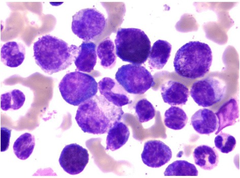
  • Myeloid or lymphoid blasts proliferate in an uncontrolled manner.
  • Presence of ≥ 1 of the following:
    • > 20% of peripheral blood or bone marrow Bone marrow The soft tissue filling the cavities of bones. Bone marrow exists in two types, yellow and red. Yellow marrow is found in the large cavities of large bones and consists mostly of fat cells and a few primitive blood cells. Red marrow is a hematopoietic tissue and is the site of production of erythrocytes and granular leukocytes. Bone marrow is made up of a framework of connective tissue containing branching fibers with the frame being filled with marrow cells. Bone Marrow: Composition and Hematopoiesis blasts Blasts Injuries resulting when a person is struck by particles impelled with violent force from an explosion. Blast causes pulmonary contusion and hemorrhage, laceration of other thoracic and abdominal viscera, ruptured ear drums, and minor effects in the central nervous system. Blunt Chest Trauma
    • Large clusters of blasts Blasts Injuries resulting when a person is struck by particles impelled with violent force from an explosion. Blast causes pulmonary contusion and hemorrhage, laceration of other thoracic and abdominal viscera, ruptured ear drums, and minor effects in the central nervous system. Blunt Chest Trauma on bone marrow Bone marrow The soft tissue filling the cavities of bones. Bone marrow exists in two types, yellow and red. Yellow marrow is found in the large cavities of large bones and consists mostly of fat cells and a few primitive blood cells. Red marrow is a hematopoietic tissue and is the site of production of erythrocytes and granular leukocytes. Bone marrow is made up of a framework of connective tissue containing branching fibers with the frame being filled with marrow cells. Bone Marrow: Composition and Hematopoiesis biopsy Biopsy Removal and pathologic examination of specimens from the living body. Ewing Sarcoma
    • Extramedullary blast proliferation (other tissues/organs beyond the marrow)
  • Indistinguishable from acute leukemia: fever Fever Fever is defined as a measured body temperature of at least 38°C (100.4°F). Fever is caused by circulating endogenous and/or exogenous pyrogens that increase levels of prostaglandin E2 in the hypothalamus. Fever is commonly associated with chills, rigors, sweating, and flushing of the skin. Fever, bleeding, petechiae Petechiae Primary Skin Lesions, ecchymosis Ecchymosis Extravasation of blood into the skin, resulting in a nonelevated, rounded or irregular, blue or purplish patch, larger than a petechia. Orbital Fractures, pallor, and progressive splenomegaly Splenomegaly Splenomegaly is pathologic enlargement of the spleen that is attributable to numerous causes, including infections, hemoglobinopathies, infiltrative processes, and outflow obstruction of the portal vein. Splenomegaly
  • Response to therapy is poor.

Diagnosis

Hematologic and biochemical findings

  • Hematologic tests:
    • CBC: elevated WBC
    • Peripheral smear:
  • Biochemical findings:
    • Low leukocyte alkaline phosphatase Alkaline Phosphatase An enzyme that catalyzes the conversion of an orthophosphoric monoester and water to an alcohol and orthophosphate. Osteosarcoma (LAP) score
    • Uric acid Uric acid An oxidation product, via xanthine oxidase, of oxypurines such as xanthine and hypoxanthine. It is the final oxidation product of purine catabolism in humans and primates, whereas in most other mammals urate oxidase further oxidizes it to allantoin. Nephrolithiasis, ↑ LDH LDH Osteosarcoma

Bone marrow Bone marrow The soft tissue filling the cavities of bones. Bone marrow exists in two types, yellow and red. Yellow marrow is found in the large cavities of large bones and consists mostly of fat cells and a few primitive blood cells. Red marrow is a hematopoietic tissue and is the site of production of erythrocytes and granular leukocytes. Bone marrow is made up of a framework of connective tissue containing branching fibers with the frame being filled with marrow cells. Bone Marrow: Composition and Hematopoiesis and cytogenetic findings

  • Bone marrow Bone marrow The soft tissue filling the cavities of bones. Bone marrow exists in two types, yellow and red. Yellow marrow is found in the large cavities of large bones and consists mostly of fat cells and a few primitive blood cells. Red marrow is a hematopoietic tissue and is the site of production of erythrocytes and granular leukocytes. Bone marrow is made up of a framework of connective tissue containing branching fibers with the frame being filled with marrow cells. Bone Marrow: Composition and Hematopoiesis biopsy Biopsy Removal and pathologic examination of specimens from the living body. Ewing Sarcoma:
    • Hypercellular (↑ granulocytic precursors)
    • Myeloid hyperplasia Hyperplasia An increase in the number of cells in a tissue or organ without tumor formation. It differs from hypertrophy, which is an increase in bulk without an increase in the number of cells. Cellular Adaptation with myeloid:erythroid ratio of 15:1–20:1
    • Sea-blue histiocytes Histiocytes Macrophages found in the tissues, as opposed to those found in the blood (monocytes) or serous cavities (serous membrane). Chronic Granulomatous Disease (scattered macrophages Macrophages The relatively long-lived phagocytic cell of mammalian tissues that are derived from blood monocytes. Main types are peritoneal macrophages; alveolar macrophages; histiocytes; kupffer cells of the liver; and osteoclasts. They may further differentiate within chronic inflammatory lesions to epithelioid cells or may fuse to form foreign body giant cells or langhans giant cells. Innate Immunity: Phagocytes and Antigen Presentation with increased green-blue cytoplasm)
    • ↑ Reticulin deposition
  • Cytogenetic studies:
    • Test for:
      • Philadelphia chromosome Philadelphia chromosome An aberrant form of human chromosome 22 characterized by translocation of the distal end of chromosome 9 from 9q34, to the long arm of chromosome 22 at 22q11. It is present in the bone marrow cells of 80 to 90 percent of patients with chronic myelocytic leukemia. Acute Lymphoblastic Leukemia 
      • BCR-ABL1 fusion gene Gene A category of nucleic acid sequences that function as units of heredity and which code for the basic instructions for the development, reproduction, and maintenance of organisms. Basic Terms of Genetics 
      • Fusion mRNA mRNA RNA sequences that serve as templates for protein synthesis. Bacterial mRNAs are generally primary transcripts in that they do not require post-transcriptional processing. Eukaryotic mRNA is synthesized in the nucleus and must be exported to the cytoplasm for translation. Most eukaryotic mRNAs have a sequence of polyadenylic acid at the 3′ end, referred to as the poly(a) tail. The function of this tail is not known for certain, but it may play a role in the export of mature mRNA from the nucleus as well as in helping stabilize some mRNA molecules by retarding their degradation in the cytoplasm. RNA Types and Structure gene Gene A category of nucleic acid sequences that function as units of heredity and which code for the basic instructions for the development, reproduction, and maintenance of organisms. Basic Terms of Genetics product Product A molecule created by the enzymatic reaction. Basics of Enzymes 
    • Techniques:
      • Karyotyping Karyotyping Mapping of the karyotype of a cell. Chromosome Testing
      • FISH FISH A type of in situ hybridization in which target sequences are stained with fluorescent dye so their location and size can be determined using fluorescence microscopy. This staining is sufficiently distinct that the hybridization signal can be seen both in metaphase spreads and in interphase nuclei. Chromosome Testing
      • RT-PCR RT-PCR A variation of the pcr technique in which cDNA is made from RNA via reverse transcription. The resultant cDNA is then amplified using standard pcr protocols. Polymerase Chain Reaction (PCR)

Comparison with other myeloproliferative neoplasms Neoplasms New abnormal growth of tissue. Malignant neoplasms show a greater degree of anaplasia and have the properties of invasion and metastasis, compared to benign neoplasms. Benign Bone Tumors

Myeloproliferative neoplasms Neoplasms New abnormal growth of tissue. Malignant neoplasms show a greater degree of anaplasia and have the properties of invasion and metastasis, compared to benign neoplasms. Benign Bone Tumors can be compared with the following WHO classification:

Table: Classic types of myeloproliferative neoplasms Neoplasms New abnormal growth of tissue. Malignant neoplasms show a greater degree of anaplasia and have the properties of invasion and metastasis, compared to benign neoplasms. Benign Bone Tumors
Disease Mutations Key points
CML BCR-ABL1 ( Philadelphia chromosome Philadelphia chromosome An aberrant form of human chromosome 22 characterized by translocation of the distal end of chromosome 9 from 9q34, to the long arm of chromosome 22 at 22q11. It is present in the bone marrow cells of 80 to 90 percent of patients with chronic myelocytic leukemia. Acute Lymphoblastic Leukemia) Proliferation of mature and maturing granulocytes Granulocytes Leukocytes with abundant granules in the cytoplasm. They are divided into three groups according to the staining properties of the granules: neutrophilic, eosinophilic, and basophilic. Mature granulocytes are the neutrophils; eosinophils; and basophils. White Myeloid Cells: Histology
Essential thrombocythemia Essential thrombocythemia Essential thrombocythemia (ET) is a type of myeloproliferative neoplasm characterized by the clonal thrombocytosis linked to somatic mutations involving Janus kinase 2 (JAK2), calreticulin (CALR), and myeloproliferative leukemia virus oncogene (MPL). Patients can be asymptomatic or present with vasomotor symptoms. Essential Thrombocythemia JAK2, CALR, or MPL Excessive clonal platelet production
Polycythemia Polycythemia An increase in the total red cell mass of the blood. Renal Cell Carcinoma vera JAK2 Elevated RBC mass Mass Three-dimensional lesion that occupies a space within the breast Imaging of the Breast
Primary myelofibrosis Primary myelofibrosis Primary myelofibrosis (PMF) is a myeloproliferative neoplasm characterized by chronic myeloproliferation with nonclonal fibroblastic deposition, resulting in bone marrow fibrosis. The abnormality stems from genetic mutations of the hematopoietic stem cells (typically, JAK2 mutation). Primary symptoms are anemia and extramedullary hematopoiesis,. Primary Myelofibrosis JAK2, CALR, or MPL Obliterative bone marrow fibrosis Bone Marrow Fibrosis Primary Myelofibrosis
JAK: Janus kinase
CALR: calreticulin

Other types:

  • Chronic neutrophilic leukemia (CNL)
  • Chronic eosinophilic leukemia Chronic eosinophilic leukemia Chronic eosinophilic leukemia (CEL) is a chronic myeloproliferative neoplasm caused by autonomous clonal proliferation of normal-appearing eosinophils, resulting in increased eosinophils in the peripheral blood and bone marrow. The disorder is a myeloid variant of hypereosinophilic syndrome (HES) and is associated with tissue infiltration leading to end-organ damage. Chronic Eosinophilic Leukemia ( CEL CEL Chronic eosinophilic leukemia (CEL) is a chronic myeloproliferative neoplasm caused by autonomous clonal proliferation of normal-appearing eosinophils, resulting in increased eosinophils in the peripheral blood and bone marrow. The disorder is a myeloid variant of hypereosinophilic syndrome (HES) and is associated with tissue infiltration leading to end-organ damage. Chronic Eosinophilic Leukemia), not otherwise specified
  • Myeloproliferative neoplasm, unclassifiable

Management

Treatment goals and options

  • Goals of therapy:
    • Hematologic remission Remission A spontaneous diminution or abatement of a disease over time, without formal treatment. Cluster Headaches: achieving normal blood counts and spleen Spleen The spleen is the largest lymphoid organ in the body, located in the LUQ of the abdomen, superior to the left kidney and posterior to the stomach at the level of the 9th-11th ribs just below the diaphragm. The spleen is highly vascular and acts as an important blood filter, cleansing the blood of pathogens and damaged erythrocytes. Spleen: Anatomy size
    • Cytogenetic remission Remission A spontaneous diminution or abatement of a disease over time, without formal treatment. Cluster Headaches: no Philadelphia chromosome Philadelphia chromosome An aberrant form of human chromosome 22 characterized by translocation of the distal end of chromosome 9 from 9q34, to the long arm of chromosome 22 at 22q11. It is present in the bone marrow cells of 80 to 90 percent of patients with chronic myelocytic leukemia. Acute Lymphoblastic Leukemia–positive cells
    • Molecular remission Remission A spontaneous diminution or abatement of a disease over time, without formal treatment. Cluster Headaches: negative BCR-ABL1 fusion gene Gene A category of nucleic acid sequences that function as units of heredity and which code for the basic instructions for the development, reproduction, and maintenance of organisms. Basic Terms of Genetics on PCR PCR Polymerase chain reaction (PCR) is a technique that amplifies DNA fragments exponentially for analysis. The process is highly specific, allowing for the targeting of specific genomic sequences, even with minuscule sample amounts. The PCR cycles multiple times through 3 phases: denaturation of the template DNA, annealing of a specific primer to the individual DNA strands, and synthesis/elongation of new DNA molecules. Polymerase Chain Reaction (PCR)
  • Treatment is based on the specific disease phase, and options include:
    • Tyrosine Tyrosine A non-essential amino acid. In animals it is synthesized from phenylalanine. It is also the precursor of epinephrine; thyroid hormones; and melanin. Synthesis of Nonessential Amino Acids kinase inhibitors (TKIs):
      • Inhibit the constitutively active tyrosine Tyrosine A non-essential amino acid. In animals it is synthesized from phenylalanine. It is also the precursor of epinephrine; thyroid hormones; and melanin. Synthesis of Nonessential Amino Acids kinase and cause apoptosis Apoptosis A regulated cell death mechanism characterized by distinctive morphologic changes in the nucleus and cytoplasm, including the endonucleolytic cleavage of genomic DNA, at regularly spaced, internucleosomal sites, I.e., DNA fragmentation. It is genetically-programmed and serves as a balance to mitosis in regulating the size of animal tissues and in mediating pathologic processes associated with tumor growth. Ischemic Cell Damage of neoplastic cells
      • Preferred initial treatment
      • Imatinib Imatinib A tyrosine kinase inhibitor and antineoplastic agent that inhibits the bcr-abl kinase created by chromosome rearrangements in chronic myeloid leukemia and acute lymphoblastic leukemia, as well as pdg-derived tyrosine kinases that are overexpressed in gastrointestinal stromal tumors. Targeted and Other Nontraditional Antineoplastic Therapy, dasatinib Dasatinib A pyrimidine and thiazole derived antineoplastic agent and protein kinase inhibitor of bcr-abl kinase. It is used in the treatment of patients with chronic myeloid leukemia who are resistant or intolerant to imatinib. Targeted and Other Nontraditional Antineoplastic Therapy, nilotinib Nilotinib Targeted and Other Nontraditional Antineoplastic Therapy, bosutinib, ponatinib
      • Adverse effects: nausea Nausea An unpleasant sensation in the stomach usually accompanied by the urge to vomit. Common causes are early pregnancy, sea and motion sickness, emotional stress, intense pain, food poisoning, and various enteroviruses. Antiemetics, diarrhea Diarrhea Diarrhea is defined as ≥ 3 watery or loose stools in a 24-hour period. There are a multitude of etiologies, which can be classified based on the underlying mechanism of disease. The duration of symptoms (acute or chronic) and characteristics of the stools (e.g., watery, bloody, steatorrheic, mucoid) can help guide further diagnostic evaluation. Diarrhea, fluid retention, myelosuppression Myelosuppression Oxazolidinones, QT prolongation
    • Allogeneic hematopoietic cell transplantation (HCT)
    • Other agents (used if refractory to initial treatment and/or patient is not a transplant candidate):
      • Hydroxyurea Hydroxyurea An antineoplastic agent that inhibits DNA synthesis through the inhibition of ribonucleoside diphosphate reductase. Antimetabolite Chemotherapy: to reduce WBC
      • Interferon alfa Interferon Alfa Antivirals for Hepatitis C (with/without cytarabine Cytarabine A pyrimidine nucleoside analog that is used mainly in the treatment of leukemia, especially acute non-lymphoblastic leukemia. Cytarabine is an antimetabolite antineoplastic agent that inhibits the synthesis of DNA. Its actions are specific for the s phase of the cell cycle. It also has antiviral and immunosuppressant properties. Antimetabolite Chemotherapy)
      • Busulfan Busulfan An alkylating agent having a selective immunosuppressive effect on bone marrow. It has been used in the palliative treatment of chronic myeloid leukemia, but although symptomatic relief is provided, no permanent remission is brought about. According to the fourth annual report on carcinogens, busulfan is listed as a known carcinogen. Bronchiolitis Obliterans
      • Omacetaxine mepesuccinate (inhibitor of protein synthesis Synthesis Polymerase Chain Reaction (PCR))

Treatment approach

Chronic stable phase:

  • TKIs:
    • Treatment of choice for almost all newly diagnosed patients Patients Individuals participating in the health care system for the purpose of receiving therapeutic, diagnostic, or preventive procedures. Clinician–Patient Relationship
    • To achieve long-term control in the majority of patients Patients Individuals participating in the health care system for the purpose of receiving therapeutic, diagnostic, or preventive procedures. Clinician–Patient Relationship, with cytogenetic and molecular responses
  • The prognosis Prognosis A prediction of the probable outcome of a disease based on a individual’s condition and the usual course of the disease as seen in similar situations. Non-Hodgkin Lymphomas is good.

Accelerated phase:

  • Tends to be difficult to control
  • TKIs:
    • An initial response may be seen but is generally short-lived.
    • Significant relapse Relapse Relapsing Fever rate even after successful treatment with TKIs
  • Allogeneic HCT: 
    • Potentially curative
    • Referral for transplantation (with search for a donor) is also initiated during TKI treatment for all eligible patients Patients Individuals participating in the health care system for the purpose of receiving therapeutic, diagnostic, or preventive procedures. Clinician–Patient Relationship.
  • The prognosis Prognosis A prediction of the probable outcome of a disease based on a individual’s condition and the usual course of the disease as seen in similar situations. Non-Hodgkin Lymphomas is poor.

Blast crisis:

  • Aggressive disease
  • All therapies discussed thus far are viable options.
  • Manage as acute leukemia.
  • Palliative treatment to relieve symptoms
  • The prognosis Prognosis A prediction of the probable outcome of a disease based on a individual’s condition and the usual course of the disease as seen in similar situations. Non-Hodgkin Lymphomas is dismal.

Monitoring and supportive management

  • Monitor the following:
    • CBC to determine hematologic response
    • Chromosome Chromosome In a prokaryotic cell or in the nucleus of a eukaryotic cell, a structure consisting of or containing DNA which carries the genetic information essential to the cell. Basic Terms of Genetics banding analysis (percentage of Philadelphia chromosome Philadelphia chromosome An aberrant form of human chromosome 22 characterized by translocation of the distal end of chromosome 9 from 9q34, to the long arm of chromosome 22 at 22q11. It is present in the bone marrow cells of 80 to 90 percent of patients with chronic myelocytic leukemia. Acute Lymphoblastic Leukemia–positive cell) to determine cytogenetic response
    • BCR-ABL1 by quantitative PCR Quantitative PCR Methods used for detecting the amplified DNA products from the polymerase chain reaction as they accumulate instead of at the end of the reaction. Polymerase Chain Reaction (PCR) to determine the molecular response
  • Follow-up of response is critical in predicting when therapies other than TKIs should be considered.
  • Supportive therapies:
    • Blood transfusions Blood transfusions The introduction of whole blood or blood component directly into the bloodstream. Transfusion Products for anemia Anemia Anemia is a condition in which individuals have low Hb levels, which can arise from various causes. Anemia is accompanied by a reduced number of RBCs and may manifest with fatigue, shortness of breath, pallor, and weakness. Subtypes are classified by the size of RBCs, chronicity, and etiology. Anemia: Overview and Types
    • Platelet transfusions for thrombocytopenia Thrombocytopenia Thrombocytopenia occurs when the platelet count is < 150,000 per microliter. The normal range for platelets is usually 150,000-450,000/µL of whole blood. Thrombocytopenia can be a result of decreased production, increased destruction, or splenic sequestration of platelets. Patients are often asymptomatic until platelet counts are < 50,000/µL. Thrombocytopenia
  • Tumor lysis Tumor Lysis Tumor Lysis Syndrome syndrome:
    • Medical emergency caused by metabolic disturbances that occurs when large numbers of cancer cells are killed rapidly
    • Lysis of tumor Tumor Inflammation cells leads to the release Release Release of a virus from the host cell following virus assembly and maturation. Egress can occur by host cell lysis, exocytosis, or budding through the plasma membrane. Virology of intracellular contents into the bloodstream:
      • Electrolyte imbalances (↑ potassium Potassium An element in the alkali group of metals with an atomic symbol k, atomic number 19, and atomic weight 39. 10. It is the chief cation in the intracellular fluid of muscle and other cells. Potassium ion is a strong electrolyte that plays a significant role in the regulation of fluid volume and maintenance of the water-electrolyte balance. Hyperkalemia, ↑ phosphate Phosphate Inorganic salts of phosphoric acid. Electrolytes, ↓ calcium Calcium A basic element found in nearly all tissues. It is a member of the alkaline earth family of metals with the atomic symbol ca, atomic number 20, and atomic weight 40. Calcium is the most abundant mineral in the body and combines with phosphorus to form calcium phosphate in the bones and teeth. It is essential for the normal functioning of nerves and muscles and plays a role in blood coagulation (as factor IV) and in many enzymatic processes. Electrolytes) → neurologic, cardiac, and GI complications
      • Increase in nucleic acid breakdown → hyperuricemia Hyperuricemia Excessive uric acid or urate in blood as defined by its solubility in plasma at 37 degrees c; greater than 0. 42 mmol per liter (7. 0 mg/dl) in men or 0. 36 mmol per liter (6. 0 mg/dl) in women. Gout, which can lead to AKI AKI Acute kidney injury refers to sudden and often reversible loss of renal function, which develops over days or weeks. Azotemia refers to elevated levels of nitrogen-containing substances in the blood that accompany AKI, which include BUN and creatinine. Acute Kidney Injury
    • Management:

Differential Diagnosis

Other chronic myeloproliferative neoplasms Neoplasms New abnormal growth of tissue. Malignant neoplasms show a greater degree of anaplasia and have the properties of invasion and metastasis, compared to benign neoplasms. Benign Bone Tumors

  • Primary myelofibrosis Primary myelofibrosis Primary myelofibrosis (PMF) is a myeloproliferative neoplasm characterized by chronic myeloproliferation with nonclonal fibroblastic deposition, resulting in bone marrow fibrosis. The abnormality stems from genetic mutations of the hematopoietic stem cells (typically, JAK2 mutation). Primary symptoms are anemia and extramedullary hematopoiesis,. Primary Myelofibrosis: chronic myeloproliferative neoplasm characterized by fibrosis Fibrosis Any pathological condition where fibrous connective tissue invades any organ, usually as a consequence of inflammation or other injury. Bronchiolitis Obliterans of the bone marrow Bone marrow The soft tissue filling the cavities of bones. Bone marrow exists in two types, yellow and red. Yellow marrow is found in the large cavities of large bones and consists mostly of fat cells and a few primitive blood cells. Red marrow is a hematopoietic tissue and is the site of production of erythrocytes and granular leukocytes. Bone marrow is made up of a framework of connective tissue containing branching fibers with the frame being filled with marrow cells. Bone Marrow: Composition and Hematopoiesis and extramedullary hematopoiesis Extramedullary Hematopoiesis Primary Myelofibrosis in the spleen Spleen The spleen is the largest lymphoid organ in the body, located in the LUQ of the abdomen, superior to the left kidney and posterior to the stomach at the level of the 9th-11th ribs just below the diaphragm. The spleen is highly vascular and acts as an important blood filter, cleansing the blood of pathogens and damaged erythrocytes. Spleen: Anatomy and liver Liver The liver is the largest gland in the human body. The liver is found in the superior right quadrant of the abdomen and weighs approximately 1.5 kilograms. Its main functions are detoxification, metabolism, nutrient storage (e.g., iron and vitamins), synthesis of coagulation factors, formation of bile, filtration, and storage of blood. Liver: Anatomy: Primary myelofibrosis Primary myelofibrosis Primary myelofibrosis (PMF) is a myeloproliferative neoplasm characterized by chronic myeloproliferation with nonclonal fibroblastic deposition, resulting in bone marrow fibrosis. The abnormality stems from genetic mutations of the hematopoietic stem cells (typically, JAK2 mutation). Primary symptoms are anemia and extramedullary hematopoiesis,. Primary Myelofibrosis is linked to mutations in JAK2, CALR, and MPL. There is no BCR-ABL1 fusion gene Gene A category of nucleic acid sequences that function as units of heredity and which code for the basic instructions for the development, reproduction, and maintenance of organisms. Basic Terms of Genetics. Clinical findings are severe fatigue Fatigue The state of weariness following a period of exertion, mental or physical, characterized by a decreased capacity for work and reduced efficiency to respond to stimuli. Fibromyalgia, splenomegaly Splenomegaly Splenomegaly is pathologic enlargement of the spleen that is attributable to numerous causes, including infections, hemoglobinopathies, infiltrative processes, and outflow obstruction of the portal vein. Splenomegaly, hepatomegaly, and anemia Anemia Anemia is a condition in which individuals have low Hb levels, which can arise from various causes. Anemia is accompanied by a reduced number of RBCs and may manifest with fatigue, shortness of breath, pallor, and weakness. Subtypes are classified by the size of RBCs, chronicity, and etiology. Anemia: Overview and Types. The peripheral blood smear Peripheral Blood Smear Anemia: Overview and Types is leukoerythroblastic and contains precursors of WBCs and RBCs RBCs Erythrocytes, or red blood cells (RBCs), are the most abundant cells in the blood. While erythrocytes in the fetus are initially produced in the yolk sac then the liver, the bone marrow eventually becomes the main site of production. Erythrocytes: Histology, nucleated RBCs RBCs Erythrocytes, or red blood cells (RBCs), are the most abundant cells in the blood. While erythrocytes in the fetus are initially produced in the yolk sac then the liver, the bone marrow eventually becomes the main site of production. Erythrocytes: Histology, and teardrop cells Teardrop Cells Primary Myelofibrosis. Diagnosis is made by bone marrow Bone marrow The soft tissue filling the cavities of bones. Bone marrow exists in two types, yellow and red. Yellow marrow is found in the large cavities of large bones and consists mostly of fat cells and a few primitive blood cells. Red marrow is a hematopoietic tissue and is the site of production of erythrocytes and granular leukocytes. Bone marrow is made up of a framework of connective tissue containing branching fibers with the frame being filled with marrow cells. Bone Marrow: Composition and Hematopoiesis examination and molecular testing. Management includes allogeneic HCT and therapy with ruxolitinib Ruxolitinib Targeted and Other Nontraditional Antineoplastic Therapy and fedratinib.
  • Polycythemia Polycythemia An increase in the total red cell mass of the blood. Renal Cell Carcinoma vera: unregulated overproduction of hematopoietic stem cells Hematopoietic stem cells Progenitor cells from which all blood cells derived. They are found primarily in the bone marrow and also in small numbers in the peripheral blood. Bone Marrow: Composition and Hematopoiesis, mainly RBCs RBCs Erythrocytes, or red blood cells (RBCs), are the most abundant cells in the blood. While erythrocytes in the fetus are initially produced in the yolk sac then the liver, the bone marrow eventually becomes the main site of production. Erythrocytes: Histology, despite low levels of EPO EPO Glycoprotein hormone, secreted chiefly by the kidney in the adult and the liver in the fetus, that acts on erythroid stem cells of the bone marrow to stimulate proliferation and differentiation. Erythrocytes: Histology: In polycythemia Polycythemia An increase in the total red cell mass of the blood. Renal Cell Carcinoma vera, there is a gain-of-function mutation Mutation Genetic mutations are errors in DNA that can cause protein misfolding and dysfunction. There are various types of mutations, including chromosomal, point, frameshift, and expansion mutations. Types of Mutations in the JAK2 gene Gene A category of nucleic acid sequences that function as units of heredity and which code for the basic instructions for the development, reproduction, and maintenance of organisms. Basic Terms of Genetics. The majority of the patients Patients Individuals participating in the health care system for the purpose of receiving therapeutic, diagnostic, or preventive procedures. Clinician–Patient Relationship present with hyperviscosity Hyperviscosity Hypercoagulable States symptoms. Management includes phlebotomy Phlebotomy The techniques used to draw blood from a vein for diagnostic purposes or for treatment of certain blood disorders such as erythrocytosis, hemochromatosis, polycythemia vera, and porphyria cutanea tarda. Hereditary Hemochromatosis, low-dose aspirin Aspirin The prototypical analgesic used in the treatment of mild to moderate pain. It has anti-inflammatory and antipyretic properties and acts as an inhibitor of cyclooxygenase which results in the inhibition of the biosynthesis of prostaglandins. Aspirin also inhibits platelet aggregation and is used in the prevention of arterial and venous thrombosis. Nonsteroidal Antiinflammatory Drugs (NSAIDs), and myelosuppressive therapies.
  • Essential thrombocythemia Essential thrombocythemia Essential thrombocythemia (ET) is a type of myeloproliferative neoplasm characterized by the clonal thrombocytosis linked to somatic mutations involving Janus kinase 2 (JAK2), calreticulin (CALR), and myeloproliferative leukemia virus oncogene (MPL). Patients can be asymptomatic or present with vasomotor symptoms. Essential Thrombocythemia: proliferation of megakaryocytes, usually due to JAK2, CALR, and MPL mutations, that results in thrombocytosis: The clinical manifestations of essential thrombocythemia Essential thrombocythemia Essential thrombocythemia (ET) is a type of myeloproliferative neoplasm characterized by the clonal thrombocytosis linked to somatic mutations involving Janus kinase 2 (JAK2), calreticulin (CALR), and myeloproliferative leukemia virus oncogene (MPL). Patients can be asymptomatic or present with vasomotor symptoms. Essential Thrombocythemia are headaches, visual disturbances, and erythromelalgia Erythromelalgia A peripheral arterial disease that is characterized by the triad of erythema, burning pain, and increased skin temperature of the extremities (or red, painful extremities). Erythromelalgia may be classified as primary or idiopathic, familial or non-familial. Secondary erythromelalgia is associated with other diseases, the most common being myeloproliferative disorders. Polycythemia Vera. Excessive platelet counts Platelet counts The number of platelets per unit volume in a sample of venous blood. Coagulation Studies result in both thrombosis Thrombosis Formation and development of a thrombus or blood clot in the blood vessel. Epidemic Typhus and bleeding. Management is aimed at reducing the platelet count (with hydroxyurea Hydroxyurea An antineoplastic agent that inhibits DNA synthesis through the inhibition of ribonucleoside diphosphate reductase. Antimetabolite Chemotherapy) and decreasing the risk of thrombosis Thrombosis Formation and development of a thrombus or blood clot in the blood vessel. Epidemic Typhus (by systemic anticoagulation Anticoagulation Pulmonary Hypertension Drugs and/or antiplatelet agents Antiplatelet agents Antiplatelet agents are medications that inhibit platelet aggregation, a critical step in the formation of the initial platelet plug. Abnormal, or inappropriate, platelet aggregation is a key step in the pathophysiology of arterial ischemic events. The primary categories of antiplatelet agents include aspirin, ADP inhibitors, phosphodiesterase/adenosine uptake inhibitors, and glycoprotein IIb/IIIa inhibitors. Antiplatelet Drugs).
  • CNL: rare disorder characterized by the proliferation of mature granulocytes Granulocytes Leukocytes with abundant granules in the cytoplasm. They are divided into three groups according to the staining properties of the granules: neutrophilic, eosinophilic, and basophilic. Mature granulocytes are the neutrophils; eosinophils; and basophils. White Myeloid Cells: Histology in the blood and marrow: Organ infiltration can occur in CNL, leading to hepatosplenomegaly Hepatosplenomegaly Cytomegalovirus. There is no Philadelphia chromosome Philadelphia chromosome An aberrant form of human chromosome 22 characterized by translocation of the distal end of chromosome 9 from 9q34, to the long arm of chromosome 22 at 22q11. It is present in the bone marrow cells of 80 to 90 percent of patients with chronic myelocytic leukemia. Acute Lymphoblastic Leukemia, but an increased LAP score is noted in CNL. Chronic neutrophilic leukemia does not usually progress to AML AML Acute myeloid leukemia (AML) is a hematologic malignancy characterized by the uncontrolled proliferation of myeloid precursor cells. Seen predominantly in older adults, AML includes an accumulation of myeloblasts and a replacement of normal marrow by malignant cells, which leads to impaired hematopoiesis. Acute Myeloid Leukemia. Management options are not well defined, but the JAK1/2 inhibitor, ruxolitinib Ruxolitinib Targeted and Other Nontraditional Antineoplastic Therapy, is an option.
  • CEL CEL Chronic eosinophilic leukemia (CEL) is a chronic myeloproliferative neoplasm caused by autonomous clonal proliferation of normal-appearing eosinophils, resulting in increased eosinophils in the peripheral blood and bone marrow. The disorder is a myeloid variant of hypereosinophilic syndrome (HES) and is associated with tissue infiltration leading to end-organ damage. Chronic Eosinophilic Leukemia, not otherwise specified: rare clonal chronic myeloproliferative disorder characterized by excessive production of eosinophils Eosinophils Granular leukocytes with a nucleus that usually has two lobes connected by a slender thread of chromatin, and cytoplasm containing coarse, round granules that are uniform in size and stainable by eosin. Innate Immunity: Phagocytes and Antigen Presentation in the bone marrow Bone marrow The soft tissue filling the cavities of bones. Bone marrow exists in two types, yellow and red. Yellow marrow is found in the large cavities of large bones and consists mostly of fat cells and a few primitive blood cells. Red marrow is a hematopoietic tissue and is the site of production of erythrocytes and granular leukocytes. Bone marrow is made up of a framework of connective tissue containing branching fibers with the frame being filled with marrow cells. Bone Marrow: Composition and Hematopoiesis leading to proliferation in the blood and organ infiltration (hepatomegaly/ splenomegaly Splenomegaly Splenomegaly is pathologic enlargement of the spleen that is attributable to numerous causes, including infections, hemoglobinopathies, infiltrative processes, and outflow obstruction of the portal vein. Splenomegaly): According to cytogenetics in CEL CEL Chronic eosinophilic leukemia (CEL) is a chronic myeloproliferative neoplasm caused by autonomous clonal proliferation of normal-appearing eosinophils, resulting in increased eosinophils in the peripheral blood and bone marrow. The disorder is a myeloid variant of hypereosinophilic syndrome (HES) and is associated with tissue infiltration leading to end-organ damage. Chronic Eosinophilic Leukemia, CEL CEL Chronic eosinophilic leukemia (CEL) is a chronic myeloproliferative neoplasm caused by autonomous clonal proliferation of normal-appearing eosinophils, resulting in increased eosinophils in the peripheral blood and bone marrow. The disorder is a myeloid variant of hypereosinophilic syndrome (HES) and is associated with tissue infiltration leading to end-organ damage. Chronic Eosinophilic Leukemia does not exhibit the Philadelphia chromosome Philadelphia chromosome An aberrant form of human chromosome 22 characterized by translocation of the distal end of chromosome 9 from 9q34, to the long arm of chromosome 22 at 22q11. It is present in the bone marrow cells of 80 to 90 percent of patients with chronic myelocytic leukemia. Acute Lymphoblastic Leukemia or the BCR-ABL1 fusion gene Gene A category of nucleic acid sequences that function as units of heredity and which code for the basic instructions for the development, reproduction, and maintenance of organisms. Basic Terms of Genetics. Management is supportive and involves imatinib Imatinib A tyrosine kinase inhibitor and antineoplastic agent that inhibits the bcr-abl kinase created by chromosome rearrangements in chronic myeloid leukemia and acute lymphoblastic leukemia, as well as pdg-derived tyrosine kinases that are overexpressed in gastrointestinal stromal tumors. Targeted and Other Nontraditional Antineoplastic Therapy therapy.

Other disorders that resemble CML

  • Leukemoid reaction Leukemoid reaction A peripheral blood picture resembling that of leukemia or indistinguishable from it on the basis of morphologic appearance alone. Shigella: condition characterized by WBC > 50,000/µL from causes other than leukemia: Neutrophilia and prominent left shift Left Shift Yersinia pestis/Plague, usually in response to infection, are typical. Findings include toxic granulation Granulation Wound Healing in the neutrophils Neutrophils Granular leukocytes having a nucleus with three to five lobes connected by slender threads of chromatin, and cytoplasm containing fine inconspicuous granules and stainable by neutral dyes. Innate Immunity: Phagocytes and Antigen Presentation, a high LAP score, lack of a myelocyte Myelocyte The classes of bone marrow-derived blood cells in the monocytic series (monocytes and their precursors) and granulocytic series (granulocytes and their precursors). White Myeloid Cells: Histology bulge, and most importantly, the presence of an obvious cause for neutrophilia. Cytogenetic or molecular testing is performed to rule out CML if the distinction cannot be made. Management is supportive and involves treatment of the underlying etiology. Leukapheresis is indicated in the case of hyperviscosity Hyperviscosity Hypercoagulable States.
  • Chronic myelomonocytic leukemia (CMML): rare myelodysplastic/myeloproliferative neoplasm with increased production of maturing monocytic cells and, sometimes, dysplastic neutrophils Neutrophils Granular leukocytes having a nucleus with three to five lobes connected by slender threads of chromatin, and cytoplasm containing fine inconspicuous granules and stainable by neutral dyes. Innate Immunity: Phagocytes and Antigen Presentation. Chronic myelomonocytic leukemia is often accompanied by constitutional symptoms Constitutional Symptoms Antineutrophil Cytoplasmic Antibody (ANCA)-Associated Vasculitis, splenomegaly Splenomegaly Splenomegaly is pathologic enlargement of the spleen that is attributable to numerous causes, including infections, hemoglobinopathies, infiltrative processes, and outflow obstruction of the portal vein. Splenomegaly, anemia Anemia Anemia is a condition in which individuals have low Hb levels, which can arise from various causes. Anemia is accompanied by a reduced number of RBCs and may manifest with fatigue, shortness of breath, pallor, and weakness. Subtypes are classified by the size of RBCs, chronicity, and etiology. Anemia: Overview and Types, and/or thrombocytopenia Thrombocytopenia Thrombocytopenia occurs when the platelet count is < 150,000 per microliter. The normal range for platelets is usually 150,000-450,000/µL of whole blood. Thrombocytopenia can be a result of decreased production, increased destruction, or splenic sequestration of platelets. Patients are often asymptomatic until platelet counts are < 50,000/µL. Thrombocytopenia. Bone marrow Bone marrow The soft tissue filling the cavities of bones. Bone marrow exists in two types, yellow and red. Yellow marrow is found in the large cavities of large bones and consists mostly of fat cells and a few primitive blood cells. Red marrow is a hematopoietic tissue and is the site of production of erythrocytes and granular leukocytes. Bone marrow is made up of a framework of connective tissue containing branching fibers with the frame being filled with marrow cells. Bone Marrow: Composition and Hematopoiesis morphology shows prominent dysplasia in at least 2 of the 3 myeloid lineages. There is no Philadelphia chromosome Philadelphia chromosome An aberrant form of human chromosome 22 characterized by translocation of the distal end of chromosome 9 from 9q34, to the long arm of chromosome 22 at 22q11. It is present in the bone marrow cells of 80 to 90 percent of patients with chronic myelocytic leukemia. Acute Lymphoblastic Leukemia or BCR-ABL1 gene Gene A category of nucleic acid sequences that function as units of heredity and which code for the basic instructions for the development, reproduction, and maintenance of organisms. Basic Terms of Genetics. Management includes supportive care, hydroxyurea Hydroxyurea An antineoplastic agent that inhibits DNA synthesis through the inhibition of ribonucleoside diphosphate reductase. Antimetabolite Chemotherapy, a hypomethylating agent, and allogeneic HCT.

References

  1. Chen, Y., Wang, H., Kantarjian, H., Cortes, J. (2013). Trends in chronic myeloid leukemia incidence and survival in the United States from 1975 to 2009. Leuk Lymphoma 54:1411–1417. https://pubmed.ncbi.nlm.nih.gov/23121646/
  2. Cortes, J.E., Talpaz, M., O’Brien, S., et al. (2006). Staging of chronic myeloid leukemia in the imatinib era: an evaluation of the World Health Organization proposal. Cancer 106:1306–1315. https://pubmed.ncbi.nlm.nih.gov/16463391/
  3. Konopka, J.B., Witte, O.N. (1985). Detection of c-abl tyrosine kinase activity in vitro permits direct comparison of normal and altered abl gene products. Mol Cell Biol 5:3116–3123. https://pubmed.ncbi.nlm.nih.gov/3879812/
  4. Schiffer, C., Atallah, E. (2021). Overview of the treatment of chronic myeloid leukemia. In Larson, R. (Ed.), UpToDate. Retrieved March 27, 2021, from https://www.uptodate.com/contents/overview-of-the-treatment-of-chronic-myeloid-leukemia
  5. Van Etten, R. (2021). Clinical manifestations and diagnosis of chronic myeloid leukemia. In Larson, R. (Ed.), UpToDate. Retrieved March 27, 2021, from https://www.uptodate.com/contents/clinical-manifestations-and-diagnosis-of-chronic-myeloid-leukemia
  6. Van Etten, R. (2021). Molecular genetics of chronic myeloid leukemia. In Larson, R. (Ed.), UpToDate. Retrieved March 27, 2021, from https://www.uptodate.com/contents/molecular-genetics-of-chronic-myeloid-leukemia
  7. Van Etten, R. (2003). c-Abl regulation: a tail of two lipids. Curr Biol 13:R608–R610. https://pubmed.ncbi.nlm.nih.gov/12906815/
  8. Vardiman, J.W., Harris, N.L., Brunning, R.D. (2002). The World Health Organization (WHO) classification of the myeloid neoplasms. Blood 100:2292–2302. https://pubmed.ncbi.nlm.nih.gov/12239137/

Create your free account or log in to continue reading!

Sign up now and get free access to Lecturio with concept pages, medical videos, and questions for your medical education.

User Reviews

Details