Advertisement

Advertisement

Advertisement

Advertisement

Chronic Granulomatous Disease

Chronic granulomatous disease Granulomatous disease A defect of leukocyte function in which phagocytic cells ingest but fail to digest bacteria, resulting in recurring bacterial infections with granuloma formation. When chronic granulomatous disease is caused by mutations in the cybb gene, the condition is inherited in an X-linked recessive pattern. When chronic granulomatous disease is caused by cyba, ncf1, ncf2, or ncf4 gene mutations, the condition is inherited in an autosomal recessive pattern. Common Variable Immunodeficiency (CVID) (CGD), as the name implies, is a chronic disorder that is characterized by granuloma formation. This disorder is a consequence of defective phagocytic cells that are unable to produce bactericidal Bactericidal Penicillins superoxide because of a defect in nicotinamide adenine dinucleotide Nicotinamide adenine dinucleotide A coenzyme composed of ribosylnicotinamide 5'-diphosphate coupled to adenosine 5'-phosphate by pyrophosphate linkage. It is found widely in nature and is involved in numerous enzymatic reactions in which it serves as an electron carrier by being alternately oxidized (NAD+) and reduced (NADH). Pentose Phosphate Pathway phosphate Phosphate Inorganic salts of phosphoric acid. Electrolytes ( NADPH NADPH Nicotinamide adenine dinucleotide phosphate. A coenzyme composed of ribosylnicotinamide 5'-phosphate (nmn) coupled by pyrophosphate linkage to the 5'-phosphate adenosine 2. Pentose Phosphate Pathway), the oxidase Oxidase Neisseria responsible for the respiratory burst Respiratory burst A large increase in oxygen uptake by neutrophils and most types of tissue macrophages through activation of an NADPH-cytochrome b-dependent oxidase that reduces oxygen to a superoxide. Individuals with an inherited defect in which the oxidase that reduces oxygen to superoxide is decreased or absent often die as a result of recurrent bacterial infections. Leukocyte Adhesion Deficiency Type 1 in phagocytic leukocytes Leukocytes White blood cells. These include granular leukocytes (basophils; eosinophils; and neutrophils) as well as non-granular leukocytes (lymphocytes and monocytes). White Myeloid Cells: Histology. The diagnosis is made by testing neutrophil function for superoxide production. Patients Patients Individuals participating in the health care system for the purpose of receiving therapeutic, diagnostic, or preventive procedures. Clinician–Patient Relationship with CGD are at increased risk of life-threatening infections with fungi Fungi A kingdom of eukaryotic, heterotrophic organisms that live parasitically as saprobes, including mushrooms; yeasts; smuts, molds, etc. They reproduce either sexually or asexually, and have life cycles that range from simple to complex. Filamentous fungi, commonly known as molds, refer to those that grow as multicellular colonies. Mycology and catalase-positive bacteria Bacteria Bacteria are prokaryotic single-celled microorganisms that are metabolically active and divide by binary fission. Some of these organisms play a significant role in the pathogenesis of diseases. Bacteriology. Inflammatory complications such as CGD colitis Colitis Inflammation of the colon section of the large intestine, usually with symptoms such as diarrhea (often with blood and mucus), abdominal pain, and fever. Pseudomembranous Colitis are also possible. The introduction of antimicrobial prophylaxis Prophylaxis Cephalosporins and the use of azole antifungals has increased the overall life expectancy Life expectancy Based on known statistical data, the number of years which any person of a given age may reasonably expected to live. Population Pyramids of these patients Patients Individuals participating in the health care system for the purpose of receiving therapeutic, diagnostic, or preventive procedures. Clinician–Patient Relationship.

Last updated: Nov 6, 2024

Editorial responsibility: Stanley Oiseth, Lindsay Jones, Evelin Maza

Advertisement

Advertisement

Advertisement

Advertisement

Advertisement

Advertisement

Epidemiology and Etiology

Epidemiology

  • Equally identified in all ethnicities and races
  • Estimated incidence Incidence The number of new cases of a given disease during a given period in a specified population. It also is used for the rate at which new events occur in a defined population. It is differentiated from prevalence, which refers to all cases in the population at a given time. Measures of Disease Frequency: approximately 1 case per 200,000 live births per year
  • Predominant in males (65%–70% of cases are from an X-linked X-linked Genetic diseases that are linked to gene mutations on the X chromosome in humans or the X chromosome in other species. Included here are animal models of human X-linked diseases. Common Variable Immunodeficiency (CVID) mutation Mutation Genetic mutations are errors in DNA that can cause protein misfolding and dysfunction. There are various types of mutations, including chromosomal, point, frameshift, and expansion mutations. Types of Mutations)
  • Mortality Mortality All deaths reported in a given population. Measures of Health Status:

Etiology

Chronic granulomatous disease Granulomatous disease A defect of leukocyte function in which phagocytic cells ingest but fail to digest bacteria, resulting in recurring bacterial infections with granuloma formation. When chronic granulomatous disease is caused by mutations in the cybb gene, the condition is inherited in an X-linked recessive pattern. When chronic granulomatous disease is caused by cyba, ncf1, ncf2, or ncf4 gene mutations, the condition is inherited in an autosomal recessive pattern. Common Variable Immunodeficiency (CVID) (CGD) is caused by mutations in the genes Genes A category of nucleic acid sequences that function as units of heredity and which code for the basic instructions for the development, reproduction, and maintenance of organisms. DNA Types and Structure encoding for proteins Proteins Linear polypeptides that are synthesized on ribosomes and may be further modified, crosslinked, cleaved, or assembled into complex proteins with several subunits. The specific sequence of amino acids determines the shape the polypeptide will take, during protein folding, and the function of the protein. Energy Homeostasis that make up the nicotinamide adenine dinucleotide Nicotinamide adenine dinucleotide A coenzyme composed of ribosylnicotinamide 5′-diphosphate coupled to adenosine 5′-phosphate by pyrophosphate linkage. It is found widely in nature and is involved in numerous enzymatic reactions in which it serves as an electron carrier by being alternately oxidized (NAD+) and reduced (NADH). Pentose Phosphate Pathway phosphate Phosphate Inorganic salts of phosphoric acid. Electrolytes ( NADPH NADPH Nicotinamide adenine dinucleotide phosphate. A coenzyme composed of ribosylnicotinamide 5′-phosphate (nmn) coupled by pyrophosphate linkage to the 5′-phosphate adenosine 2. Pentose Phosphate Pathway) oxidase Oxidase Neisseria

  • Gp91phox ( X-linked X-linked Genetic diseases that are linked to gene mutations on the X chromosome in humans or the X chromosome in other species. Included here are animal models of human X-linked diseases. Common Variable Immunodeficiency (CVID) disorder)
  • P47phox (NCF1 gene Gene A category of nucleic acid sequences that function as units of heredity and which code for the basic instructions for the development, reproduction, and maintenance of organisms. Basic Terms of Genetics found on chromosome Chromosome In a prokaryotic cell or in the nucleus of a eukaryotic cell, a structure consisting of or containing DNA which carries the genetic information essential to the cell. Basic Terms of Genetics 7, autosomal inheritance)
  • P22phox ( autosomal recessive Autosomal recessive Autosomal inheritance, both dominant and recessive, refers to the transmission of genes from the 22 autosomal chromosomes. Autosomal recessive diseases are only expressed when 2 copies of the recessive allele are inherited. Autosomal Recessive and Autosomal Dominant Inheritance)
  • P67phox ( autosomal recessive Autosomal recessive Autosomal inheritance, both dominant and recessive, refers to the transmission of genes from the 22 autosomal chromosomes. Autosomal recessive diseases are only expressed when 2 copies of the recessive allele are inherited. Autosomal Recessive and Autosomal Dominant Inheritance)
  • P40phox ( autosomal recessive Autosomal recessive Autosomal inheritance, both dominant and recessive, refers to the transmission of genes from the 22 autosomal chromosomes. Autosomal recessive diseases are only expressed when 2 copies of the recessive allele are inherited. Autosomal Recessive and Autosomal Dominant Inheritance)

Pathophysiology

Note: This animation does not have sound.

Normal phagocytosis Phagocytosis The engulfing and degradation of microorganisms; other cells that are dead, dying, or pathogenic; and foreign particles by phagocytic cells (phagocytes). Innate Immunity: Phagocytes and Antigen Presentation pathway

  • Neutrophils Neutrophils Granular leukocytes having a nucleus with three to five lobes connected by slender threads of chromatin, and cytoplasm containing fine inconspicuous granules and stainable by neutral dyes. Innate Immunity: Phagocytes and Antigen Presentation phagocytose bacteria Bacteria Bacteria are prokaryotic single-celled microorganisms that are metabolically active and divide by binary fission. Some of these organisms play a significant role in the pathogenesis of diseases. Bacteriology and other pathogens
  • ↑ Oxygen consumption 
  • Produce different reactive oxidant species, which are bactericidal Bactericidal Penicillins 

Pathophysiology of CGD

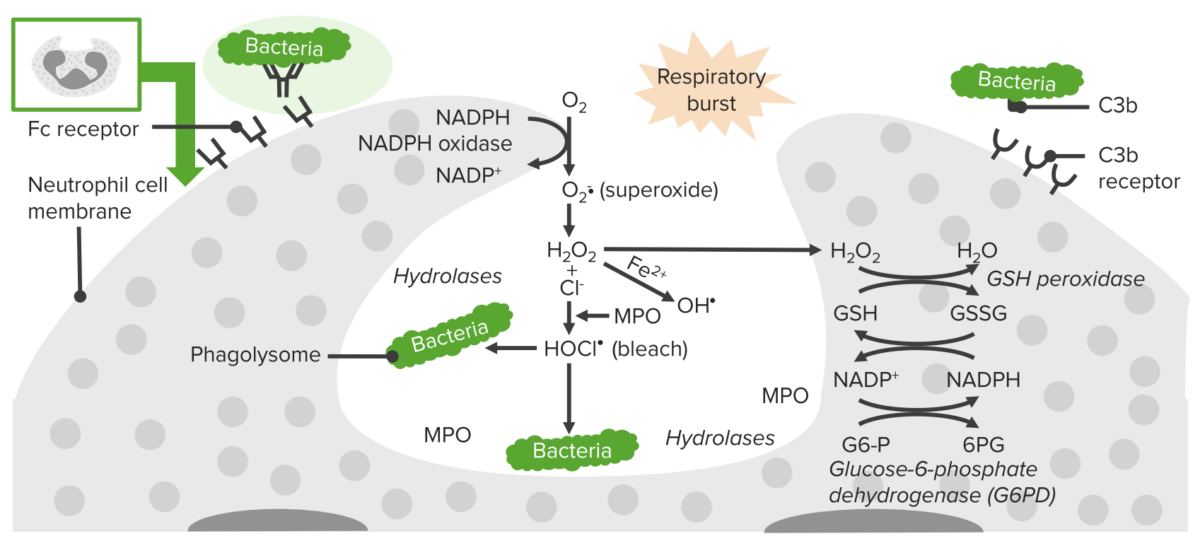
The NADPH-oxidase enzyme is impaired in phagocytic cell → ↓ production of superoxide anion O2, which affects other oxidants. This enzyme is responsible for the inability to fight infections.
  • Catalase-positive microorganisms are implicated in life-threatening infections:
    • Burkholderia Burkholderia Burkholderia species are gram-negative bacilli with 2 clinically relevant pathogens: B. pseudomallei (causing melioidosis) and B. cepacia complex (causing opportunistic infections). Melioidosis is commonly seen in Asia and Australia. Infection is transmitted by contact with contaminated soil or water (via skin wounds). Burkholderia cepacia
    • Aspergillus Aspergillus A genus of mitosporic fungi containing about 100 species and eleven different teleomorphs in the family trichocomaceae. Echinocandins
    • Candida albicans Candida albicans A unicellular budding fungus which is the principal pathogenic species causing candidiasis (moniliasis). Candida/Candidiasis
    • Staphylococcus aureus Staphylococcus aureus Potentially pathogenic bacteria found in nasal membranes, skin, hair follicles, and perineum of warm-blooded animals. They may cause a wide range of infections and intoxications. Brain Abscess
    • Nocardia Nocardia Nocardia is a branching, filamentous, gram-positive bacilli. It is partially acid fast due to the presence of mycolic acids in the cell wall. Nocardia is a ubiquitous soil organism that most commonly affects immunocompromised patients. Nocardia is transmitted via inhalation of aerosolized bacteria or less commonly, via direct contact with wounds. Nocardia/Nocardiosis spp.
    • Serratia marcescens Serratia Marcescens A species of gram-negative, facultatively anaerobic, rod-shaped bacteria found in soil, water, food, and clinical specimens. It is a prominent opportunistic pathogen for hospitalized patients. Aminoglycosides
  • Although Pseudomonas aeruginosa Pseudomonas aeruginosa A species of gram-negative, aerobic, rod-shaped bacteria commonly isolated from clinical specimens (wound, burn, and urinary tract infections). It is also found widely distributed in soil and water. P. Aeruginosa is a major agent of nosocomial infection. Pseudomonas is catalase-positive, it is killed by neutrophils Neutrophils Granular leukocytes having a nucleus with three to five lobes connected by slender threads of chromatin, and cytoplasm containing fine inconspicuous granules and stainable by neutral dyes. Innate Immunity: Phagocytes and Antigen Presentation in CGD patients Patients Individuals participating in the health care system for the purpose of receiving therapeutic, diagnostic, or preventive procedures. Clinician–Patient Relationship.
  • Catalase-negative microorganisms can still be killed by phagocytes.
Process of phagocytosis in neutrophils

Process of phagocytosis in neutrophils:
A deficiency in NADPH oxidase results in an inability to produce superoxide, which diminishes the cell’s ability to destroy catalase-positive microorganisms.

Image by Lecturio. License: CC BY-NC-SA 4.0

Clinical Presentation

General presentation

Most CGD patients Patients Individuals participating in the health care system for the purpose of receiving therapeutic, diagnostic, or preventive procedures. Clinician–Patient Relationship are < 5 years of age, and the severity is variable Variable Variables represent information about something that can change. The design of the measurement scales, or of the methods for obtaining information, will determine the data gathered and the characteristics of that data. As a result, a variable can be qualitative or quantitative, and may be further classified into subgroups. Types of Variables (depending on the genetic defect Genetic Defect Ion Channel Myopathy). Clinical presentation can include:

  • Growth failure
  • Abnormal wound healing Wound healing Wound healing is a physiological process involving tissue repair in response to injury. It involves a complex interaction of various cell types, cytokines, and inflammatory mediators. Wound healing stages include hemostasis, inflammation, granulation, and remodeling. Wound Healing
  • Infections

Infections

Patients Patients Individuals participating in the health care system for the purpose of receiving therapeutic, diagnostic, or preventive procedures. Clinician–Patient Relationship may experience repeated infections caused by bacterial and fungal pathogens.
  • Patients Patients Individuals participating in the health care system for the purpose of receiving therapeutic, diagnostic, or preventive procedures. Clinician–Patient Relationship can exhibit few clinical symptoms and signs despite the severity of infection
  • Viral infection response is normal
  • An infectious etiology should always be searched for in any child presenting with a fever Fever Fever is defined as a measured body temperature of at least 38°C (100.4°F). Fever is caused by circulating endogenous and/or exogenous pyrogens that increase levels of prostaglandin E2 in the hypothalamus. Fever is commonly associated with chills, rigors, sweating, and flushing of the skin. Fever.
  • Types of infections include:
    • Pneumonia Pneumonia Pneumonia or pulmonary inflammation is an acute or chronic inflammation of lung tissue. Causes include infection with bacteria, viruses, or fungi. In more rare cases, pneumonia can also be caused through toxic triggers through inhalation of toxic substances, immunological processes, or in the course of radiotherapy. Pneumonia
    • Abscesses (perianal, perirectal, liver Liver The liver is the largest gland in the human body. The liver is found in the superior right quadrant of the abdomen and weighs approximately 1.5 kilograms. Its main functions are detoxification, metabolism, nutrient storage (e.g., iron and vitamins), synthesis of coagulation factors, formation of bile, filtration, and storage of blood. Liver: Anatomy)
    • Suppurative adenitis
    • Osteomyelitis Osteomyelitis Osteomyelitis is an infection of the bone that results from the spread of microorganisms from the blood (hematogenous), nearby infected tissue, or open wounds (non-hematogenous). Infections are most commonly caused by Staphylococcus aureus. Osteomyelitis
    • Bacteremia Bacteremia The presence of viable bacteria circulating in the blood. Fever, chills, tachycardia, and tachypnea are common acute manifestations of bacteremia. The majority of cases are seen in already hospitalized patients, most of whom have underlying diseases or procedures which render their bloodstreams susceptible to invasion. Glycopeptides/fungemia
    • Superficial skin Skin The skin, also referred to as the integumentary system, is the largest organ of the body. The skin is primarily composed of the epidermis (outer layer) and dermis (deep layer). The epidermis is primarily composed of keratinocytes that undergo rapid turnover, while the dermis contains dense layers of connective tissue. Skin: Structure and Functions infections ( cellulitis Cellulitis Cellulitis is a common infection caused by bacteria that affects the dermis and subcutaneous tissue of the skin. It is frequently caused by Staphylococcus aureus and Streptococcus pyogenes. The skin infection presents as an erythematous and edematous area with warmth and tenderness. Cellulitis/ impetigo Impetigo Impetigo is a highly contagious superficial bacterial infection typically caused by Staphylococcus aureus (most common) and Streptococcus pyogenes. Impetigo most commonly presents in children aged 2 to 5 years with lesions that evolve from papules to vesicles to pustules, which eventually break down to form characteristic “honey-colored” crusts. Impetigo)

Inflammatory and other manifestations

  • Granulomata: mostly in GI and genitourinary tracts
  • GI manifestations:
    • Inflammatory bowel disease (IBD)
    • Diarrhea Diarrhea Diarrhea is defined as ≥ 3 watery or loose stools in a 24-hour period. There are a multitude of etiologies, which can be classified based on the underlying mechanism of disease. The duration of symptoms (acute or chronic) and characteristics of the stools (e.g., watery, bloody, steatorrheic, mucoid) can help guide further diagnostic evaluation. Diarrhea 
    • Colitis Colitis Inflammation of the colon section of the large intestine, usually with symptoms such as diarrhea (often with blood and mucus), abdominal pain, and fever. Pseudomembranous Colitis
    • Proctitis
    • Fistulae 
    • Vomiting Vomiting The forcible expulsion of the contents of the stomach through the mouth. Hypokalemia
    • Constipation Constipation Constipation is common and may be due to a variety of causes. Constipation is generally defined as bowel movement frequency < 3 times per week. Patients who are constipated often strain to pass hard stools. The condition is classified as primary (also known as idiopathic or functional constipation) or secondary, and as acute or chronic. Constipation
  • Hepatic:
    • Elevated liver Liver The liver is the largest gland in the human body. The liver is found in the superior right quadrant of the abdomen and weighs approximately 1.5 kilograms. Its main functions are detoxification, metabolism, nutrient storage (e.g., iron and vitamins), synthesis of coagulation factors, formation of bile, filtration, and storage of blood. Liver: Anatomy enzymes Enzymes Enzymes are complex protein biocatalysts that accelerate chemical reactions without being consumed by them. Due to the body’s constant metabolic needs, the absence of enzymes would make life unsustainable, as reactions would occur too slowly without these molecules. Basics of Enzymes
    • Hepatitis
    • Liver Liver The liver is the largest gland in the human body. The liver is found in the superior right quadrant of the abdomen and weighs approximately 1.5 kilograms. Its main functions are detoxification, metabolism, nutrient storage (e.g., iron and vitamins), synthesis of coagulation factors, formation of bile, filtration, and storage of blood. Liver: Anatomy abscesses 
    • Portal hypertension Portal hypertension Portal hypertension is increased pressure in the portal venous system. This increased pressure can lead to splanchnic vasodilation, collateral blood flow through portosystemic anastomoses, and increased hydrostatic pressure. There are a number of etiologies, including cirrhosis, right-sided congestive heart failure, schistosomiasis, portal vein thrombosis, hepatitis, and Budd-Chiari syndrome. Portal Hypertension
  • Ophthalmic:
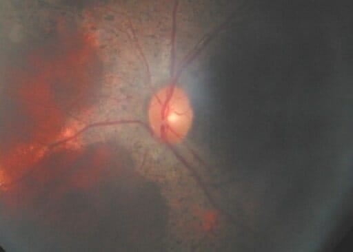
    • Chorioretinal lesions (mostly X-linked X-linked Genetic diseases that are linked to gene mutations on the X chromosome in humans or the X chromosome in other species. Included here are animal models of human X-linked diseases. Common Variable Immunodeficiency (CVID) CGD)
  • Pulmonary:
    • Bronchiectasis Bronchiectasis Bronchiectasis is a chronic disease of the airways that results from permanent bronchial distortion. This results from a continuous cycle of inflammation, bronchial damage and dilation, impaired clearance of secretions, and recurrent infections. Bronchiectasis
    • Chronic fibrosis Fibrosis Any pathological condition where fibrous connective tissue invades any organ, usually as a consequence of inflammation or other injury. Bronchiolitis Obliterans
The fundus of an individual with cgd showing a retinal mass

The fundus of an individual with chronic granulomatous disease (CGD) showing a retinal mass

Image: “F3: Brother’s fundus” by Mansour A. M. et al. License: CC BY 2.0

Diagnosis

General evaluation

  • Hypergammaglobulinemia Hypergammaglobulinemia An excess of gamma-globulins in the serum due to chronic infections or paraproteinemias. Autoimmune Hepatitis chronic inflammation Chronic Inflammation Inflammation
  • ↓ Circulating B cells B cells Lymphoid cells concerned with humoral immunity. They are short-lived cells resembling bursa-derived lymphocytes of birds in their production of immunoglobulin upon appropriate stimulation. B cells: Types and Functions
  • CD4 T-cell lymphocytopenia
  • Anemia Anemia Anemia is a condition in which individuals have low Hb levels, which can arise from various causes. Anemia is accompanied by a reduced number of RBCs and may manifest with fatigue, shortness of breath, pallor, and weakness. Subtypes are classified by the size of RBCs, chronicity, and etiology. Anemia: Overview and Types of chronic disease
  • ↑ Markers of inflammation Inflammation Inflammation is a complex set of responses to infection and injury involving leukocytes as the principal cellular mediators in the body’s defense against pathogenic organisms. Inflammation is also seen as a response to tissue injury in the process of wound healing. The 5 cardinal signs of inflammation are pain, heat, redness, swelling, and loss of function. Inflammation ( erythrocyte sedimentation rate Erythrocyte Sedimentation Rate Soft Tissue Abscess ( ESR ESR Soft Tissue Abscess) and CRP)
  • Hypoalbuminemia Hypoalbuminemia A condition in which albumin level in blood (serum albumin) is below the normal range. Hypoalbuminemia may be due to decreased hepatic albumin synthesis, increased albumin catabolism, altered albumin distribution, or albumin loss through the urine (albuminuria). Nephrotic Syndrome in Children
  • Imaging studies are dictated according to the presenting infectious process.

Specific laboratory evaluation

Neutrophil function tests should be done:

  • Nitroblue tetrazolium test: normal phagocytes reduce NBT to blue formazan 
  • Dihydrorhodamine (DHR) test:
    • More accurate than nitroblue tetrazolium test for the diagnosis of CGD
    • DHR results: phagocytes are unable to ↓ DHR in the fluorescent active compound, rhodamine
    • The extent of conversion varies based on the genetic mutation Mutation Genetic mutations are errors in DNA that can cause protein misfolding and dysfunction. There are various types of mutations, including chromosomal, point, frameshift, and expansion mutations. Types of Mutations.

Molecular genetic testing Genetic Testing Detection of a mutation; genotype; karyotype; or specific alleles associated with genetic traits, heritable diseases, or predisposition to a disease, or that may lead to the disease in descendants. It includes prenatal genetic testing. Myotonic Dystrophies and genotyping Genotyping Methods used to determine individuals’ specific alleles or snps (single nucleotide polymorphisms). Polymerase Chain Reaction (PCR):
  • Indicated in familial cases of CGD
  • Can be used on chorionic villus sampling for previously identified mutations

Biopsy Biopsy Removal and pathologic examination of specimens from the living body. Ewing Sarcoma

  • Performed when granulomas Granulomas A relatively small nodular inflammatory lesion containing grouped mononuclear phagocytes, caused by infectious and noninfectious agents. Sarcoidosis form
  • Histologic examination:
    • Necrotic centers
    • Layers of histiocytes and macrophages Macrophages The relatively long-lived phagocytic cell of mammalian tissues that are derived from blood monocytes. Main types are peritoneal macrophages; alveolar macrophages; histiocytes; kupffer cells of the liver; and osteoclasts. They may further differentiate within chronic inflammatory lesions to epithelioid cells or may fuse to form foreign body giant cells or langhans giant cells. Innate Immunity: Phagocytes and Antigen Presentation
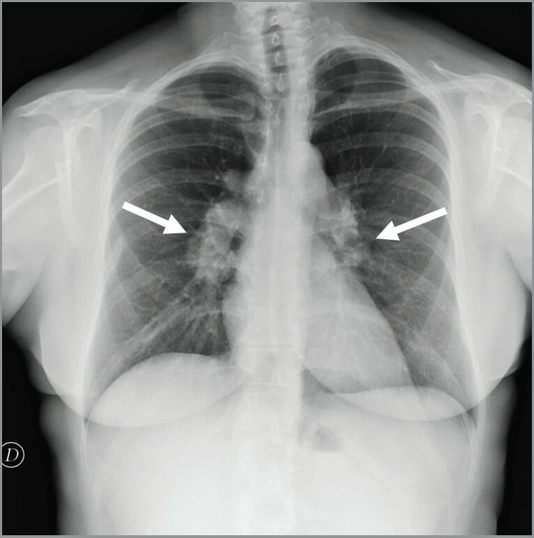
Chest x-ray showing bilateral hilar lymphadenopathy in a patient with cgd

Chest X-ray showing bilateral hilar lymphadenopathy in an individual with chronic granulomatous disease (CGD)

Image: “Chest x-ray showed bilateral hilar lymphadenopathies (arrows) suggestive for chronic granulomatous disease” by Conte G. et al. License: CC BY 3.0

Management

  • Hematopoietic stem cell transplantation Hematopoietic stem cell transplantation Transfer of hematopoietic stem cells from bone marrow or blood between individuals within the same species (homologous transplantation) or transfer within the same individual (autologous transplantation). Hematopoietic stem cell transplantation has been used as an alternative to bone marrow transplantation in the treatment of a variety of neoplasms. Organ Transplantation from a HLA-compatible donor
  • Antimicrobial prophylaxis Prophylaxis Cephalosporins ( trimethoprim Trimethoprim The sulfonamides are a class of antimicrobial drugs inhibiting folic acid synthesize in pathogens. The prototypical drug in the class is sulfamethoxazole. Although not technically sulfonamides, trimethoprim, dapsone, and pyrimethamine are also important antimicrobial agents inhibiting folic acid synthesis. The agents are often combined with sulfonamides, resulting in a synergistic effect. Sulfonamides and Trimethoprim sulfamethoxazole Sulfamethoxazole A bacteriostatic antibacterial agent that interferes with folic acid synthesis in susceptible bacteria. Its broad spectrum of activity has been limited by the development of resistance. Sulfonamides and Trimethoprim is considered 1st-line treatment)
  • Aggressive treatment of current infections
    • Skin Skin The skin, also referred to as the integumentary system, is the largest organ of the body. The skin is primarily composed of the epidermis (outer layer) and dermis (deep layer). The epidermis is primarily composed of keratinocytes that undergo rapid turnover, while the dermis contains dense layers of connective tissue. Skin: Structure and Functions infections: IV ciprofloxacin Ciprofloxacin A broad-spectrum antimicrobial carboxyfluoroquinoline. Fluoroquinolones
    • Fungal pneumonia Pneumonia Pneumonia or pulmonary inflammation is an acute or chronic inflammation of lung tissue. Causes include infection with bacteria, viruses, or fungi. In more rare cases, pneumonia can also be caused through toxic triggers through inhalation of toxic substances, immunological processes, or in the course of radiotherapy. Pneumonia: voriconazole Voriconazole A triazole antifungal agent that specifically inhibits sterol 14-alpha-demethylase and cytochrome p-450 cyp3a. Azoles or posaconazole Posaconazole Azoles for up to 6 months
  • Administration of interferon-gamma
  • Surgical drainage of abscesses
  • Gene Gene A category of nucleic acid sequences that function as units of heredity and which code for the basic instructions for the development, reproduction, and maintenance of organisms. Basic Terms of Genetics therapy is undergoing extensive research Research Critical and exhaustive investigation or experimentation, having for its aim the discovery of new facts and their correct interpretation, the revision of accepted conclusions, theories, or laws in the light of newly discovered facts, or the practical application of such new or revised conclusions, theories, or laws. Conflict of Interest and development.

Differential Diagnosis

  • Cystic Cystic Fibrocystic Change fibrosis Fibrosis Any pathological condition where fibrous connective tissue invades any organ, usually as a consequence of inflammation or other injury. Bronchiolitis Obliterans (CF): autosomal recessive Autosomal recessive Autosomal inheritance, both dominant and recessive, refers to the transmission of genes from the 22 autosomal chromosomes. Autosomal recessive diseases are only expressed when 2 copies of the recessive allele are inherited. Autosomal Recessive and Autosomal Dominant Inheritance disorder. Most carriers Carriers The Cell: Cell Membrane of the CF gene Gene A category of nucleic acid sequences that function as units of heredity and which code for the basic instructions for the development, reproduction, and maintenance of organisms. Basic Terms of Genetics are asymptomatic, and the disease manifests as an exocrine gland dysfunction that involves multiple organ systems but usually results in chronic respiratory infections, pancreatic enzyme insufficiency, and associated complications in untreated patients Patients Individuals participating in the health care system for the purpose of receiving therapeutic, diagnostic, or preventive procedures. Clinician–Patient Relationship. Pulmonary involvement Pulmonary involvement Coccidioides/Coccidioidomycosis occurs in 90% of patients Patients Individuals participating in the health care system for the purpose of receiving therapeutic, diagnostic, or preventive procedures. Clinician–Patient Relationship who survive the neonatal period. End-stage lung disease is the principal cause of death
  • Hyperimmunoglobulin E syndrome (HIES): first described as Job syndrome in 1966. Hyperimmunoglobulin E syndrome is characterized by recurrent skin Skin The skin, also referred to as the integumentary system, is the largest organ of the body. The skin is primarily composed of the epidermis (outer layer) and dermis (deep layer). The epidermis is primarily composed of keratinocytes that undergo rapid turnover, while the dermis contains dense layers of connective tissue. Skin: Structure and Functions abscesses, recurrent pneumonia Pneumonia Pneumonia or pulmonary inflammation is an acute or chronic inflammation of lung tissue. Causes include infection with bacteria, viruses, or fungi. In more rare cases, pneumonia can also be caused through toxic triggers through inhalation of toxic substances, immunological processes, or in the course of radiotherapy. Pneumonia with pneumatocele, eczematous dermatitis Dermatitis Any inflammation of the skin. Atopic Dermatitis (Eczema), and elevated serum IgE IgE An immunoglobulin associated with mast cells. Overexpression has been associated with allergic hypersensitivity. Immunoglobulins: Types and Functions levels. HIES was initially reported to have an autosomal dominant inheritance Autosomal dominant inheritance Autosomal Recessive and Autosomal Dominant Inheritance pattern, but cases with autosomal recessive inheritance Autosomal recessive inheritance Autosomal Recessive and Autosomal Dominant Inheritance and sporadic Sporadic Selective IgA Deficiency cases have been reported
  • Crohn’s disease (in patients Patients Individuals participating in the health care system for the purpose of receiving therapeutic, diagnostic, or preventive procedures. Clinician–Patient Relationship with inflammation Inflammation Inflammation is a complex set of responses to infection and injury involving leukocytes as the principal cellular mediators in the body’s defense against pathogenic organisms. Inflammation is also seen as a response to tissue injury in the process of wound healing. The 5 cardinal signs of inflammation are pain, heat, redness, swelling, and loss of function. Inflammation limited to the rectum Rectum The rectum and anal canal are the most terminal parts of the lower GI tract/large intestine that form a functional unit and control defecation. Fecal continence is maintained by several important anatomic structures including rectal folds, anal valves, the sling-like puborectalis muscle, and internal and external anal sphincters. Rectum and Anal Canal: Anatomy): idiopathic Idiopathic Dermatomyositis, chronic regional enteritis Enteritis Inflammation of any segment of the small intestine. Lactose Intolerance that most commonly affects the terminal ileum Ileum The distal and narrowest portion of the small intestine, between the jejunum and the ileocecal valve of the large intestine. Small Intestine: Anatomy but has the potential to affect any part of the GI tract from mouth to anus. CD is defined by transmural inflammation Transmural inflammation Crohn’s Disease that lacks continuity along the intestine (skip areas). In some cases, CD has nonnecrotizing granulomas Granulomas A relatively small nodular inflammatory lesion containing grouped mononuclear phagocytes, caused by infectious and noninfectious agents. Sarcoidosis containing epithelioid histiocytes. Patients Patients Individuals participating in the health care system for the purpose of receiving therapeutic, diagnostic, or preventive procedures. Clinician–Patient Relationship present with typical signs of CD including abdominal pain Abdominal Pain Acute Abdomen, bloody diarrhea Bloody diarrhea Diarrhea, and in severe cases by intestinal fistulas, intramural abscesses, and bowel obstruction Bowel obstruction Any impairment, arrest, or reversal of the normal flow of intestinal contents toward the anal canal. Ascaris/Ascariasis.

References

  1. Zerbe CS, Marciano BE, Holland SM. (2023). Chronic granulomatous disease: pathogenesis, clinical manifestations, and diagnosis. UpToDate. Retrieved November 6, 2024, from https://www.uptodate.com/contents/chronic-granulomatous-disease-pathogenesis-clinical-manifestations-and-diagnosis
  2. Marciano BE, Zerbe CS, Holland SM. (2023). Chronic granulomatous disease: Treatment and prognosis. UpToDate. Retrieved November 6, 2024, from https://www.uptodate.com/contents/chronic-granulomatous-disease-treatment-and-prognosis
  3. Nowicki RJ. (2019). Chronic Granulomatous Disease. eMedicine. Retrieved November 6, 2024, from https://emedicine.medscape.com/article/1116022
  4. Fischer, A. (2018). Primary Immune Deficiency Diseases. In Jameson JL, et al. (Ed.), Harrison’s Principles of Internal Medicine (20th ed., ch. 344).

Create your free account or log in to continue reading!

Sign up now and get free access to Lecturio with concept pages, medical videos, and questions for your medical education.

User Reviews

Details