Chédiak-Higashi Syndrome

Chédiak-Higashi syndrome ( CHS CHS Cannabinoids) is an autosomal recessive Autosomal recessive Autosomal inheritance, both dominant and recessive, refers to the transmission of genes from the 22 autosomal chromosomes. Autosomal recessive diseases are only expressed when 2 copies of the recessive allele are inherited. Autosomal Recessive and Autosomal Dominant Inheritance disorder caused by mutations affecting a lysosomal trafficking regulator protein. This protein plays a crucial role in the inability of neutrophils Neutrophils Granular leukocytes having a nucleus with three to five lobes connected by slender threads of chromatin, and cytoplasm containing fine inconspicuous granules and stainable by neutral dyes. Innate Immunity: Phagocytes and Antigen Presentation to kill phagocytosed microbes. Patients Patients Individuals participating in the health care system for the purpose of receiving therapeutic, diagnostic, or preventive procedures. Clinician–Patient Relationship with CHS CHS Cannabinoids exhibit recurrent pyogenic infections Infections Invasion of the host organism by microorganisms or their toxins or by parasites that can cause pathological conditions or diseases. Chronic Granulomatous Disease, easy bleeding and bruising, and neurologic manifestations. The syndrome is also associated with oculocutaneous albinism Oculocutaneous albinism Heterogeneous group of autosomal recessive disorders comprising at least four recognized types, all having in common varying degrees of hypopigmentation of the skin, hair, and eyes. The two most common are the tyrosinase-positive and tyrosinase-negative types. Albinism. The diagnosis is made based on analysis of the patient’s blood or bone marrow Bone marrow The soft tissue filling the cavities of bones. Bone marrow exists in two types, yellow and red. Yellow marrow is found in the large cavities of large bones and consists mostly of fat cells and a few primitive blood cells. Red marrow is a hematopoietic tissue and is the site of production of erythrocytes and granular leukocytes. Bone marrow is made up of a framework of connective tissue containing branching fibers with the frame being filled with marrow cells. Bone Marrow: Composition and Hematopoiesis smear and genetic testing Genetic Testing Detection of a mutation; genotype; karyotype; or specific alleles associated with genetic traits, heritable diseases, or predisposition to a disease, or that may lead to the disease in descendants. It includes prenatal genetic testing. Myotonic Dystrophies. The treatment of choice is allogeneic hematopoietic cell transplantation.

Last updated: Nov 6, 2024

Editorial responsibility: Stanley Oiseth, Lindsay Jones, Evelin Maza

Overview

Epidemiology

  • Incidence Incidence The number of new cases of a given disease during a given period in a specified population. It also is used for the rate at which new events occur in a defined population. It is differentiated from prevalence, which refers to all cases in the population at a given time. Measures of Disease Frequency: < 1 in 1 million people
  • < 500 cases reported worldwide
  • Most patients Patients Individuals participating in the health care system for the purpose of receiving therapeutic, diagnostic, or preventive procedures. Clinician–Patient Relationship die before 7 years of age.

Etiology

  • Mode of inheritance: autosomal recessive Autosomal recessive Autosomal inheritance, both dominant and recessive, refers to the transmission of genes from the 22 autosomal chromosomes. Autosomal recessive diseases are only expressed when 2 copies of the recessive allele are inherited. Autosomal Recessive and Autosomal Dominant Inheritance
  • Gene Gene A category of nucleic acid sequences that function as units of heredity and which code for the basic instructions for the development, reproduction, and maintenance of organisms. Basic Terms of Genetics: mutations in CHS1 (or LYST) gene Gene A category of nucleic acid sequences that function as units of heredity and which code for the basic instructions for the development, reproduction, and maintenance of organisms. Basic Terms of Genetics
    • Encodes for a lysosomal trafficking regulator, which plays a role in trafficking materials into lysosomes Lysosomes A class of morphologically heterogeneous cytoplasmic particles in animal and plant tissues characterized by their content of hydrolytic enzymes and the structure-linked latency of these enzymes. The intracellular functions of lysosomes depend on their lytic potential. The single unit membrane of the lysosome acts as a barrier between the enzymes enclosed in the lysosome and the external substrate. The activity of the enzymes contained in lysosomes is limited or nil unless the vesicle in which they are enclosed is ruptured or undergoes membrane fusion. The Cell: Organelles
    • The exact role lysosomal trafficking regulator proteins Proteins Linear polypeptides that are synthesized on ribosomes and may be further modified, crosslinked, cleaved, or assembled into complex proteins with several subunits. The specific sequence of amino acids determines the shape the polypeptide will take, during protein folding, and the function of the protein. Energy Homeostasis play in killing off microbes in lysosomes Lysosomes A class of morphologically heterogeneous cytoplasmic particles in animal and plant tissues characterized by their content of hydrolytic enzymes and the structure-linked latency of these enzymes. The intracellular functions of lysosomes depend on their lytic potential. The single unit membrane of the lysosome acts as a barrier between the enzymes enclosed in the lysosome and the external substrate. The activity of the enzymes contained in lysosomes is limited or nil unless the vesicle in which they are enclosed is ruptured or undergoes membrane fusion. The Cell: Organelles is unknown.

Pathophysiology

  • Disruption in lysosomal function, maintenance, and trafficking → failure in phagolysosome formation → phagocytosed bacteria Bacteria Bacteria are prokaryotic single-celled microorganisms that are metabolically active and divide by binary fission. Some of these organisms play a significant role in the pathogenesis of diseases. Bacteriology are not destroyed by lysosomal enzymes Enzymes Enzymes are complex protein biocatalysts that accelerate chemical reactions without being consumed by them. Due to the body’s constant metabolic needs, the absence of enzymes would make life unsustainable, as reactions would occur too slowly without these molecules. Basics of Enzymes
  • Additional effects:
    • Impaired chemotaxis Chemotaxis The movement of leukocytes in response to a chemical concentration gradient or to products formed in an immunologic reaction. Leukocyte Adhesion Deficiency Type 1, reduced natural killer cell function, and poor cytolysis → increased infection susceptibility
    • Melanocytes Melanocytes Mammalian pigment cells that produce melanins, pigments found mainly in the epidermis, but also in the eyes and the hair, by a process called melanogenesis. Coloration can be altered by the number of melanocytes or the amount of pigment produced and stored in the organelles called melanosomes. The large non-mammalian melanin-containing cells are called melanophores. Skin: Structure and Functions are unable to transfer melanin Melanin Insoluble polymers of tyrosine derivatives found in and causing darkness in skin (skin pigmentation), hair, and feathers providing protection against sunburn induced by sunlight. Carotenes contribute yellow and red coloration. Seborrheic Keratosis to keratinocytes Keratinocytes Epidermal cells which synthesize keratin and undergo characteristic changes as they move upward from the basal layers of the epidermis to the cornified (horny) layer of the skin. Successive stages of differentiation of the keratinocytes forming the epidermal layers are basal cell, spinous or prickle cell, and the granular cell. Skin: Structure and Functions albinism Albinism Albinism refers to a group of inherited disorders that result in the disruption of melanin production, causing hypopigmentation and visual impairment. The condition is classified according to the clinical phenotype. Oculocutaneous albinism results in hypopigmentation of the skin, eyes, and hair. Ocular albinism affects only the eyes. Albinism
    • ↓ Platelet storage granules → impaired platelet aggregation Platelet aggregation The attachment of platelets to one another. This clumping together can be induced by a number of agents (e.g., thrombin; collagen) and is part of the mechanism leading to the formation of a thrombus. Hemostasis → bleeding

Clinical Presentation

General signs and symptoms

Signs and symptoms that usually appear soon after birth include the following:

  • Oculocutaneous albinism Oculocutaneous albinism Heterogeneous group of autosomal recessive disorders comprising at least four recognized types, all having in common varying degrees of hypopigmentation of the skin, hair, and eyes. The two most common are the tyrosinase-positive and tyrosinase-negative types. Albinism (nonpigmented skin Skin The skin, also referred to as the integumentary system, is the largest organ of the body. The skin is primarily composed of the epidermis (outer layer) and dermis (deep layer). The epidermis is primarily composed of keratinocytes that undergo rapid turnover, while the dermis contains dense layers of connective tissue. Skin: Structure and Functions, eyes, and hair)
  • Lymphadenopathy Lymphadenopathy Lymphadenopathy is lymph node enlargement (> 1 cm) and is benign and self-limited in most patients. Etiologies include malignancy, infection, and autoimmune disorders, as well as iatrogenic causes such as the use of certain medications. Generalized lymphadenopathy often indicates underlying systemic disease. Lymphadenopathy
  • Gingivitis and periodontal disease
  • Oral ulcers
  • Bleeding:
  • Immunodeficiency:
    • Severe and recurrent pyogenic sinopulmonary infections Infections Invasion of the host organism by microorganisms or their toxins or by parasites that can cause pathological conditions or diseases. Chronic Granulomatous Disease
    • Common organisms:
      • Staphylococcus aureus Staphylococcus aureus Potentially pathogenic bacteria found in nasal membranes, skin, hair follicles, and perineum of warm-blooded animals. They may cause a wide range of infections and intoxications. Brain Abscess (most common)
      • Streptococcus Streptococcus Streptococcus is one of the two medically important genera of gram-positive cocci, the other being Staphylococcus. Streptococci are identified as different species on blood agar on the basis of their hemolytic pattern and sensitivity to optochin and bacitracin. There are many pathogenic species of streptococci, including S. pyogenes, S. agalactiae, S. pneumoniae, and the viridans streptococci. Streptococcus
      • Fungi Fungi A kingdom of eukaryotic, heterotrophic organisms that live parasitically as saprobes, including mushrooms; yeasts; smuts, molds, etc. They reproduce either sexually or asexually, and have life cycles that range from simple to complex. Filamentous fungi, commonly known as molds, refer to those that grow as multicellular colonies. Mycology

Late-onset neurological manifestations that appear in individuals who survive early childhood with curative treatment:

  • Central and peripheral neuropathies
  • Sensory Sensory Neurons which conduct nerve impulses to the central nervous system. Nervous System: Histology loss
  • Muscle weakness
  • Parkinsonism Parkinsonism West Nile Virus
  • Cerebellar ataxia Cerebellar ataxia Incoordination of voluntary movements that occur as a manifestation of cerebellar diseases. Characteristic features include a tendency for limb movements to overshoot or undershoot a target (dysmetria), a tremor that occurs during attempted movements (intention tremor), impaired force and rhythm of diadochokinesis (rapidly alternating movements), and gait ataxia. Cerebellar Disorders
  • Cognitive impairment
Hypopigmented eyes chediak-higashi syndrome

Hypopigmented eyes and eyelashes in a patient with oculocutaneous albinism:
This may be seen in Chédiak-Higashi syndrome.

Image: “Eyes from a patient with OCA1A” by Karen G et al. License: CC BY 2.0

Accelerated phase Accelerated phase The phase of chronic myeloid leukemia following the chronic phase, where there are increased systemic symptoms, worsening cytopenias, and refractory leukocytosis. Chronic Myeloid Leukemia

Chédiak-Higashi syndrome ( CHS CHS Cannabinoids) can progress to the accelerated phase Accelerated phase The phase of chronic myeloid leukemia following the chronic phase, where there are increased systemic symptoms, worsening cytopenias, and refractory leukocytosis. Chronic Myeloid Leukemia characterized by hemophagocytic lymphohistiocytosis Hemophagocytic lymphohistiocytosis A group of related disorders characterized by lymphocytosis; histiocytosis; and hemophagocytosis. The two major forms are familial and reactive. Epstein-Barr Virus (HLH), where hyperactive WBCs cause excessive inflammation Inflammation Inflammation is a complex set of responses to infection and injury involving leukocytes as the principal cellular mediators in the body’s defense against pathogenic organisms. Inflammation is also seen as a response to tissue injury in the process of wound healing. The 5 cardinal signs of inflammation are pain, heat, redness, swelling, and loss of function. Inflammation and tissue damage. It develops in 80-85% of CHS CHS Cannabinoids patients Patients Individuals participating in the health care system for the purpose of receiving therapeutic, diagnostic, or preventive procedures. Clinician–Patient Relationship and is usually lethal. Signs and symptoms include:
  • Fever Fever Fever is defined as a measured body temperature of at least 38°C (100.4°F). Fever is caused by circulating endogenous and/or exogenous pyrogens that increase levels of prostaglandin E2 in the hypothalamus. Fever is commonly associated with chills, rigors, sweating, and flushing of the skin. Fever
  • Jaundice Jaundice Jaundice is the abnormal yellowing of the skin and/or sclera caused by the accumulation of bilirubin. Hyperbilirubinemia is caused by either an increase in bilirubin production or a decrease in the hepatic uptake, conjugation, or excretion of bilirubin. Jaundice
  • Hepatosplenomegaly Hepatosplenomegaly Cytomegalovirus
  • Lymphadenopathy Lymphadenopathy Lymphadenopathy is lymph node enlargement (> 1 cm) and is benign and self-limited in most patients. Etiologies include malignancy, infection, and autoimmune disorders, as well as iatrogenic causes such as the use of certain medications. Generalized lymphadenopathy often indicates underlying systemic disease. Lymphadenopathy
  • Pancytopenia Pancytopenia Deficiency of all three cell elements of the blood, erythrocytes, leukocytes and platelets. Aplastic Anemia
  • Abnormal bleeding
  • Profound immunodeficiency
  • Organ failure

Diagnosis

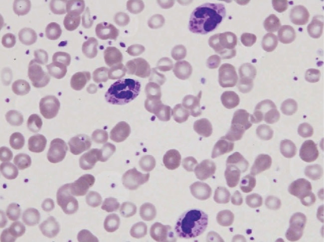
  • Peripheral blood smear Peripheral Blood Smear Anemia: Overview and Types: giant azurophilic granules Azurophilic granules Myeloperoxidase Deficiency can be seen in
    • Neutrophils Neutrophils Granular leukocytes having a nucleus with three to five lobes connected by slender threads of chromatin, and cytoplasm containing fine inconspicuous granules and stainable by neutral dyes. Innate Immunity: Phagocytes and Antigen Presentation
    • Eosinophils Eosinophils Granular leukocytes with a nucleus that usually has two lobes connected by a slender thread of chromatin, and cytoplasm containing coarse, round granules that are uniform in size and stainable by eosin. Innate Immunity: Phagocytes and Antigen Presentation
    • Granulocytes Granulocytes Leukocytes with abundant granules in the cytoplasm. They are divided into three groups according to the staining properties of the granules: neutrophilic, eosinophilic, and basophilic. Mature granulocytes are the neutrophils; eosinophils; and basophils. White Myeloid Cells: Histology
  • Bone marrow Bone marrow The soft tissue filling the cavities of bones. Bone marrow exists in two types, yellow and red. Yellow marrow is found in the large cavities of large bones and consists mostly of fat cells and a few primitive blood cells. Red marrow is a hematopoietic tissue and is the site of production of erythrocytes and granular leukocytes. Bone marrow is made up of a framework of connective tissue containing branching fibers with the frame being filled with marrow cells. Bone Marrow: Composition and Hematopoiesis smear:
    • Shows “giant inclusion bodies” in leukocyte precursor cells 
    • Granules are peroxidase positive and contain lysosomal enzymes Enzymes Enzymes are complex protein biocatalysts that accelerate chemical reactions without being consumed by them. Due to the body’s constant metabolic needs, the absence of enzymes would make life unsustainable, as reactions would occur too slowly without these molecules. Basics of Enzymes.
  • Microscopic examination of hair:
    • Clumped melanin Melanin Insoluble polymers of tyrosine derivatives found in and causing darkness in skin (skin pigmentation), hair, and feathers providing protection against sunburn induced by sunlight. Carotenes contribute yellow and red coloration. Seborrheic Keratosis granules that are larger than those found in normal hairs 
    • Bright and polychromatic refringence pattern
  • Genetic testing Genetic Testing Detection of a mutation; genotype; karyotype; or specific alleles associated with genetic traits, heritable diseases, or predisposition to a disease, or that may lead to the disease in descendants. It includes prenatal genetic testing. Myotonic Dystrophies for mutations in the LYST gene Gene A category of nucleic acid sequences that function as units of heredity and which code for the basic instructions for the development, reproduction, and maintenance of organisms. Basic Terms of Genetics
  • Supporting laboratory evaluation:
    • Neutropenia Neutropenia Neutrophils are an important component of the immune system and play a significant role in the eradication of infections. Low numbers of circulating neutrophils, referred to as neutropenia, predispose the body to recurrent infections or sepsis, though patients can also be asymptomatic. Neutropenia 
    • Hypergammaglobulinemia Hypergammaglobulinemia An excess of gamma-globulins in the serum due to chronic infections or paraproteinemias. Autoimmune Hepatitis

Management

  • Treatment of choice: curative allogeneic hematopoietic cell transplantation (HCT)
  • Supportive management:
    • Prophylactic antibiotics
    • Aggressive antibiotic therapy for acute bacterial infections Infections Invasion of the host organism by microorganisms or their toxins or by parasites that can cause pathological conditions or diseases. Chronic Granulomatous Disease
    • Routine immunizations
    • Platelet transfusions for serious bleeding
    • Management of the accelerated phase Accelerated phase The phase of chronic myeloid leukemia following the chronic phase, where there are increased systemic symptoms, worsening cytopenias, and refractory leukocytosis. Chronic Myeloid Leukemia:
      • Corticosteroids Corticosteroids Chorioretinitis
      • Intravenous immunoglobulin ( IVIG IVIG Dermatomyositis)
      • Chemotherapy Chemotherapy Osteosarcoma (e.g., etoposide Etoposide A semisynthetic derivative of podophyllotoxin that exhibits antitumor activity. Etoposide inhibits DNA synthesis by forming a complex with topoisomerase II and DNA. This complex induces breaks in double stranded DNA and prevents repair by topoisomerase II binding. Accumulated breaks in DNA prevent entry into the mitotic phase of cell division, and lead to cell death. Etoposide acts primarily in the g2 and s phases of the cell cycle. Microtubule and Topoisomerase Inhibitors, methotrexate Methotrexate An antineoplastic antimetabolite with immunosuppressant properties. It is an inhibitor of tetrahydrofolate dehydrogenase and prevents the formation of tetrahydrofolate, necessary for synthesis of thymidylate, an essential component of DNA. Antimetabolite Chemotherapy)
      • Splenectomy Splenectomy Surgical procedure involving either partial or entire removal of the spleen. Rupture of the Spleen

Differential Diagnosis

  • Leukocyte adhesion Leukocyte adhesion Leukocyte Adhesion Deficiency Type 1 deficiency type 1 Type 1 Spinal Muscular Atrophy: an inherited condition resulting in a lack of CD18 CD18 Cell-surface glycoprotein beta-chains that are non-covalently linked to specific alpha-chains of the cd11 family of leukocyte-adhesion molecules (receptors, leukocyte-adhesion). A defect in the gene encoding cd18 causes leukocyte-adhesion deficiency syndrome. Leukocyte Adhesion Deficiency Type 1 expression on neutrophils Neutrophils Granular leukocytes having a nucleus with three to five lobes connected by slender threads of chromatin, and cytoplasm containing fine inconspicuous granules and stainable by neutral dyes. Innate Immunity: Phagocytes and Antigen Presentation leading to a decrease in the ability of neutrophils Neutrophils Granular leukocytes having a nucleus with three to five lobes connected by slender threads of chromatin, and cytoplasm containing fine inconspicuous granules and stainable by neutral dyes. Innate Immunity: Phagocytes and Antigen Presentation to migrate from the blood vessels. Patients Patients Individuals participating in the health care system for the purpose of receiving therapeutic, diagnostic, or preventive procedures. Clinician–Patient Relationship with this condition will have recurrent infections Recurrent infections Common Variable Immunodeficiency (CVID), delayed wound healing Wound healing Wound healing is a physiological process involving tissue repair in response to injury. It involves a complex interaction of various cell types, cytokines, and inflammatory mediators. Wound healing stages include hemostasis, inflammation, granulation, and remodeling. Wound Healing, and an elevated neutrophil count. The deficiency is not associated with oculocutaneous albinism Oculocutaneous albinism Heterogeneous group of autosomal recessive disorders comprising at least four recognized types, all having in common varying degrees of hypopigmentation of the skin, hair, and eyes. The two most common are the tyrosinase-positive and tyrosinase-negative types. Albinism. The diagnosis is confirmed with flow cytometry Flow cytometry Technique using an instrument system for making, processing, and displaying one or more measurements on individual cells obtained from a cell suspension. Cells are usually stained with one or more fluorescent dyes specific to cell components of interest, e.g., DNA, and fluorescence of each cell is measured as it rapidly transverses the excitation beam (laser or mercury arc lamp). Fluorescence provides a quantitative measure of various biochemical and biophysical properties of the cell, as well as a basis for cell sorting. Other measurable optical parameters include light absorption and light scattering, the latter being applicable to the measurement of cell size, shape, density, granularity, and stain uptake. X-linked Agammaglobulinemia demonstrating a deficiency in CD18 CD18 Cell-surface glycoprotein beta-chains that are non-covalently linked to specific alpha-chains of the cd11 family of leukocyte-adhesion molecules (receptors, leukocyte-adhesion). A defect in the gene encoding cd18 causes leukocyte-adhesion deficiency syndrome. Leukocyte Adhesion Deficiency Type 1. Hematopoietic stem cell transplant is the treatment of choice.
  • Chronic granulomatous disease Granulomatous disease A defect of leukocyte function in which phagocytic cells ingest but fail to digest bacteria, resulting in recurring bacterial infections with granuloma formation. When chronic granulomatous disease is caused by mutations in the cybb gene, the condition is inherited in an X-linked recessive pattern. When chronic granulomatous disease is caused by cyba, ncf1, ncf2, or ncf4 gene mutations, the condition is inherited in an autosomal recessive pattern. Common Variable Immunodeficiency (CVID): a consequence of defective phagocytic cells. Patients Patients Individuals participating in the health care system for the purpose of receiving therapeutic, diagnostic, or preventive procedures. Clinician–Patient Relationship will have recurrent infections Recurrent infections Common Variable Immunodeficiency (CVID), abscesses, and granulomatous lesions of multiple organs. Hypergammaglobulinemia Hypergammaglobulinemia An excess of gamma-globulins in the serum due to chronic infections or paraproteinemias. Autoimmune Hepatitis may be seen. The diagnosis is made with neutrophil function testing for superoxide production. Antimicrobial prophylaxis Prophylaxis Cephalosporins, interferon gamma, granulocyte transfusion Granulocyte transfusion Severe Congenital Neutropenia, and hematopoietic stem cell transplantation Hematopoietic stem cell transplantation Transfer of hematopoietic stem cells from bone marrow or blood between individuals within the same species (homologous transplantation) or transfer within the same individual (autologous transplantation). Hematopoietic stem cell transplantation has been used as an alternative to bone marrow transplantation in the treatment of a variety of neoplasms. Organ Transplantation are potential management options.
  • Severe congenital neutropenia Congenital neutropenia Severe Congenital Neutropenia: a rare disease resulting from genetic mutations Genetic Mutations Carcinogenesis that affect myelopoiesis Myelopoiesis Formation of myeloid cells from the pluripotent hematopoietic stem cells in the bone marrow via myeloid stem cells. Myelopoiesis generally refers to the production of leukocytes in blood, such as monocytes and granulocytes. This process also produces precursor cells for macrophage and dendritic cells found in the lymphoid tissue. White Myeloid Cells: Histology. The condition manifests in infancy with life-threatening bacterial infections Infections Invasion of the host organism by microorganisms or their toxins or by parasites that can cause pathological conditions or diseases. Chronic Granulomatous Disease. Neutropenia Neutropenia Neutrophils are an important component of the immune system and play a significant role in the eradication of infections. Low numbers of circulating neutrophils, referred to as neutropenia, predispose the body to recurrent infections or sepsis, though patients can also be asymptomatic. Neutropenia is found on laboratory evaluation, often with an elevated monocyte count. A bone marrow Bone marrow The soft tissue filling the cavities of bones. Bone marrow exists in two types, yellow and red. Yellow marrow is found in the large cavities of large bones and consists mostly of fat cells and a few primitive blood cells. Red marrow is a hematopoietic tissue and is the site of production of erythrocytes and granular leukocytes. Bone marrow is made up of a framework of connective tissue containing branching fibers with the frame being filled with marrow cells. Bone Marrow: Composition and Hematopoiesis biopsy Biopsy Removal and pathologic examination of specimens from the living body. Ewing Sarcoma will also aid in the diagnosis. Management includes granulocyte colony-stimulating factor Granulocyte colony-stimulating factor A glycoprotein of mw 25 kda containing internal disulfide bonds. It induces the survival, proliferation, and differentiation of neutrophilic granulocyte precursor cells and functionally activates mature blood neutrophils. Among the family of colony-stimulating factors, G-CSF is the most potent inducer of terminal differentiation to granulocytes and macrophages of leukemic myeloid cell lines. White Myeloid Cells: Histology and hematopoietic stem cell transplantation Hematopoietic stem cell transplantation Transfer of hematopoietic stem cells from bone marrow or blood between individuals within the same species (homologous transplantation) or transfer within the same individual (autologous transplantation). Hematopoietic stem cell transplantation has been used as an alternative to bone marrow transplantation in the treatment of a variety of neoplasms. Organ Transplantation..
  • Myeloperoxidase Myeloperoxidase Acute Myeloid Leukemia deficiency: an autosomal recessive Autosomal recessive Autosomal inheritance, both dominant and recessive, refers to the transmission of genes from the 22 autosomal chromosomes. Autosomal recessive diseases are only expressed when 2 copies of the recessive allele are inherited. Autosomal Recessive and Autosomal Dominant Inheritance disorder that causes impaired microbial killing by phagocytes. The majority of patients Patients Individuals participating in the health care system for the purpose of receiving therapeutic, diagnostic, or preventive procedures. Clinician–Patient Relationship are asymptomatic, while others can have recurrent, severe fungal infections Infections Invasion of the host organism by microorganisms or their toxins or by parasites that can cause pathological conditions or diseases. Chronic Granulomatous Disease. The diagnosis is made with histochemical staining Histochemical staining Myeloperoxidase Deficiency for myeloperoxidase Myeloperoxidase Acute Myeloid Leukemia in neutrophils Neutrophils Granular leukocytes having a nucleus with three to five lobes connected by slender threads of chromatin, and cytoplasm containing fine inconspicuous granules and stainable by neutral dyes. Innate Immunity: Phagocytes and Antigen Presentation. There is no specific management for this condition, but patients Patients Individuals participating in the health care system for the purpose of receiving therapeutic, diagnostic, or preventive procedures. Clinician–Patient Relationship should be treated for any infections Infections Invasion of the host organism by microorganisms or their toxins or by parasites that can cause pathological conditions or diseases. Chronic Granulomatous Disease.

References

  1. Nowicki, R.J. (2019). Chediak-Higashi syndrome. In Elston, D.M. (Ed.), Medscape. Retrieved November 6, 2024, from  https://emedicine.medscape.com/article/1114607-overview
  2. Boztug, K. (2023). Chediak-Higashi syndrome. In TePas, E. (Ed.), UpToDate. Retrieved November 6, 2024, from https://www.uptodate.com/contents/chediak-higashi-syndrome
  3. Fernandez, J. (2024). Chediak-Higashi syndrome. [online] MSD Manual Professional Version. Retrieved November 6, 2024, from https://www.msdmanuals.com/professional/immunology-allergic-disorders/immunodeficiency-disorders/ch%C3%A9diak-higashi-syndrome
  4. Ajitkumar, A., Yarrarapu, S.N.S., and Ramphul, K. (2023). Chediak Higashi syndrome. [online] StatPearls. Retrieved November 6, 2024, from https://www.ncbi.nlm.nih.gov/books/NBK507881/

Create your free account or log in to continue reading!

Sign up now and get free access to Lecturio with concept pages, medical videos, and questions for your medical education.

User Reviews

Details