The cerebellum, Latin for “little brain Brain The part of central nervous system that is contained within the skull (cranium). Arising from the neural tube, the embryonic brain is comprised of three major parts including prosencephalon (the forebrain); mesencephalon (the midbrain); and rhombencephalon (the hindbrain). The developed brain consists of cerebrum; cerebellum; and other structures in the brain stem. Nervous System: Anatomy, Structure, and Classification,” is located in the posterior cranial fossa Posterior cranial fossa The infratentorial compartment that contains the cerebellum and brain stem. It is formed by the posterior third of the superior surface of the body of the sphenoid (sphenoid bone), by the occipital, the petrous, and mastoid portions of the temporal bone, and the posterior inferior angle of the parietal bone. Skull: Anatomy, dorsal to the pons Pons The front part of the hindbrain (rhombencephalon) that lies between the medulla and the midbrain (mesencephalon) ventral to the cerebellum. It is composed of two parts, the dorsal and the ventral. The pons serves as a relay station for neural pathways between the cerebellum to the cerebrum. Brain Stem: Anatomy and midbrain Midbrain The middle of the three primitive cerebral vesicles of the embryonic brain. Without further subdivision, midbrain develops into a short, constricted portion connecting the pons and the diencephalon. Midbrain contains two major parts, the dorsal tectum mesencephali and the ventral tegmentum mesencephali, housing components of auditory, visual, and other sensorimotor systems. Brain Stem: Anatomy, and its principal role is in the coordination Coordination Cerebellar Disorders of movements. The cerebellum consists of 3 lobes on either side of its 2 hemispheres and is connected in the middle by the vermis. Three paired peduncles link the cerebellum to the brainstem and diencephalon Diencephalon The paired caudal parts of the prosencephalon from which the thalamus; hypothalamus; epithalamus; and subthalamus are derived. Development of the Nervous System and Face. Much like the cerebral cortex Cerebral cortex The cerebral cortex is the largest and most developed part of the human brain and CNS. Occupying the upper part of the cranial cavity, the cerebral cortex has 4 lobes and is divided into 2 hemispheres that are joined centrally by the corpus callosum. Cerebral Cortex: Anatomy, the cerebellum has a cortex of gray matter Gray matter Region of central nervous system that appears darker in color than the other type, white matter. It is composed of neuronal cell bodies; neuropil; glial cells and capillaries but few myelinated nerve fibers. Cerebral Cortex: Anatomy on the surface.
Last updated: Nov 19, 2024
Cerebellar peduncles and their connections:
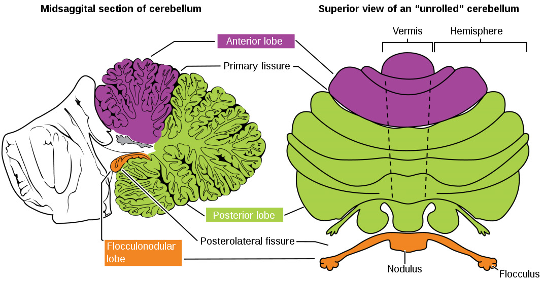
Lobes of the cerebellum: anterior in purple, posterior in green, and flocculonodular in orange. The image on the left shows the midsagittal section of the cerebellum and the image on the right illustrates a superior view of an “unrolled” cerebellum, such that the vermis is in 1 plane.
Image: “1613 Major Regions of the Cerebellum-02” by OpenStax College. License: CC BY 3.0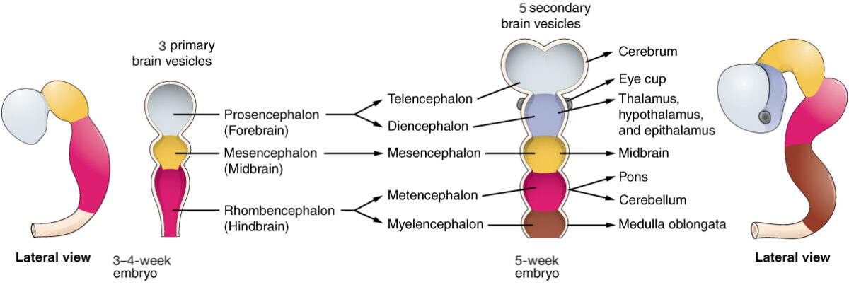
Development of the cerebellum from the rhombencephalon and, subsequently, the metencephalon
Image: “Primary and Secondary Vesicle Stages of Development ” by Phil Schatz. License: CC BY 4.0, edited by Lecturio.The majority of neuronal cell bodies are located in the cortex, which can be divided into 3 layers:
Granular cell layer:
Purkinje cell layer:
Molecular layer:
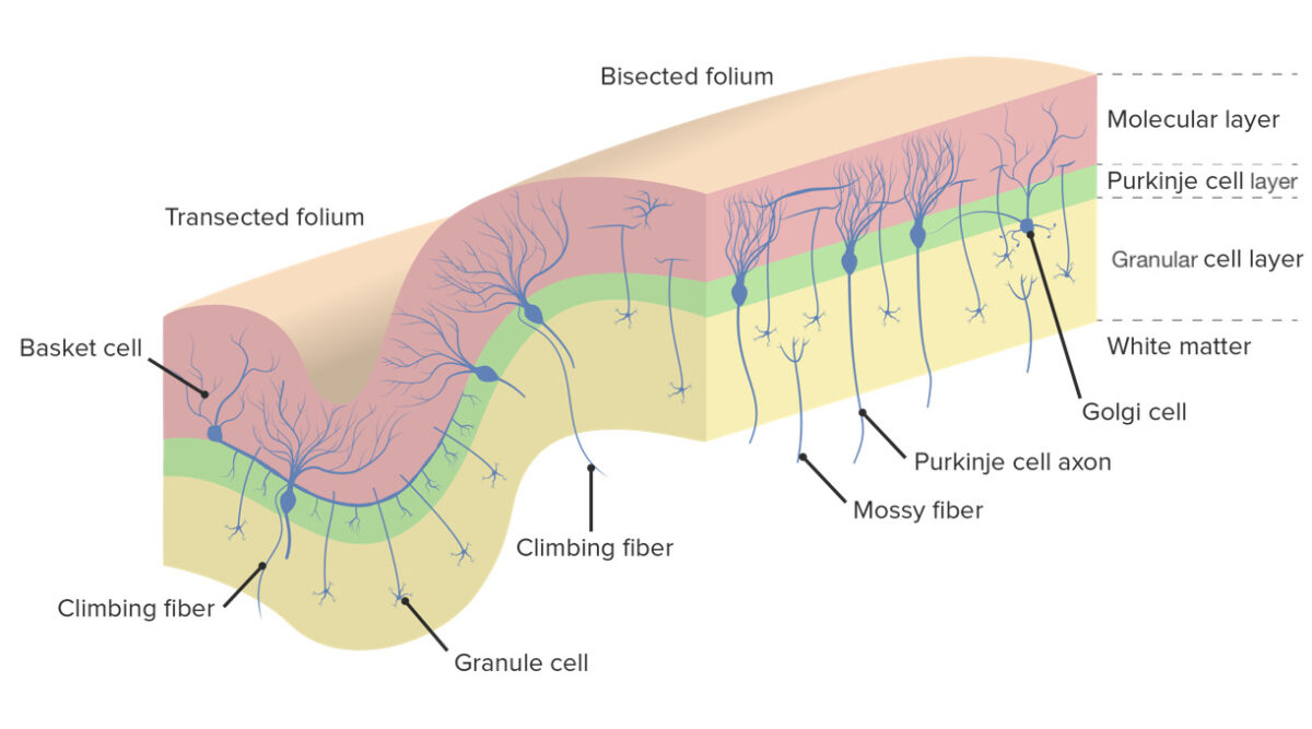
Cerebellar cortex and the 3 layers, namely the molecular layer, Purkinje cell layer, and granular cell layer. Note the bodies of Purkinje cells situated in the Purkinje layer while their dendrites project into the molecular layer.
Image by Lecturio.Cortical interneurons can be divided into 2 categories:
Cerebellar nuclei consist of 4 pairs of areas:
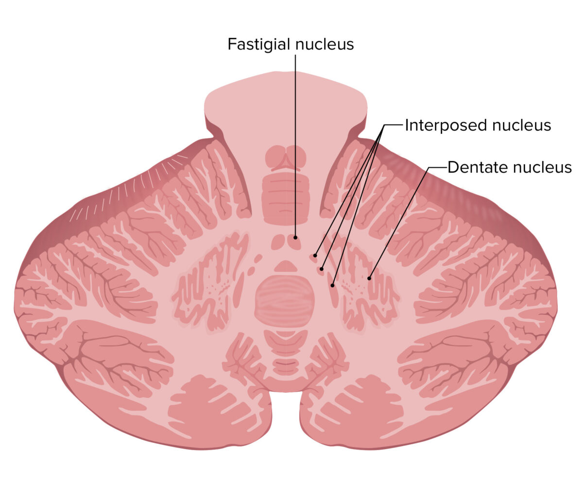
Location of the nuclei within the cerebellum
From lateral to medial: dentate nucleus, interposed nucleus (consisting of the emboliform and globose nuclei), and most medially, the fastigial nucleus
Many afferents pass through the 3 cerebellar peduncles to the cerebellar cortex. There are 2 extracerebellar excitatory glutamatergic afferent Afferent Neurons which conduct nerve impulses to the central nervous system. Nervous System: Histology systems:
Efferent Efferent Neurons which send impulses peripherally to activate muscles or secretory cells. Nervous System: Histology pathways of the cerebellum pass from the cerebellar nuclei to the following:
The cerebellum can also be divided into 3 regions based on connections:

Functional divisions of the cerebellum from an anterior view: The vestibulocerebellum is in the flocculonodular lobe, the spinocerebellum is in the intermediate hemisphere (IH), and the pontocerebellum is in the lateral hemisphere (LH).
Image by Lecturio.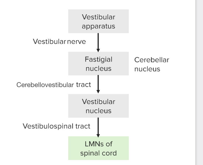
Progression of signals from the vestibular apparatus (semicircular canals and otoliths) through the cerebellum and to the lower motor neurons (LMNs) of the spinal cord
Image by Lecturio.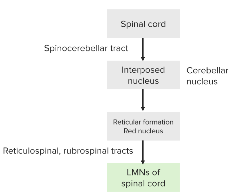
Flow of signals via the spinocerebellar tract: Note the signals coming from the spinal cord to the cerebellum via the spinocerebellar tract and finally to the reticular formation and red nucleus to the LMNs of the spinal cord.
Image by Lecturio.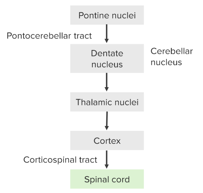
Flow of signals in the pontocerebellar tract: information comes from the pontine nuclei → dentate nucleus of the cerebellum → thalamus → cortex, and then back down through the spinal cord
Image by Lecturio.