Advertisement

Advertisement

Advertisement

Advertisement

Cephalosporins

Cephalosporins are a group of bactericidal Bactericidal Penicillins beta-lactam Beta-Lactam Penicillins antibiotics (similar to penicillins Penicillins Beta-lactam antibiotics contain a beta-lactam ring as a part of their chemical structure. Drugs in this class include penicillin G and V, penicillinase-sensitive and penicillinase-resistant penicillins, cephalosporins, carbapenems, and aztreonam. Penicillins) that exert their effects by preventing bacteria Bacteria Bacteria are prokaryotic single-celled microorganisms that are metabolically active and divide by binary fission. Some of these organisms play a significant role in the pathogenesis of diseases. Bacteriology from producing their cell walls, ultimately leading to cell death Cell death Injurious stimuli trigger the process of cellular adaptation, whereby cells respond to withstand the harmful changes in their environment. Overwhelmed adaptive mechanisms lead to cell injury. Mild stimuli produce reversible injury. If the stimulus is severe or persistent, injury becomes irreversible. Apoptosis is programmed cell death, a mechanism with both physiologic and pathologic effects. Cell Injury and Death. Cephalosporins are categorized by generation and all drug names begin with “cef-” or “ceph-.” Cephalosporins have expanded antimicrobial activity compared with most penicillins Penicillins Beta-lactam antibiotics contain a beta-lactam ring as a part of their chemical structure. Drugs in this class include penicillin G and V, penicillinase-sensitive and penicillinase-resistant penicillins, cephalosporins, carbapenems, and aztreonam. Penicillins, and are more effective against Enterobacteriaceae; some drugs are active against Pseudomonas Pseudomonas Pseudomonas is a non-lactose-fermenting, gram-negative bacillus that produces pyocyanin, which gives it a characteristic blue-green color. Pseudomonas is found ubiquitously in the environment, as well as in moist reservoirs, such as hospital sinks and respiratory equipment. Pseudomonas and/or anaerobic species as well. Cephalosporins are often used to treat skin Skin The skin, also referred to as the integumentary system, is the largest organ of the body. The skin is primarily composed of the epidermis (outer layer) and dermis (deep layer). The epidermis is primarily composed of keratinocytes that undergo rapid turnover, while the dermis contains dense layers of connective tissue. Skin: Structure and Functions, soft tissue Soft Tissue Soft Tissue Abscess, bone Bone Bone is a compact type of hardened connective tissue composed of bone cells, membranes, an extracellular mineralized matrix, and central bone marrow. The 2 primary types of bone are compact and spongy. Bones: Structure and Types, lung, urinary tract Urinary tract The urinary tract is located in the abdomen and pelvis and consists of the kidneys, ureters, urinary bladder, and urethra. The structures permit the excretion of urine from the body. Urine flows from the kidneys through the ureters to the urinary bladder and out through the urethra. Urinary Tract: Anatomy, intraabdominal, and pelvic infections Infections Invasion of the host organism by microorganisms or their toxins or by parasites that can cause pathological conditions or diseases. Chronic Granulomatous Disease caused by susceptible organisms.

Last updated: Oct 6, 2022

Editorial responsibility: Stanley Oiseth, Lindsay Jones, Evelin Maza

Advertisement

Advertisement

Advertisement

Advertisement

Advertisement

Advertisement

Chemistry

Cephalosporin Cephalosporin Multidrug-resistant Organisms and Nosocomial Infections structure

Cephalosporins are members of the beta-lactam Beta-Lactam Penicillins drug family and consist of:
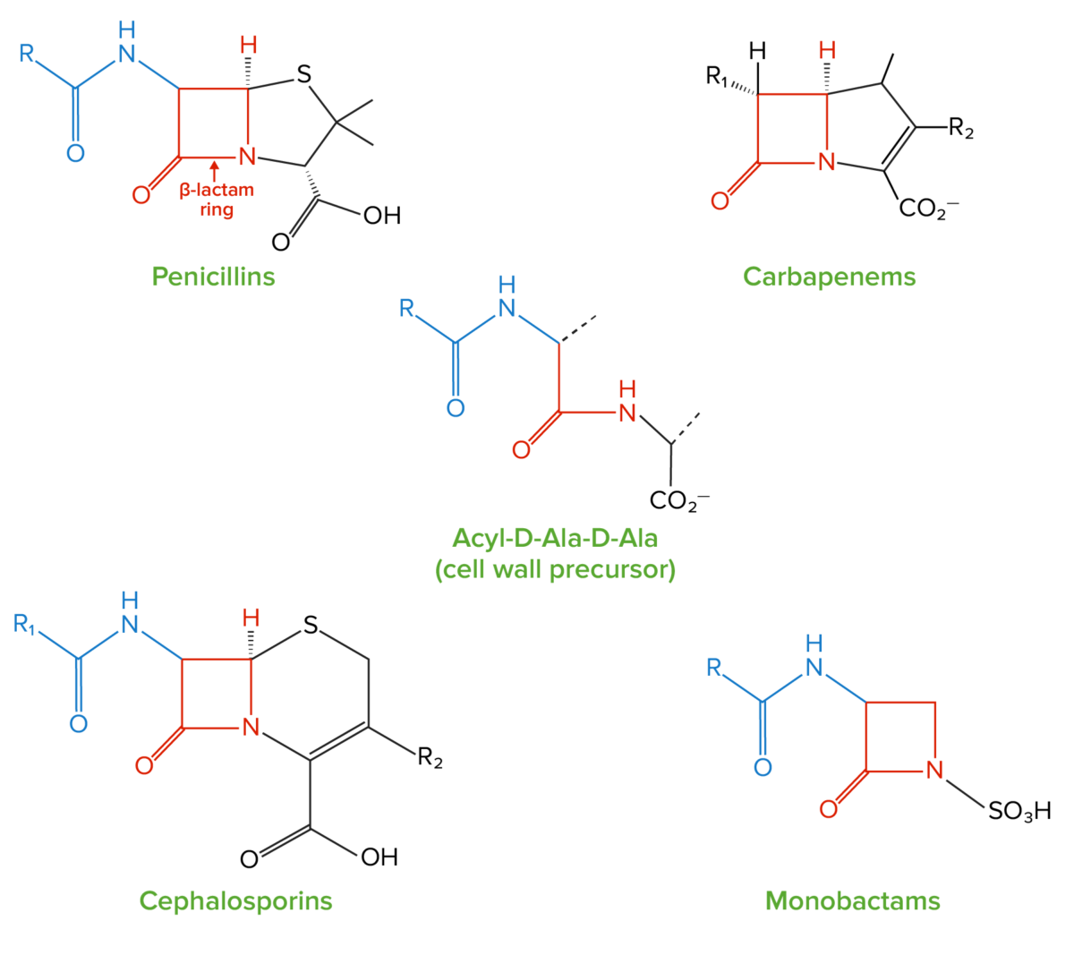
  • A beta-lactam Beta-Lactam Penicillins ring: a 4-membered ring containing 2 carbons (α and β carbons), a nitrogen Nitrogen An element with the atomic symbol n, atomic number 7, and atomic weight [14. 00643; 14. 00728]. Nitrogen exists as a diatomic gas and makes up about 78% of the earth’s atmosphere by volume. It is a constituent of proteins and nucleic acids and found in all living cells. Urea Cycle, and a carbonyl group (a carbon double-bonded to oxygen)
    • The group in the compound responsible for antibacterial Antibacterial Penicillins activity
    • Can be hydrolyzed (i.e., broken down) by beta-lactamases, which are produced by certain resistant bacteria Bacteria Bacteria are prokaryotic single-celled microorganisms that are metabolically active and divide by binary fission. Some of these organisms play a significant role in the pathogenesis of diseases. Bacteriology
    • If the beta-lactam Beta-Lactam Penicillins ring is broken, the drug loses its antibacterial Antibacterial Penicillins properties.
    • All beta-lactams contain a beta-lactam Beta-Lactam Penicillins ring.
  • Side chain (R group): 
    • Bound to the α-carbon in the beta-lactam Beta-Lactam Penicillins ring
    • Differentiates cephalosporins from each other
    • Responsible for their unique pharmacokinetics Pharmacokinetics Pharmacokinetics is the science that analyzes how the human body interacts with a drug. Pharmacokinetics examines how the drug is absorbed, distributed, metabolized, and excreted by the body. Pharmacokinetics and Pharmacodynamics and spectra of activity
    • Certain structures can sterically inhibit the hydrolysis Hydrolysis The process of cleaving a chemical compound by the addition of a molecule of water. Proteins and Peptides of the beta-lactam Beta-Lactam Penicillins ring by beta-lactamases. 
  • A 6-membered ring containing both sulfur and nitrogen Nitrogen An element with the atomic symbol n, atomic number 7, and atomic weight [14. 00643; 14. 00728]. Nitrogen exists as a diatomic gas and makes up about 78% of the earth’s atmosphere by volume. It is a constituent of proteins and nucleic acids and found in all living cells. Urea Cycle with a 2nd R group
Structure of beta-lactams

Structure of beta-lactams:
All beta-lactam antibiotics contain the same core 4-membered “beta-lactam” ring (highlighted in red). This ring is responsible for the antibacterial properties of the drug because it is the region that binds to and inhibits the penicillin-binding proteins (PBPs). The PBPs catalyze the formation of the cell wall by generating crosslinks between the peptide chains in the peptidoglycan molecules; PBPs form these crosslinks between acyl-D-Ala-D-Ala peptides, which have a structure similar to the beta-lactam ring.

Image by Lecturio. License: CC BY-NC-SA 4.0

Mechanisms of Action and Resistance

All beta-lactams, including cephalosporins, exert their effects by inhibiting bacterial cell wall Cell wall The outermost layer of a cell in most plants; bacteria; fungi; and algae. The cell wall is usually a rigid structure that lies external to the cell membrane, and provides a protective barrier against physical or chemical agents. Cell Types: Eukaryotic versus Prokaryotic synthesis Synthesis Polymerase Chain Reaction (PCR).
Background: understanding cell walls

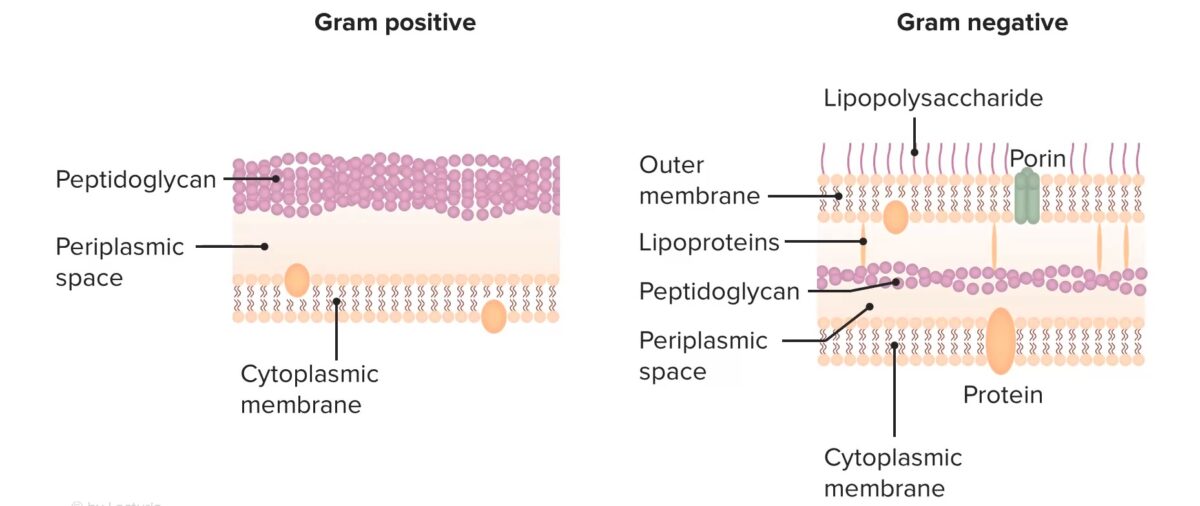
  • Bacterial cell walls contain peptidoglycan Peptidoglycan Penicillins chains (large, thick layers in gram-positive Gram-Positive Penicillins organisms, and relatively smaller/thinner layers in gram-negative organisms).
  • Peptidoglycan Peptidoglycan Penicillins chains are composed of:
    • A sugar backbone with 2 alternating sugars: 
    • Short, peptide side chains branching off the NAM sugars
  • The short peptides form crosslinked bridges between the adjacent peptidoglycan Peptidoglycan Penicillins chains and create a fishnet structure.
    • Crosslinked bridges are necessary for the peptidoglycan Peptidoglycan Penicillins (and thus cell wall Cell wall The outermost layer of a cell in most plants; bacteria; fungi; and algae. The cell wall is usually a rigid structure that lies external to the cell membrane, and provides a protective barrier against physical or chemical agents. Cell Types: Eukaryotic versus Prokaryotic) structure.
    • Penicillin-binding proteins Penicillin-Binding Proteins Bacterial proteins that share the property of binding irreversibly to penicillins and other antibacterial agents derived from lactams. The penicillin-binding proteins are primarily enzymes involved in cell wall biosynthesis including muramoylpentapeptide carboxypeptidase; peptide synthases; transpeptidases; and hexosyltransferases. Penicillins ( PBPs PBPs Bacterial proteins that share the property of binding irreversibly to penicillins and other antibacterial agents derived from lactams. The penicillin-binding proteins are primarily enzymes involved in cell wall biosynthesis including muramoylpentapeptide carboxypeptidase; peptide synthases; transpeptidases; and hexosyltransferases. Penicillins) are enzymes Enzymes Enzymes are complex protein biocatalysts that accelerate chemical reactions without being consumed by them. Due to the body’s constant metabolic needs, the absence of enzymes would make life unsustainable, as reactions would occur too slowly without these molecules. Basics of Enzymes that create these crosslinked bridges.
Structure of bacterial cell walls cephalosporins

Structure of bacterial cell walls

Image by Lecturio. License: CC BY-NC-SA 4.0

Mechanism of action

All beta-lactams work by irreversibly binding to and inhibiting the PBPs PBPs Bacterial proteins that share the property of binding irreversibly to penicillins and other antibacterial agents derived from lactams. The penicillin-binding proteins are primarily enzymes involved in cell wall biosynthesis including muramoylpentapeptide carboxypeptidase; peptide synthases; transpeptidases; and hexosyltransferases. Penicillins beta-lactam Beta-Lactam Penicillins antibiotics inhibit cell wall Cell wall The outermost layer of a cell in most plants; bacteria; fungi; and algae. The cell wall is usually a rigid structure that lies external to the cell membrane, and provides a protective barrier against physical or chemical agents. Cell Types: Eukaryotic versus Prokaryotic synthesis Synthesis Polymerase Chain Reaction (PCR).
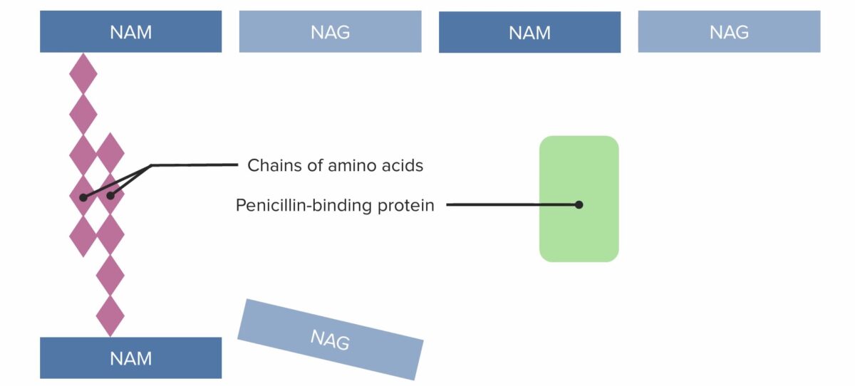
Bactericidal Bactericidal Penicillins activity

Beta-lactams, including cephalosporins, have a bactericidal Bactericidal Penicillins (rather than bacteriostatic Bacteriostatic Sulfonamides and Trimethoprim) effect. 

  • Bacterial cell wall Cell wall The outermost layer of a cell in most plants; bacteria; fungi; and algae. The cell wall is usually a rigid structure that lies external to the cell membrane, and provides a protective barrier against physical or chemical agents. Cell Types: Eukaryotic versus Prokaryotic is necessary for survival → if lacking, cell death Cell death Injurious stimuli trigger the process of cellular adaptation, whereby cells respond to withstand the harmful changes in their environment. Overwhelmed adaptive mechanisms lead to cell injury. Mild stimuli produce reversible injury. If the stimulus is severe or persistent, injury becomes irreversible. Apoptosis is programmed cell death, a mechanism with both physiologic and pathologic effects. Cell Injury and Death is initiated
  • When bacteria Bacteria Bacteria are prokaryotic single-celled microorganisms that are metabolically active and divide by binary fission. Some of these organisms play a significant role in the pathogenesis of diseases. Bacteriology attempt to replicate, they shed their cell walls.
  • In the presence of beta-lactams, however, bacteria Bacteria Bacteria are prokaryotic single-celled microorganisms that are metabolically active and divide by binary fission. Some of these organisms play a significant role in the pathogenesis of diseases. Bacteriology are unable to form a new cell wall Cell wall The outermost layer of a cell in most plants; bacteria; fungi; and algae. The cell wall is usually a rigid structure that lies external to the cell membrane, and provides a protective barrier against physical or chemical agents. Cell Types: Eukaryotic versus Prokaryotic.
  • Bacteria Bacteria Bacteria are prokaryotic single-celled microorganisms that are metabolically active and divide by binary fission. Some of these organisms play a significant role in the pathogenesis of diseases. Bacteriology are unable to effectively divide, and the remaining cell autocatalyzes and dies.
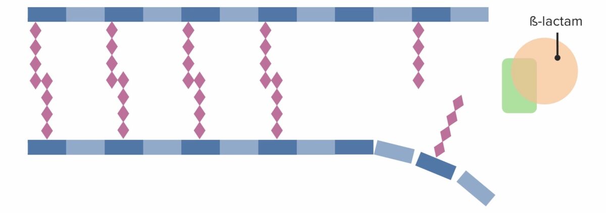
Bacteria attempting to divide in the presence of penicillin

Bacterium attempting to divide in the presence of penicillin:
The bacterium sheds its wall and becomes a spheroplast. The spheroplast is unable to survive and autocatalyzes (dies).

Image by Lecturio. License: CC BY-NC-SA 4.0
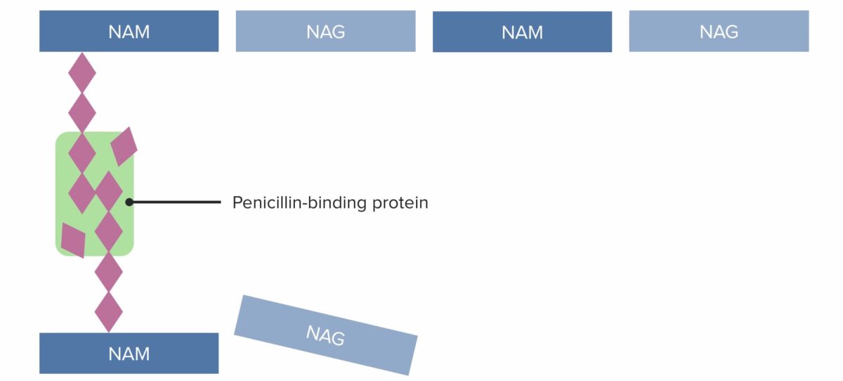
Mechanisms of resistance Resistance Physiologically, the opposition to flow of air caused by the forces of friction. As a part of pulmonary function testing, it is the ratio of driving pressure to the rate of air flow. Ventilation: Mechanics of Breathing

Bacteria Bacteria Bacteria are prokaryotic single-celled microorganisms that are metabolically active and divide by binary fission. Some of these organisms play a significant role in the pathogenesis of diseases. Bacteriology use 3 primary mechanisms to resist cephalosporins:

  • Beta-lactamase resistance Beta-Lactamase Resistance Penicillins (cephalosporins are ineffective): 
  • PBP-mediated resistance Pbp-Mediated Resistance Penicillins (↓ cephalosporin Cephalosporin Multidrug-resistant Organisms and Nosocomial Infections binding to PBPs PBPs Bacterial proteins that share the property of binding irreversibly to penicillins and other antibacterial agents derived from lactams. The penicillin-binding proteins are primarily enzymes involved in cell wall biosynthesis including muramoylpentapeptide carboxypeptidase; peptide synthases; transpeptidases; and hexosyltransferases. Penicillins)
    • Mutations in PBPs PBPs Bacterial proteins that share the property of binding irreversibly to penicillins and other antibacterial agents derived from lactams. The penicillin-binding proteins are primarily enzymes involved in cell wall biosynthesis including muramoylpentapeptide carboxypeptidase; peptide synthases; transpeptidases; and hexosyltransferases. Penicillins → ↓ affinity of cephalosporins to PBP 
    • Despite the mutations, these PBPs PBPs Bacterial proteins that share the property of binding irreversibly to penicillins and other antibacterial agents derived from lactams. The penicillin-binding proteins are primarily enzymes involved in cell wall biosynthesis including muramoylpentapeptide carboxypeptidase; peptide synthases; transpeptidases; and hexosyltransferases. Penicillins can produce a cell wall Cell wall The outermost layer of a cell in most plants; bacteria; fungi; and algae. The cell wall is usually a rigid structure that lies external to the cell membrane, and provides a protective barrier against physical or chemical agents. Cell Types: Eukaryotic versus Prokaryotic.
  • Porin-mediated resistance Porin-Mediated Resistance Penicillins (↓ cephalosporin Cephalosporin Multidrug-resistant Organisms and Nosocomial Infections uptake): 
    • Cephalosporins enter bacteria Bacteria Bacteria are prokaryotic single-celled microorganisms that are metabolically active and divide by binary fission. Some of these organisms play a significant role in the pathogenesis of diseases. Bacteriology through channels Channels The Cell: Cell Membrane called porins in the cell walls.
    • Bacteria Bacteria Bacteria are prokaryotic single-celled microorganisms that are metabolically active and divide by binary fission. Some of these organisms play a significant role in the pathogenesis of diseases. Bacteriology can ↓ production of porins → ↓ antibiotic within the cell → antibiotic resistance Resistance Physiologically, the opposition to flow of air caused by the forces of friction. As a part of pulmonary function testing, it is the ratio of driving pressure to the rate of air flow. Ventilation: Mechanics of Breathing
    • Common mechanism of resistance Resistance Physiologically, the opposition to flow of air caused by the forces of friction. As a part of pulmonary function testing, it is the ratio of driving pressure to the rate of air flow. Ventilation: Mechanics of Breathing in Pseudomonas aeruginosa Pseudomonas aeruginosa A species of gram-negative, aerobic, rod-shaped bacteria commonly isolated from clinical specimens (wound, burn, and urinary tract infections). It is also found widely distributed in soil and water. P. Aeruginosa is a major agent of nosocomial infection. Pseudomonas

Beta-lactamase-resistant medications

  • Some medications can overcome beta-lactamases by acting as beta-lactamase Beta-Lactamase Penicillins inhibitors.
  • Beta-lactamase Beta-Lactamase Penicillins inhibitors can be combined with beta-lactamase-sensitive cephalosporins to enhance their activity.
  • Beta-lactamase Beta-Lactamase Penicillins inhibitors include:
    • Tazobactam
    • Avibactam
    • Clavulanic acid (not available in combination with cephalosporins)
    • Sulbactam (not available in combination with cephalosporins)

Pharmacokinetics

Absorption Absorption Absorption involves the uptake of nutrient molecules and their transfer from the lumen of the GI tract across the enterocytes and into the interstitial space, where they can be taken up in the venous or lymphatic circulation. Digestion and Absorption and half-life Half-Life The time it takes for a substance (drug, radioactive nuclide, or other) to lose half of its pharmacologic, physiologic, or radiologic activity. Pharmacokinetics and Pharmacodynamics

  • Oral cephalosporins are rapidly absorbed.
  • Half-lives tend to be short.
    • Most cephalosporins have a half-life Half-Life The time it takes for a substance (drug, radioactive nuclide, or other) to lose half of its pharmacologic, physiologic, or radiologic activity. Pharmacokinetics and Pharmacodynamics < 1 hour and should be dosed every 4 hours in patients Patients Individuals participating in the health care system for the purpose of receiving therapeutic, diagnostic, or preventive procedures. Clinician–Patient Relationship with normal renal function.
    • Cephalosporins with longer half-lives:
      • Ceftriaxone (dosed q24 hours)
      • Cefazolin (dosed q8 hours)

Distribution

  • All cephalosporins achieve therapeutic levels in:
    • Pleural fluid
    • Pericardial fluid Pericardial fluid Watery fluid produced in the serous and visceral pericardium surrounding the surface of the heart. Heart: Anatomy
    • Peritoneal fluid
    • Synovial fluid
    • Urine
  • Penetration Penetration X-rays across the blood-brain barrier Blood-brain barrier Specialized non-fenestrated tightly-joined endothelial cells with tight junctions that form a transport barrier for certain substances between the cerebral capillaries and the brain tissue. Systemic and Special Circulations into the CSF:
  • Protein binding varies significantly between drugs:
    • < 20%: cefalexin, ceftazidime, cefepime, ceftolozane-tazobactam, ceftaroline
    • 20%‒80%: cefuroxime, cefoxitin
    • > 80%: cefazolin, cefotetan, ceftriaxone

Excretion

  • Primarily renal excretion
  • All cephalosporins except ceftriaxone require dose modification in renal failure Renal failure Conditions in which the kidneys perform below the normal level in the ability to remove wastes, concentrate urine, and maintain electrolyte balance; blood pressure; and calcium metabolism. Renal insufficiency can be classified by the degree of kidney damage (as measured by the level of proteinuria) and reduction in glomerular filtration rate. Crush Syndrome.

Ceftriaxone precipitation

  • Ceftriaxone can precipitate in the presence of calcium Calcium A basic element found in nearly all tissues. It is a member of the alkaline earth family of metals with the atomic symbol ca, atomic number 20, and atomic weight 40. Calcium is the most abundant mineral in the body and combines with phosphorus to form calcium phosphate in the bones and teeth. It is essential for the normal functioning of nerves and muscles and plays a role in blood coagulation (as factor IV) and in many enzymatic processes. Electrolytes, especially in the lungs Lungs Lungs are the main organs of the respiratory system. Lungs are paired viscera located in the thoracic cavity and are composed of spongy tissue. The primary function of the lungs is to oxygenate blood and eliminate CO2. Lungs: Anatomy and kidneys Kidneys The kidneys are a pair of bean-shaped organs located retroperitoneally against the posterior wall of the abdomen on either side of the spine. As part of the urinary tract, the kidneys are responsible for blood filtration and excretion of water-soluble waste in the urine. Kidneys: Anatomy.

Thus, ceftriaxone should not be reconstituted/directly mixed with calcium-containing products such as lactated Ringers or total parenteral nutrition Parenteral nutrition The administering of nutrients for assimilation and utilization by a patient who cannot maintain adequate nutrition by enteral feeding alone. Nutrients are administered by a route other than the alimentary canal (e.g., intravenously, subcutaneously). Central Venous Catheter (TPN).

Classification

Cephalosporins are generally classified by generation, with the 1st generation drugs being the oldest in this class. Some of the most commonly used drugs are shown in the table below.

Table: Classification of cephalosporins
Generation Parental agents Oral agents
1st generation Cefazolin (Ancef) Cephalexin (Keflex)
2nd generation
  • Cefuroxime
  • Cephamycins (subgroup):
    • Cefotetan
    • Cefoxitin
Cefaclor (Ceclor)
3rd generation
  • Ceftriaxone (Rocephin)
  • Cefotaxime
  • Ceftazidime
  • Cefdinir (Omnicef)
  • Cefixime (Suprax)
  • Cefpodoxime
4th generation Cefepime None
5th generation and advanced combinations
  • Ceftaroline
  • Ceftolozane
  • Ceftolozane-tazobactam
  • Ceftazidime-avibactam
None

Indications

Activity spectrum

Table: Spectrum of activity of cephalosporins
Drugs Gram-positive Gram-Positive Penicillins cocci Cocci Bacteriology Gram-negative bacilli Bacilli Shigella Gram-negative cocci Cocci Bacteriology Anaerobes Anaerobes Lincosamides
Streptococcus Streptococcus Streptococcus is one of the two medically important genera of gram-positive cocci, the other being Staphylococcus. Streptococci are identified as different species on blood agar on the basis of their hemolytic pattern and sensitivity to optochin and bacitracin. There are many pathogenic species of streptococci, including S. pyogenes, S. agalactiae, S. pneumoniae, and the viridans streptococci. Streptococcus, MSSA
  • Escherichia coli Escherichia coli The gram-negative bacterium Escherichia coli is a key component of the human gut microbiota. Most strains of E. coli are avirulent, but occasionally they escape the GI tract, infecting the urinary tract and other sites. Less common strains of E. coli are able to cause disease within the GI tract, most commonly presenting as abdominal pain and diarrhea. Escherichia coli
  • Klebsiella pneumoniae Klebsiella Pneumoniae Gram-negative, non-motile, capsulated, gas-producing rods found widely in nature and associated with urinary and respiratory infections in humans. Aminoglycosides
  • Haemophilus influenzae Haemophilus Influenzae A species of Haemophilus found on the mucous membranes of humans and a variety of animals. The species is further divided into biotypes I through viii. Haemophilus
SPACE organisms Pseudomonas Pseudomonas Pseudomonas is a non-lactose-fermenting, gram-negative bacillus that produces pyocyanin, which gives it a characteristic blue-green color. Pseudomonas is found ubiquitously in the environment, as well as in moist reservoirs, such as hospital sinks and respiratory equipment. Pseudomonas Neisseria Neisseria Neisseria is a genus of bacteria commonly present on mucosal surfaces. Several species exist, but only 2 are pathogenic to humans: N. gonorrhoeae and N. meningitidis. Neisseria species are non-motile, gram-negative diplococci most commonly isolated on modified Thayer-Martin (MTM) agar. Neisseriaspp Peptostreptococcus Peptostreptococcus A genus of gram-positive, anaerobic, coccoid bacteria that is part of the normal flora of humans. Its organisms are opportunistic pathogens causing bacteremias and soft tissue infections. Perianal and Perirectal Abscess
1st generation
  • Cephalexin
  • Cefazolin
X X* X
2nd generation
  • Cefuroxime
  • Cefaclor
  • Cefoxitin
  • Cefotetan
X X X** X+
3rd generation
  • Ceftriaxone
  • Cefotaxime
  • Ceftazidime
  • Cefdinir
  • Cefixime
X X X++ X X
4th generation
  • Cefepime
X X X X X+
5th generation and combinations:
  • Ceftaroline+++
  • Ceftolozane-tazobactam
  • Ceftazidime-avibactam
X X X X X+

SPACE organisms: Serratia marcescens, Proteus mirabilis, Acinetobacter spp., Citrobacter spp., and Enterobacter spp.
*Only non-beta-lactamase-producing species
**Only cephamycins: cefoxitin and cefotetan
+Cefoxitin and ceftolozane-tazobactam are also active against “below the diaphragm anaerobes” Bacteroides
++Only ceftazidime
+++Ceftaroline is active against methicillin-resistant S. aureus (MRSA)

Clinical uses

Cephalosporins are used to treat infections Infections Invasion of the host organism by microorganisms or their toxins or by parasites that can cause pathological conditions or diseases. Chronic Granulomatous Disease caused by susceptible organisms, including:

  • Preoperative prophylaxis against surgical site infections Infections Invasion of the host organism by microorganisms or their toxins or by parasites that can cause pathological conditions or diseases. Chronic Granulomatous Disease (cefazolin)
  • Skin Skin The skin, also referred to as the integumentary system, is the largest organ of the body. The skin is primarily composed of the epidermis (outer layer) and dermis (deep layer). The epidermis is primarily composed of keratinocytes that undergo rapid turnover, while the dermis contains dense layers of connective tissue. Skin: Structure and Functions and soft tissue Soft Tissue Soft Tissue Abscess infections Infections Invasion of the host organism by microorganisms or their toxins or by parasites that can cause pathological conditions or diseases. Chronic Granulomatous Disease
  • Bone Bone Bone is a compact type of hardened connective tissue composed of bone cells, membranes, an extracellular mineralized matrix, and central bone marrow. The 2 primary types of bone are compact and spongy. Bones: Structure and Types and joint infections Infections Invasion of the host organism by microorganisms or their toxins or by parasites that can cause pathological conditions or diseases. Chronic Granulomatous Disease
  • Respiratory infections Infections Invasion of the host organism by microorganisms or their toxins or by parasites that can cause pathological conditions or diseases. Chronic Granulomatous Disease caused by S. pneumoniae and S. pyogenes
    • Upper respiratory infections Infections Invasion of the host organism by microorganisms or their toxins or by parasites that can cause pathological conditions or diseases. Chronic Granulomatous Disease, including pharyngitis Pharyngitis Pharyngitis is an inflammation of the back of the throat (pharynx). Pharyngitis is usually caused by an upper respiratory tract infection, which is viral in most cases. It typically results in a sore throat and fever. Other symptoms may include a runny nose, cough, headache, and hoarseness. Pharyngitis
    • Pneumonia Pneumonia Pneumonia or pulmonary inflammation is an acute or chronic inflammation of lung tissue. Causes include infection with bacteria, viruses, or fungi. In more rare cases, pneumonia can also be caused through toxic triggers through inhalation of toxic substances, immunological processes, or in the course of radiotherapy. Pneumonia
    • Cystic Cystic Fibrocystic Change fibrosis Fibrosis Any pathological condition where fibrous connective tissue invades any organ, usually as a consequence of inflammation or other injury. Bronchiolitis Obliterans exacerbations
  • Otitis media due to susceptible organisms
  • UTIs due to susceptible gram-negative organisms, especially Escherichia coli Escherichia coli The gram-negative bacterium Escherichia coli is a key component of the human gut microbiota. Most strains of E. coli are avirulent, but occasionally they escape the GI tract, infecting the urinary tract and other sites. Less common strains of E. coli are able to cause disease within the GI tract, most commonly presenting as abdominal pain and diarrhea. Escherichia coli, Klebsiella Klebsiella Klebsiella are encapsulated gram-negative, lactose-fermenting bacilli. They form pink colonies on MacConkey agar due to lactose fermentation. The main virulence factor is a polysaccharide capsule. Klebsiella pneumoniae is the most important pathogenic species. Klebsiella, or Proteus Proteus Proteus spp. are gram-negative, facultatively anaerobic bacilli. Different types of infection result from Proteus, but the urinary tract is the most common site. The majority of cases are caused by Proteus mirabilis (P. mirabilis). The bacteria are part of the normal intestinal flora and are also found in the environment. Proteus spp.
  • Sepsis Sepsis Systemic inflammatory response syndrome with a proven or suspected infectious etiology. When sepsis is associated with organ dysfunction distant from the site of infection, it is called severe sepsis. When sepsis is accompanied by hypotension despite adequate fluid infusion, it is called septic shock. Sepsis and Septic Shock due to susceptible organisms
  • Bacterial meningitis Bacterial meningitis Bacterial infections of the leptomeninges and subarachnoid space, frequently involving the cerebral cortex, cranial nerves, cerebral blood vessels, spinal cord, and nerve roots. Meningitis due to Enterobacteriaceae: 3rd- and 4th-generation cephalosporins (therapy of choice)
  • “Below the diaphragm Diaphragm The diaphragm is a large, dome-shaped muscle that separates the thoracic cavity from the abdominal cavity. The diaphragm consists of muscle fibers and a large central tendon, which is divided into right and left parts. As the primary muscle of inspiration, the diaphragm contributes 75% of the total inspiratory muscle force. Diaphragm: Anatomy infections Infections Invasion of the host organism by microorganisms or their toxins or by parasites that can cause pathological conditions or diseases. Chronic Granulomatous Disease: cephamycins and 3rd-generation cephalosporins
    • Pelvic inflammatory disease Pelvic inflammatory disease Pelvic inflammatory disease (PID) is defined as a polymicrobial infection of the upper female reproductive system. The disease can affect the uterus, fallopian tubes, ovaries, and adjacent structures. Pelvic inflammatory disease is closely linked with sexually transmitted diseases, most commonly caused by Chlamydia trachomatis, Neisseria gonorrhoeae, and Gardnerella vaginalis. Pelvic Inflammatory Disease
    • Endometritis Endometritis Endometritis is an inflammation of the endometrium, the inner layer of the uterus. The most common subtype is postpartum endometritis, resulting from the ascension of normal vaginal flora to the previously aseptic uterus. Postpartum Endometritis
    • Neisseria gonorrhoeae Neisseria gonorrhoeae A species of gram-negative, aerobic bacteria primarily found in purulent venereal discharges. It is the causative agent of gonorrhea. Neisseria (note: cefoxitin is ineffective against Chlamydia trachomatis Chlamydia trachomatis Type species of Chlamydia causing a variety of ocular and urogenital diseases. Chlamydia)

Adverse Effects and Contraindications

Adverse effects

Allergic reactions Allergic Reactions Type I hypersensitivity reaction against plasma proteins in donor blood Transfusion Reactions are the most common adverse effects.

  • IgE-mediated allergic reactions IgE-mediated allergic reactions Penicillins:
    • Present with pruritis, urticaria Urticaria Urticaria is raised, well-circumscribed areas (wheals) of edema (swelling) and erythema (redness) involving the dermis and epidermis with associated pruritus (itch). Urticaria is not a single disease but rather is a reaction pattern representing cutaneous mast cell degranulation. Urticaria (Hives), angioedema Angioedema Angioedema is a localized, self-limited (but potentially life-threatening), nonpitting, asymmetrical edema occurring in the deep layers of the skin and mucosal tissue. The common underlying pathophysiology involves inflammatory mediators triggering significant vasodilation and increased capillary permeability. Angioedema, hypotension Hypotension Hypotension is defined as low blood pressure, specifically < 90/60 mm Hg, and is most commonly a physiologic response. Hypotension may be mild, serious, or life threatening, depending on the cause. Hypotension, and/or anaphylaxis Anaphylaxis An acute hypersensitivity reaction due to exposure to a previously encountered antigen. The reaction may include rapidly progressing urticaria, respiratory distress, vascular collapse, systemic shock, and death. Type I Hypersensitivity Reaction
    • Patients Patients Individuals participating in the health care system for the purpose of receiving therapeutic, diagnostic, or preventive procedures. Clinician–Patient Relationship with penicillin Penicillin Rheumatic Fever allergies Allergies A medical specialty concerned with the hypersensitivity of the individual to foreign substances and protection from the resultant infection or disorder. Selective IgA Deficiency are more likely to have cephalosporin Cephalosporin Multidrug-resistant Organisms and Nosocomial Infections allergies Allergies A medical specialty concerned with the hypersensitivity of the individual to foreign substances and protection from the resultant infection or disorder. Selective IgA Deficiency due to cross-reactivity
      • Most common with 1st- and 2nd-generation cephalosporins due to similarities in their R groups
      • 3rd- to 5th-generation cephalosporins show minimal cross-reactivity.
  • Dermatologic reactions:
    • Morbilliform Morbilliform Erythema Infectiosum rash Rash Rocky Mountain Spotted Fever: a maculopapular Maculopapular Dermatologic Examination eruption due to a hypersensitivity reaction
    • Erythema Erythema Redness of the skin produced by congestion of the capillaries. This condition may result from a variety of disease processes. Chalazion multiforme: target lesions developing with acute onset
    • Stevens-Johnson syndrome Stevens-Johnson syndrome Stevens-Johnson syndrome (SJS) is a cutaneous, immune-mediated hypersensitivity reaction that is commonly triggered by medications, including antiepileptics and antibiotics. The condition runs on a spectrum with toxic epidermal necrolysis (TEN) based on the amount of body surface area (BSA) involved. Stevens-Johnson Syndrome: a desquamating skin Skin The skin, also referred to as the integumentary system, is the largest organ of the body. The skin is primarily composed of the epidermis (outer layer) and dermis (deep layer). The epidermis is primarily composed of keratinocytes that undergo rapid turnover, while the dermis contains dense layers of connective tissue. Skin: Structure and Functions condition involving mucosal surfaces
  • Neurologic reactions:
    • Seizures Seizures A seizure is abnormal electrical activity of the neurons in the cerebral cortex that can manifest in numerous ways depending on the region of the brain affected. Seizures consist of a sudden imbalance that occurs between the excitatory and inhibitory signals in cortical neurons, creating a net excitation. The 2 major classes of seizures are focal and generalized. Seizures (cefepime, especially in the setting of renal impairment)
  • GI and hepatic reactions:
    • Clostridioides difficile colitis Colitis Inflammation of the colon section of the large intestine, usually with symptoms such as diarrhea (often with blood and mucus), abdominal pain, and fever. Pseudomembranous Colitis 
    • Suppression Suppression Defense Mechanisms of gut flora leading to vitamin K deficiency Vitamin K Deficiency A nutritional condition produced by a deficiency of vitamin K in the diet, characterized by an increased tendency to hemorrhage (hemorrhagic disorders). Such bleeding episodes may be particularly severe in newborn infants. Fat-soluble Vitamins and their Deficiencies
    • Formation of biliary sludge and pseudocholelithiasis (ceftriaxone, especially in children)
  • Renal reactions:
    • Cephalosporins can potentiate the renal toxicity Toxicity Dosage Calculation of aminoglycosides Aminoglycosides Aminoglycosides are a class of antibiotics including gentamicin, tobramycin, amikacin, neomycin, plazomicin, and streptomycin. The class binds the 30S ribosomal subunit to inhibit bacterial protein synthesis. Unlike other medications with a similar mechanism of action, aminoglycosides are bactericidal. Aminoglycosides.
    • Glomerulonephritis is seen in association with allergic reactions Allergic Reactions Type I hypersensitivity reaction against plasma proteins in donor blood Transfusion Reactions.
  • Hematologic reactions:
    • Hemolytic anemia Hemolytic Anemia Hemolytic anemia (HA) is the term given to a large group of anemias that are caused by the premature destruction/hemolysis of circulating red blood cells (RBCs). Hemolysis can occur within (intravascular hemolysis) or outside the blood vessels (extravascular hemolysis). Hemolytic Anemia
    • Neutropenia Neutropenia Neutrophils are an important component of the immune system and play a significant role in the eradication of infections. Low numbers of circulating neutrophils, referred to as neutropenia, predispose the body to recurrent infections or sepsis, though patients can also be asymptomatic. Neutropenia
    • Immune thrombocytopenia Thrombocytopenia Thrombocytopenia occurs when the platelet count is < 150,000 per microliter. The normal range for platelets is usually 150,000-450,000/µL of whole blood. Thrombocytopenia can be a result of decreased production, increased destruction, or splenic sequestration of platelets. Patients are often asymptomatic until platelet counts are < 50,000/µL. Thrombocytopenia

Contraindications Contraindications A condition or factor associated with a recipient that makes the use of a drug, procedure, or physical agent improper or inadvisable. Contraindications may be absolute (life threatening) or relative (higher risk of complications in which benefits may outweigh risks). Noninvasive Ventilation

  • All cephalosporins:
    • Hypersensitivity reactions
    • Anaphylactic reactions Anaphylactic reactions An acute hypersensitivity reaction due to exposure to a previously encountered antigen. The reaction may include rapidly progressing urticaria, respiratory distress, vascular collapse, systemic shock, and death. Vaccination to penicillins Penicillins Beta-lactam antibiotics contain a beta-lactam ring as a part of their chemical structure. Drugs in this class include penicillin G and V, penicillinase-sensitive and penicillinase-resistant penicillins, cephalosporins, carbapenems, and aztreonam. Penicillins
  • Ceftriaxone-specific contraindications Contraindications A condition or factor associated with a recipient that makes the use of a drug, procedure, or physical agent improper or inadvisable. Contraindications may be absolute (life threatening) or relative (higher risk of complications in which benefits may outweigh risks). Noninvasive Ventilation:
    • Hyperbilirubinemia Hyperbilirubinemia A condition characterized by an abnormal increase of bilirubin in the blood, which may result in jaundice. Bilirubin, a breakdown product of heme, is normally excreted in the bile or further catabolized before excretion in the urine. Jaundice in neonates (especially if premature Premature Childbirth before 37 weeks of pregnancy (259 days from the first day of the mother’s last menstrual period, or 245 days after fertilization). Necrotizing Enterocolitis)
    • Infants < 28 days of age receiving any IV calcium-containing products
  • Use with caution:

Comparison of Antibiotic Coverage

Comparison based on mechanisms of action

Antibiotics can be classified in several ways. One way is to classify them based on their mechanism of action:

Table: Antibiotics classified by primary mechanism of action
Mechanism Classes of antibiotics
Bacterial cell wall Cell wall The outermost layer of a cell in most plants; bacteria; fungi; and algae. The cell wall is usually a rigid structure that lies external to the cell membrane, and provides a protective barrier against physical or chemical agents. Cell Types: Eukaryotic versus Prokaryotic synthesis Synthesis Polymerase Chain Reaction (PCR) inhibitors
  • Penicillins Penicillins Beta-lactam antibiotics contain a beta-lactam ring as a part of their chemical structure. Drugs in this class include penicillin G and V, penicillinase-sensitive and penicillinase-resistant penicillins, cephalosporins, carbapenems, and aztreonam. Penicillins
  • Cephalosporins
  • Penems
  • Miscellaneous
Bacterial protein synthesis Synthesis Polymerase Chain Reaction (PCR) inhibitors
  • Tetracyclines Tetracyclines Tetracyclines are a class of broad-spectrum antibiotics indicated for a wide variety of bacterial infections. These medications bind the 30S ribosomal subunit to inhibit protein synthesis of bacteria. Tetracyclines cover gram-positive and gram-negative organisms, as well as atypical bacteria such as chlamydia, mycoplasma, spirochetes, and even protozoa. Tetracyclines
  • Macrolides Macrolides Macrolides and ketolides are antibiotics that inhibit bacterial protein synthesis by binding to the 50S ribosomal subunit and blocking transpeptidation. These antibiotics have a broad spectrum of antimicrobial activity but are best known for their coverage of atypical microorganisms. Macrolides and Ketolides
  • Ketolide
  • Lincosamides Lincosamides The lincosamides, lincomycin and clindamycin, are inhibitors of bacterial protein synthesis. Drugs in this class share the same binding site as that of macrolides and amphenicols; however, they differ in chemical structure. Lincosamides target the 50S ribosomal subunit and interfere with transpeptidation. Lincosamides
  • Streptogramins
  • Linezolid Linezolid An oxazolidinone and acetamide derived anti-bacterial agent and protein synthesis inhibitor that is used in the treatment of gram-positive bacterial infections of the skin and respiratory tract. Oxazolidinones
Agents acting against DNA DNA A deoxyribonucleotide polymer that is the primary genetic material of all cells. Eukaryotic and prokaryotic organisms normally contain DNA in a double-stranded state, yet several important biological processes transiently involve single-stranded regions. DNA, which consists of a polysugar-phosphate backbone possessing projections of purines (adenine and guanine) and pyrimidines (thymine and cytosine), forms a double helix that is held together by hydrogen bonds between these purines and pyrimidines (adenine to thymine and guanine to cytosine). DNA Types and Structure and/or folate Folate Folate and vitamin B12 are 2 of the most clinically important water-soluble vitamins. Deficiencies can present with megaloblastic anemia, GI symptoms, neuropsychiatric symptoms, and adverse pregnancy complications, including neural tube defects. Folate and Vitamin B12
  • Sulfonamides Sulfonamides A group of compounds that contain the structure so2nh2. Sulfonamides and Trimethoprim
  • Trimethoprim Trimethoprim The sulfonamides are a class of antimicrobial drugs inhibiting folic acid synthesize in pathogens. The prototypical drug in the class is sulfamethoxazole. Although not technically sulfonamides, trimethoprim, dapsone, and pyrimethamine are also important antimicrobial agents inhibiting folic acid synthesis. The agents are often combined with sulfonamides, resulting in a synergistic effect. Sulfonamides and Trimethoprim
  • Fluoroquinolones Fluoroquinolones Fluoroquinolones are a group of broad-spectrum, bactericidal antibiotics inhibiting bacterial DNA replication. Fluoroquinolones cover gram-negative, anaerobic, and atypical organisms, as well as some gram-positive and multidrug-resistant (MDR) organisms. Fluoroquinolones
Antimycobacterial agents Antimycobacterial Agents Antimycobacterial agents represent a diverse group of compounds that have activity against mycobacterial infections, including tuberculosis, leprosy and Mycobacterium avium complex (MAC) disease. The 1st-line agents for tuberculosis are rifampin, isoniazid, pyrazinamide, and ethambutol. Antimycobacterial Drugs
  • Anti-TB agents
  • Antileprosy agents
  • Atypical mycobacterial agents

Comparison based on coverage

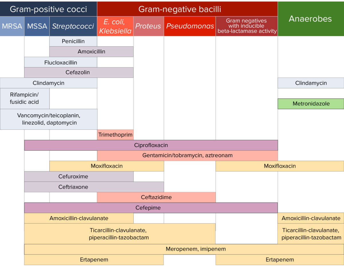
Different antibiotics have varying degrees of activity against different bacteria Bacteria Bacteria are prokaryotic single-celled microorganisms that are metabolically active and divide by binary fission. Some of these organisms play a significant role in the pathogenesis of diseases. Bacteriology. The table below outlines the antibiotics that are active against 3 important classes of bacteria Bacteria Bacteria are prokaryotic single-celled microorganisms that are metabolically active and divide by binary fission. Some of these organisms play a significant role in the pathogenesis of diseases. Bacteriology, including gram-positive Gram-Positive Penicillins cocci Cocci Bacteriology, gram-negative bacilli Bacilli Shigella, and anaerobes Anaerobes Lincosamides.

Antibiotic sensitivity chart

Antibiotic sensitivity:
Chart comparing the microbial coverage of different antibiotics for gram-positive cocci, gram-negative bacilli, and anaerobes.

Image by Lecturio. License: CC BY-NC-SA 4.0

References

  1. McCormack, J., Lalji, F. (2019). The “best” antibiotic sensitivity chart. Retrieved July 12, 2021, from https://therapeuticseducation.org/sites/therapeuticseducation.org/files/Antibiotic_Sensitivity_FINAL_Nov_2019.pdf 
  2. Letourneau, A.R. (2019). Beta-lactam antibiotics: Mechanisms of action and resistance and adverse effects. In Bloom, A. (Ed.), UpToDate. Retrieved May 20, 2021, from https://www.uptodate.com/contents/beta-lactam-antibiotics-mechanisms-of-action-and-resistance-and-adverse-effects
  3. Letourneau, A.R. (2019). Cephalosporins. In Bloom, A. (Ed.), UpToDate. Retrieved May 20, 2021, from https://www.uptodate.com/contents/cephalosporins 
  4. Bui, T., and Preuss, C.V. (2021). Cephalosporins. StatPearls. Retrieved July 12, 2021, from https://www.ncbi.nlm.nih.gov/books/NBK551517/ 
  5. Lexicomp, Inc. (2021). Drug Information Sheets, UpToDate, Retrieved July 12, 2021, from:

Create your free account or log in to continue reading!

Sign up now and get free access to Lecturio with concept pages, medical videos, and questions for your medical education.

User Reviews

Details