Central venous catheters are IV lines placed into the large central veins Veins Veins are tubular collections of cells, which transport deoxygenated blood and waste from the capillary beds back to the heart. Veins are classified into 3 types: small veins/venules, medium veins, and large veins. Each type contains 3 primary layers: tunica intima, tunica media, and tunica adventitia. Veins: Histology for monitoring of central venous pressure (CVP), prolonged drug administration, or administration of parenteral nutrition. The most common sites of insertion are the internal jugular and subclavian veins Veins Veins are tubular collections of cells, which transport deoxygenated blood and waste from the capillary beds back to the heart. Veins are classified into 3 types: small veins/venules, medium veins, and large veins. Each type contains 3 primary layers: tunica intima, tunica media, and tunica adventitia. Veins: Histology. Peripherally inserted central catheters and tunneled central venous catheters are variations that are often used when a prolonged need for central access is anticipated and are common in outpatient settings. Although often lifesaving, these catheters are associated with numerous complications and should be used judiciously.
Last updated: Dec 15, 2025
Central venous catheters are IV catheters inserted into the large central veins Veins Veins are tubular collections of cells, which transport deoxygenated blood and waste from the capillary beds back to the heart. Veins are classified into 3 types: small veins/venules, medium veins, and large veins. Each type contains 3 primary layers: tunica intima, tunica media, and tunica adventitia. Veins: Histology that are directly joining the superior or inferior venae cavae Venae Cavae The inferior and superior venae cavae. Veins: Histology.
The central veins Veins Veins are tubular collections of cells, which transport deoxygenated blood and waste from the capillary beds back to the heart. Veins are classified into 3 types: small veins/venules, medium veins, and large veins. Each type contains 3 primary layers: tunica intima, tunica media, and tunica adventitia. Veins: Histology are accessed percutaneously under local anesthesia Anesthesia A state characterized by loss of feeling or sensation. This depression of nerve function is usually the result of pharmacologic action and is induced to allow performance of surgery or other painful procedures. Anesthesiology: History and Basic Concepts. Bedside ultrasound guidance can be used and is recommended particularly for internal jugular insertion, where the vein can be easily visualized, which can help avoid inadvertent carotid artery cannulations.
The Seldinger technique is most commonly used for central venous catheter placement:
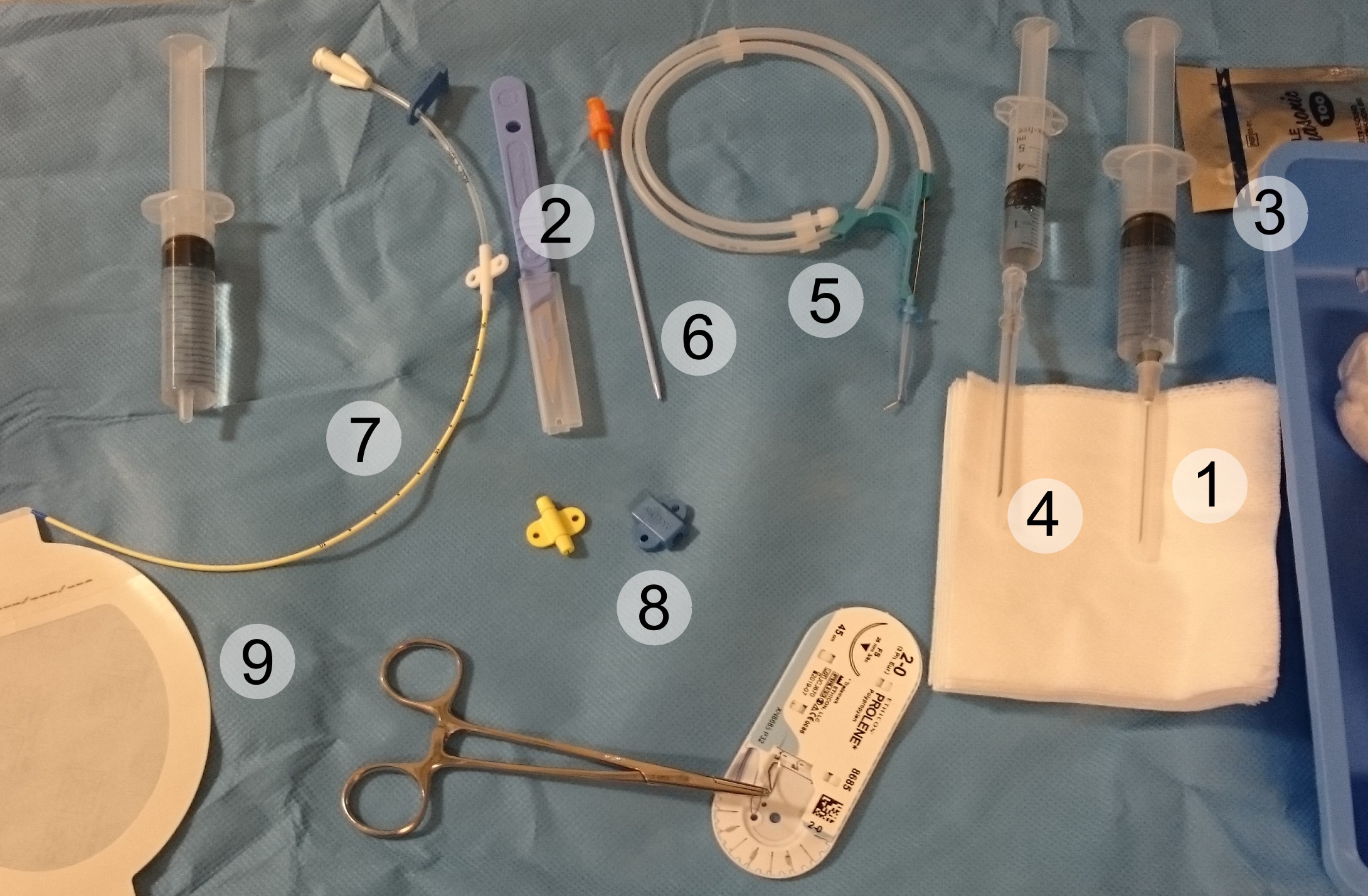
Central venous catheterization set:
1: Syringe with local anesthetic
2: Scalpel
3: Sterile gel for ultrasound guidance
4: Introducer needle (18 gauge pictured) on syringe with saline to detect backflow of blood upon vein penetration
5: Guidewire
6: Tissue dilator
7: Indwelling catheter (16 gauge pictured)
8: Additional fasteners and corresponding surgical thread
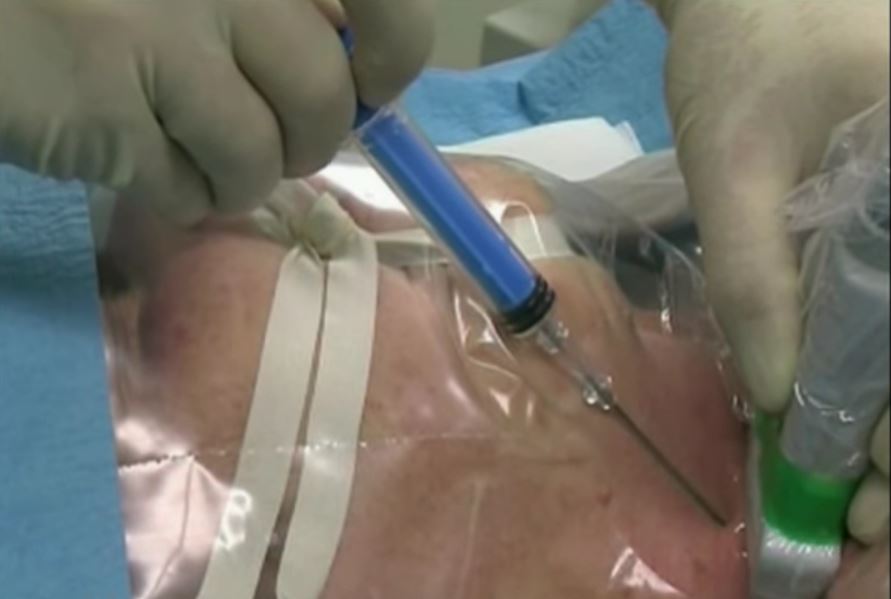
Ultrasound-guided puncture of the internal jugular vein for placement of a central venous catheter
Taken from video: “Internal jugular vein puncture with the aid of ultrasound” by MrArifnajafov. License: CC BY 3.0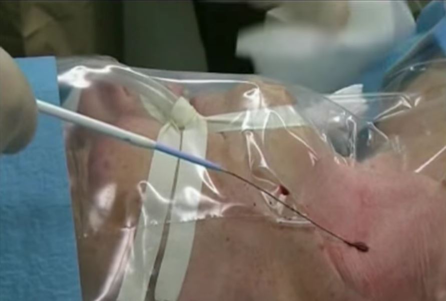
Insertion of a central venous catheter into the internal jugular vein:
Note the catheter follows the guidewire for proper insertion.

A diagram of the puncture site for a subclavian central venous catheter
Image by Lecturio. License: CC BY-NC-SA 4.0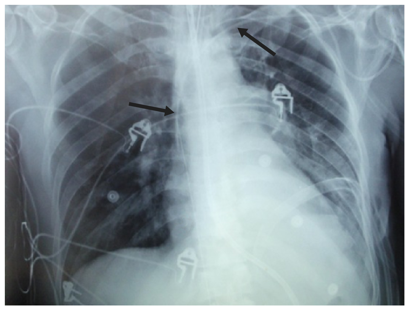
Chest X-ray showing an endotracheal tube, an orogastric tube, a left-sided subclavian central venous catheter (arrows), a left-sided chest tube, and multiple superficial leads.
Image: “Guidewire-Related Complications during Central Venous Catheter Placement: A Case Report and Review of the Literature” by Khasawneh FA, Smalligan RD. License: CC BY 3.0, edited by Lecturio.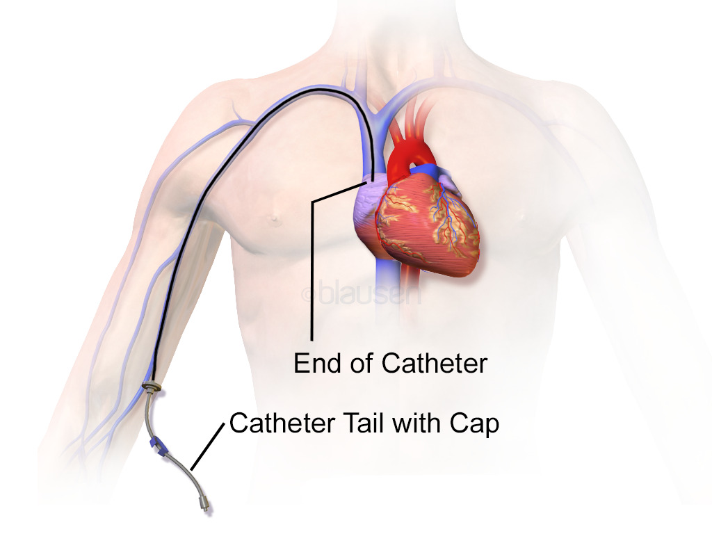
Illustration of fully inserted peripherally inserted central venous catheter
Image: “Blausen 0193 Catheter PICC” by Blausen Medical Communications, Inc. License: CC BY 3.0, edited by Lecturio.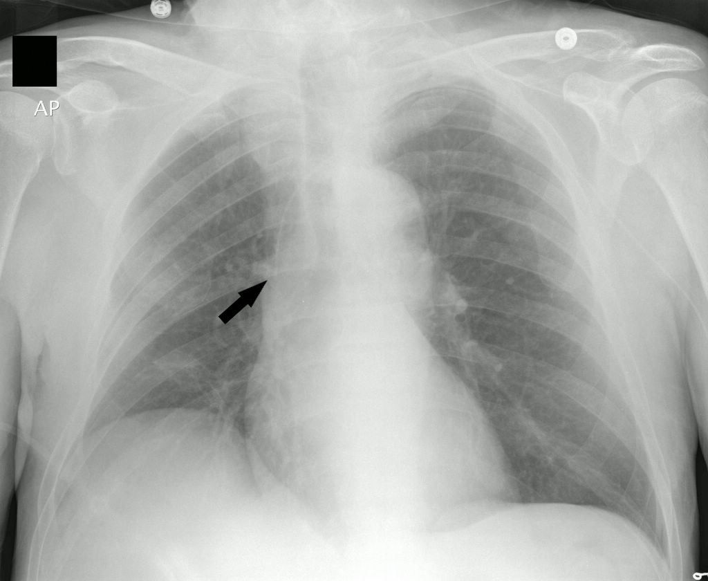
Chest X-ray of a patient with a Peripherally inserted central venous catheter:
Note the tip of the catheter in adequate position in the superior vena cava (arrow).
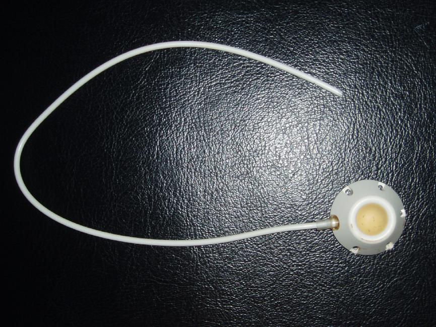
Port-a-cath:
Example of a tunneled central venous catheter. The port is placed in a subcutaneous pocket and can be easily accessed with a needle.
Placement of a central venous catheter is an invasive procedure associated with numerous complications. Therefore, these catheters should be inserted carefully and removed as early as feasible.