Advertisement

Advertisement

Advertisement

Advertisement

Carpal Tunnel Syndrome

Carpal tunnel syndrome (CTS) is a complex of signs and symptoms caused by compression Compression Blunt Chest Trauma of the median nerve Median Nerve A major nerve of the upper extremity. In humans, the fibers of the median nerve originate in the lower cervical and upper thoracic spinal cord (usually C6 to T1), travel via the brachial plexus, and supply sensory and motor innervation to parts of the forearm and hand. Cubital Fossa: Anatomy as it crosses the carpal tunnel. Presentation is with pain Pain An unpleasant sensation induced by noxious stimuli which are detected by nerve endings of nociceptive neurons. Pain: Types and Pathways and paresthesia of the dermatomal Dermatomal Dermatologic Examination target tissues innervated by the median nerve Median Nerve A major nerve of the upper extremity. In humans, the fibers of the median nerve originate in the lower cervical and upper thoracic spinal cord (usually C6 to T1), travel via the brachial plexus, and supply sensory and motor innervation to parts of the forearm and hand. Cubital Fossa: Anatomy as well as weakness and atrophy Atrophy Decrease in the size of a cell, tissue, organ, or multiple organs, associated with a variety of pathological conditions such as abnormal cellular changes, ischemia, malnutrition, or hormonal changes. Cellular Adaptation of the nerve's myotomal targets. Risk factors that cause a predisposition to CTS include obesity Obesity Obesity is a condition associated with excess body weight, specifically with the deposition of excessive adipose tissue. Obesity is considered a global epidemic. Major influences come from the western diet and sedentary lifestyles, but the exact mechanisms likely include a mixture of genetic and environmental factors. Obesity, female sex Sex The totality of characteristics of reproductive structure, functions, phenotype, and genotype, differentiating the male from the female organism. Gender Dysphoria, pregnancy Pregnancy The status during which female mammals carry their developing young (embryos or fetuses) in utero before birth, beginning from fertilization to birth. Pregnancy: Diagnosis, Physiology, and Care, diabetes Diabetes Diabetes mellitus (DM) is a metabolic disease characterized by hyperglycemia and dysfunction of the regulation of glucose metabolism by insulin. Type 1 DM is diagnosed mostly in children and young adults as the result of autoimmune destruction of β cells in the pancreas and the resulting lack of insulin. Type 2 DM has a significant association with obesity and is characterized by insulin resistance. Diabetes Mellitus, inflammatory conditions, genetic predisposition, and occupational factors. A clinical diagnosis may be made on the basis of history and physical examination and confirmed with electrodiagnostic testing. Conservative management includes splinting and physical therapy Physical Therapy Becker Muscular Dystrophy; more severe cases may require surgical correction.

Last updated: Nov 3, 2022

Editorial responsibility: Stanley Oiseth, Lindsay Jones, Evelin Maza

Advertisement

Advertisement

Advertisement

Advertisement

Advertisement

Advertisement

Overview

Anatomy

  • The median nerve Median Nerve A major nerve of the upper extremity. In humans, the fibers of the median nerve originate in the lower cervical and upper thoracic spinal cord (usually C6 to T1), travel via the brachial plexus, and supply sensory and motor innervation to parts of the forearm and hand. Cubital Fossa: Anatomy emerges from the brachial plexus Brachial Plexus The large network of nerve fibers which distributes the innervation of the upper extremity. The brachial plexus extends from the neck into the axilla. In humans, the nerves of the plexus usually originate from the lower cervical and the first thoracic spinal cord segments (c5-c8 and T1), but variations are not uncommon. Peripheral Nerve Injuries in the Cervicothoracic Region with contributions from the C6, C7, C8, and T1 nerve roots.
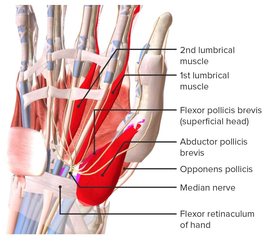
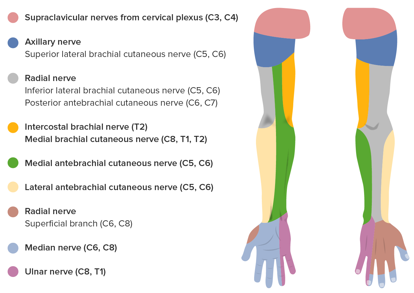
  • Roots C6 and C7 supply the median sensory Sensory Neurons which conduct nerve impulses to the central nervous system. Nervous System: Histology fibers, which in turn give sensation to the thenar eminence and the 1st 3½ digits of the hand Hand The hand constitutes the distal part of the upper limb and provides the fine, precise movements needed in activities of daily living. It consists of 5 metacarpal bones and 14 phalanges, as well as numerous muscles innervated by the median and ulnar nerves. Hand: Anatomy.
  • Roots C8 and T1 supply the motor Motor Neurons which send impulses peripherally to activate muscles or secretory cells. Nervous System: Histology fibers to the forearm Forearm The forearm is the region of the upper limb between the elbow and the wrist. The term “forearm” is used in anatomy to distinguish this area from the arm, a term that is commonly used to describe the entire upper limb. The forearm consists of 2 long bones (the radius and the ulna), the interosseous membrane, and multiple arteries, nerves, and muscles. Forearm: Anatomy and hand Hand The hand constitutes the distal part of the upper limb and provides the fine, precise movements needed in activities of daily living. It consists of 5 metacarpal bones and 14 phalanges, as well as numerous muscles innervated by the median and ulnar nerves. Hand: Anatomy musculature innervated by the median nerve Median Nerve A major nerve of the upper extremity. In humans, the fibers of the median nerve originate in the lower cervical and upper thoracic spinal cord (usually C6 to T1), travel via the brachial plexus, and supply sensory and motor innervation to parts of the forearm and hand. Cubital Fossa: Anatomy.
  • As the median nerve Median Nerve A major nerve of the upper extremity. In humans, the fibers of the median nerve originate in the lower cervical and upper thoracic spinal cord (usually C6 to T1), travel via the brachial plexus, and supply sensory and motor innervation to parts of the forearm and hand. Cubital Fossa: Anatomy passes through the antecubital fossa Antecubital Fossa Molluscum Contagiosum, it lies medial to the brachial artery Brachial Artery The continuation of the axillary artery; it branches into the radial and ulnar arteries. Cubital Fossa: Anatomy deep within the forearm Forearm The forearm is the region of the upper limb between the elbow and the wrist. The term “forearm” is used in anatomy to distinguish this area from the arm, a term that is commonly used to describe the entire upper limb. The forearm consists of 2 long bones (the radius and the ulna), the interosseous membrane, and multiple arteries, nerves, and muscles. Forearm: Anatomy
  • In the upper portion of the forearm Forearm The forearm is the region of the upper limb between the elbow and the wrist. The term “forearm” is used in anatomy to distinguish this area from the arm, a term that is commonly used to describe the entire upper limb. The forearm consists of 2 long bones (the radius and the ulna), the interosseous membrane, and multiple arteries, nerves, and muscles. Forearm: Anatomy, the median nerve Median Nerve A major nerve of the upper extremity. In humans, the fibers of the median nerve originate in the lower cervical and upper thoracic spinal cord (usually C6 to T1), travel via the brachial plexus, and supply sensory and motor innervation to parts of the forearm and hand. Cubital Fossa: Anatomy innervates:
  • The median nerve Median Nerve A major nerve of the upper extremity. In humans, the fibers of the median nerve originate in the lower cervical and upper thoracic spinal cord (usually C6 to T1), travel via the brachial plexus, and supply sensory and motor innervation to parts of the forearm and hand. Cubital Fossa: Anatomy then gives off a peripheral branch, forming the anterior interosseous nerve Anterior Interosseous Nerve Supracondylar Fracture.
  • The anterior interosseous nerve Anterior Interosseous Nerve Supracondylar Fracture innervates:
  • The median nerve Median Nerve A major nerve of the upper extremity. In humans, the fibers of the median nerve originate in the lower cervical and upper thoracic spinal cord (usually C6 to T1), travel via the brachial plexus, and supply sensory and motor innervation to parts of the forearm and hand. Cubital Fossa: Anatomy proper runs parallel to the anterior interosseous nerve Anterior Interosseous Nerve Supracondylar Fracture in the forearm Forearm The forearm is the region of the upper limb between the elbow and the wrist. The term “forearm” is used in anatomy to distinguish this area from the arm, a term that is commonly used to describe the entire upper limb. The forearm consists of 2 long bones (the radius and the ulna), the interosseous membrane, and multiple arteries, nerves, and muscles. Forearm: Anatomy.
  • Proximal to the wrist, the cutaneous sensory Sensory Neurons which conduct nerve impulses to the central nervous system. Nervous System: Histology branch of the median nerve Median Nerve A major nerve of the upper extremity. In humans, the fibers of the median nerve originate in the lower cervical and upper thoracic spinal cord (usually C6 to T1), travel via the brachial plexus, and supply sensory and motor innervation to parts of the forearm and hand. Cubital Fossa: Anatomy branches off to provide sensation to the lateral half of the palm.
  • Because the palmar branch of the median nerve Median Nerve A major nerve of the upper extremity. In humans, the fibers of the median nerve originate in the lower cervical and upper thoracic spinal cord (usually C6 to T1), travel via the brachial plexus, and supply sensory and motor innervation to parts of the forearm and hand. Cubital Fossa: Anatomy passes over, and not through, the carpal tunnel, sensory Sensory Neurons which conduct nerve impulses to the central nervous system. Nervous System: Histology changes over the thenar eminence are not typically observed with carpal tunnel syndrome (CTS).
  • The carpal tunnel is formed by the transverse carpal ligament ( flexor retinaculum Flexor Retinaculum Ankle Joint: Anatomy) superiorly and the carpal bones inferiorly.
  • The median nerve Median Nerve A major nerve of the upper extremity. In humans, the fibers of the median nerve originate in the lower cervical and upper thoracic spinal cord (usually C6 to T1), travel via the brachial plexus, and supply sensory and motor innervation to parts of the forearm and hand. Cubital Fossa: Anatomy proper and the 9 flexor tendons of the forearm Forearm The forearm is the region of the upper limb between the elbow and the wrist. The term “forearm” is used in anatomy to distinguish this area from the arm, a term that is commonly used to describe the entire upper limb. The forearm consists of 2 long bones (the radius and the ulna), the interosseous membrane, and multiple arteries, nerves, and muscles. Forearm: Anatomy musculature pass through the tunnel.
  • Upon exiting the carpal tunnel, the median nerve Median Nerve A major nerve of the upper extremity. In humans, the fibers of the median nerve originate in the lower cervical and upper thoracic spinal cord (usually C6 to T1), travel via the brachial plexus, and supply sensory and motor innervation to parts of the forearm and hand. Cubital Fossa: Anatomy provides sensory Sensory Neurons which conduct nerve impulses to the central nervous system. Nervous System: Histology and motor Motor Neurons which send impulses peripherally to activate muscles or secretory cells. Nervous System: Histology innervation to the hand Hand The hand constitutes the distal part of the upper limb and provides the fine, precise movements needed in activities of daily living. It consists of 5 metacarpal bones and 14 phalanges, as well as numerous muscles innervated by the median and ulnar nerves. Hand: Anatomy.
  • The hand Hand The hand constitutes the distal part of the upper limb and provides the fine, precise movements needed in activities of daily living. It consists of 5 metacarpal bones and 14 phalanges, as well as numerous muscles innervated by the median and ulnar nerves. Hand: Anatomy muscles innervated by the median nerve Median Nerve A major nerve of the upper extremity. In humans, the fibers of the median nerve originate in the lower cervical and upper thoracic spinal cord (usually C6 to T1), travel via the brachial plexus, and supply sensory and motor innervation to parts of the forearm and hand. Cubital Fossa: Anatomy:

Mnemonic:

Median-innervated hand Hand The hand constitutes the distal part of the upper limb and provides the fine, precise movements needed in activities of daily living. It consists of 5 metacarpal bones and 14 phalanges, as well as numerous muscles innervated by the median and ulnar nerves. Hand: Anatomy muscles (Meat-LOAF):
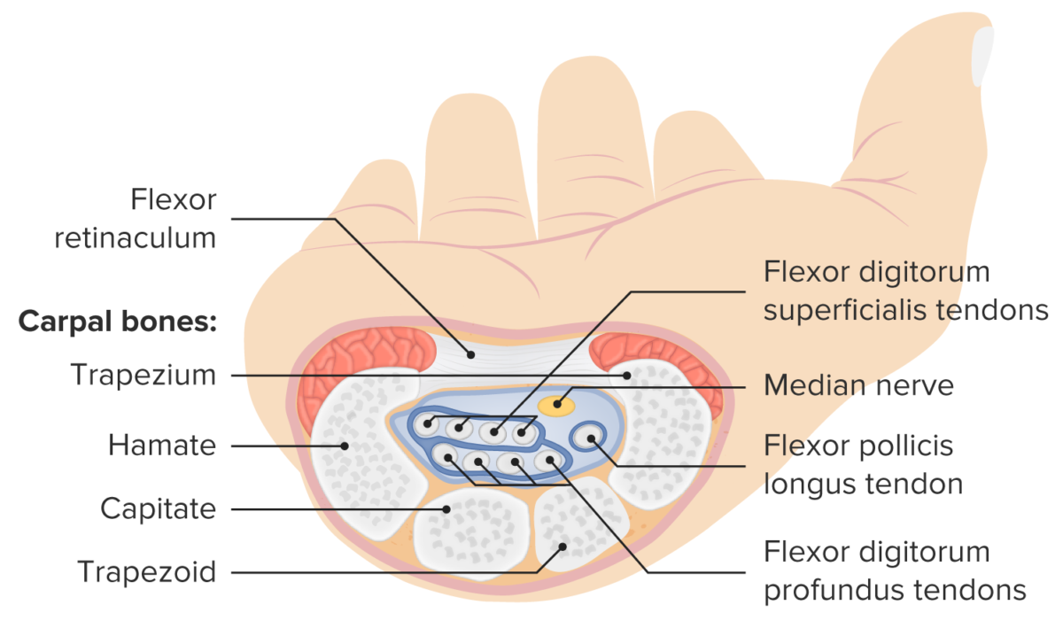
Structure and contents of the carpal tunnel

Structure and contents of the carpal tunnel

Image by Lecturio.

Epidemiology

  • Common disorder among adults
  • Prevalence Prevalence The total number of cases of a given disease in a specified population at a designated time. It is differentiated from incidence, which refers to the number of new cases in the population at a given time. Measures of Disease Frequency in general population: 1%–5%.
  • Female > male by 3:1
  • Prevalence Prevalence The total number of cases of a given disease in a specified population at a designated time. It is differentiated from incidence, which refers to the number of new cases in the population at a given time. Measures of Disease Frequency highest in obese women
  • Prevalence Prevalence The total number of cases of a given disease in a specified population at a designated time. It is differentiated from incidence, which refers to the number of new cases in the population at a given time. Measures of Disease Frequency lowest in thin and average-sized men

Risk factors

  • Obesity Obesity Obesity is a condition associated with excess body weight, specifically with the deposition of excessive adipose tissue. Obesity is considered a global epidemic. Major influences come from the western diet and sedentary lifestyles, but the exact mechanisms likely include a mixture of genetic and environmental factors. Obesity
  • Female sex Sex The totality of characteristics of reproductive structure, functions, phenotype, and genotype, differentiating the male from the female organism. Gender Dysphoria
  • Pregnancy Pregnancy The status during which female mammals carry their developing young (embryos or fetuses) in utero before birth, beginning from fertilization to birth. Pregnancy: Diagnosis, Physiology, and Care
  • Diabetes Diabetes Diabetes mellitus (DM) is a metabolic disease characterized by hyperglycemia and dysfunction of the regulation of glucose metabolism by insulin. Type 1 DM is diagnosed mostly in children and young adults as the result of autoimmune destruction of β cells in the pancreas and the resulting lack of insulin. Type 2 DM has a significant association with obesity and is characterized by insulin resistance. Diabetes Mellitus
  • Rheumatoid arthritis Arthritis Acute or chronic inflammation of joints. Osteoarthritis
  • Osteoarthritis Osteoarthritis Osteoarthritis (OA) is the most common form of arthritis, and is due to cartilage destruction and changes of the subchondral bone. The risk of developing this disorder increases with age, obesity, and repetitive joint use or trauma. Patients develop gradual joint pain, stiffness lasting < 30 minutes, and decreased range of motion. Osteoarthritis of the hand Hand The hand constitutes the distal part of the upper limb and provides the fine, precise movements needed in activities of daily living. It consists of 5 metacarpal bones and 14 phalanges, as well as numerous muscles innervated by the median and ulnar nerves. Hand: Anatomy
  • Hypothyroidism Hypothyroidism Hypothyroidism is a condition characterized by a deficiency of thyroid hormones. Iodine deficiency is the most common cause worldwide, but Hashimoto’s disease (autoimmune thyroiditis) is the leading cause in non-iodine-deficient regions. Hypothyroidism
  • Connective tissue Connective tissue Connective tissues originate from embryonic mesenchyme and are present throughout the body except inside the brain and spinal cord. The main function of connective tissues is to provide structural support to organs. Connective tissues consist of cells and an extracellular matrix. Connective Tissue: Histology disorders
  • Preexisting median mononeuropathy Mononeuropathy Disease or trauma involving a single peripheral nerve in isolation, or out of proportion to evidence of diffuse peripheral nerve dysfunction. Mononeuropathy multiplex refers to a condition characterized by multiple isolated nerve injuries. Mononeuropathies may result from a wide variety of causes, including ischemia; traumatic injury; compression; connective tissue diseases; cumulative trauma disorders; and other conditions. Mononeuropathy and Plexopathy
  • Genetic predisposition
  • Aromatase Aromatase An enzyme that catalyzes the desaturation (aromatization) of the ring a of C19 androgens and converts them to C18 estrogens. In this process, the 19-methyl is removed. This enzyme is membrane-bound, located in the endoplasmic reticulum of estrogen-producing cells of ovaries, placenta, testes, adipose, and brain tissues. Aromatase is encoded by the cyp19 gene, and functions in complex with NADPH-ferrihemoprotein reductase in the cytochrome p450 system. Adipose Tissue: Histology inhibitor use
  • Workplace factors:
    • Repetitive or forceful hand Hand The hand constitutes the distal part of the upper limb and provides the fine, precise movements needed in activities of daily living. It consists of 5 metacarpal bones and 14 phalanges, as well as numerous muscles innervated by the median and ulnar nerves. Hand: Anatomy and wrist use
  • Trauma

Pathophysiology

The pathophysiology of narrowing of the carpal tunnel and median nerve Median Nerve A major nerve of the upper extremity. In humans, the fibers of the median nerve originate in the lower cervical and upper thoracic spinal cord (usually C6 to T1), travel via the brachial plexus, and supply sensory and motor innervation to parts of the forearm and hand. Cubital Fossa: Anatomy dysfunction are multifactorial.

  • Narrowing of the carpal tunnel:
    • Congenitally small anatomic space 
    • Mass Mass Three-dimensional lesion that occupies a space within the breast Imaging of the Breast lesions encroaching on anatomic space: 
      • Cysts Cysts Any fluid-filled closed cavity or sac that is lined by an epithelium. Cysts can be of normal, abnormal, non-neoplastic, or neoplastic tissues. Fibrocystic Change
      • Neoplasms Neoplasms New abnormal growth of tissue. Malignant neoplasms show a greater degree of anaplasia and have the properties of invasion and metastasis, compared to benign neoplasms. Benign Bone Tumors
      • Persistent median artery 
    • Edema Edema Edema is a condition in which excess serous fluid accumulates in the body cavity or interstitial space of connective tissues. Edema is a symptom observed in several medical conditions. It can be categorized into 2 types, namely, peripheral (in the extremities) and internal (in an organ or body cavity). Edema or inflammation Inflammation Inflammation is a complex set of responses to infection and injury involving leukocytes as the principal cellular mediators in the body’s defense against pathogenic organisms. Inflammation is also seen as a response to tissue injury in the process of wound healing. The 5 cardinal signs of inflammation are pain, heat, redness, swelling, and loss of function. Inflammation from systemic diseases:
      • Diabetes Diabetes Diabetes mellitus (DM) is a metabolic disease characterized by hyperglycemia and dysfunction of the regulation of glucose metabolism by insulin. Type 1 DM is diagnosed mostly in children and young adults as the result of autoimmune destruction of β cells in the pancreas and the resulting lack of insulin. Type 2 DM has a significant association with obesity and is characterized by insulin resistance. Diabetes Mellitus
      • Osteoarthritis Osteoarthritis Osteoarthritis (OA) is the most common form of arthritis, and is due to cartilage destruction and changes of the subchondral bone. The risk of developing this disorder increases with age, obesity, and repetitive joint use or trauma. Patients develop gradual joint pain, stiffness lasting < 30 minutes, and decreased range of motion. Osteoarthritis
      • Rheumatoid arthritis Arthritis Acute or chronic inflammation of joints. Osteoarthritis
    • Inflammation Inflammation Inflammation is a complex set of responses to infection and injury involving leukocytes as the principal cellular mediators in the body’s defense against pathogenic organisms. Inflammation is also seen as a response to tissue injury in the process of wound healing. The 5 cardinal signs of inflammation are pain, heat, redness, swelling, and loss of function. Inflammation/thickening of the flexor tendons passing through the carpal tunnel alongside the median nerve Median Nerve A major nerve of the upper extremity. In humans, the fibers of the median nerve originate in the lower cervical and upper thoracic spinal cord (usually C6 to T1), travel via the brachial plexus, and supply sensory and motor innervation to parts of the forearm and hand. Cubital Fossa: Anatomy
    • Noninflammatory fibrosis Fibrosis Any pathological condition where fibrous connective tissue invades any organ, usually as a consequence of inflammation or other injury. Bronchiolitis Obliterans affecting the subsynovial connective tissue Connective tissue Connective tissues originate from embryonic mesenchyme and are present throughout the body except inside the brain and spinal cord. The main function of connective tissues is to provide structural support to organs. Connective tissues consist of cells and an extracellular matrix. Connective Tissue: Histology surrounding the flexor tendons
  • Mechanical compression Compression Blunt Chest Trauma of the median nerve Median Nerve A major nerve of the upper extremity. In humans, the fibers of the median nerve originate in the lower cervical and upper thoracic spinal cord (usually C6 to T1), travel via the brachial plexus, and supply sensory and motor innervation to parts of the forearm and hand. Cubital Fossa: Anatomy:
    • Increased pressure in the carpal tunnel may cause:
      • Direct nerve injury Nerve Injury Surgical Complications
      • Impaired axonal transport
      • Compression Compression Blunt Chest Trauma of vessels in the perineurium → median nerve Median Nerve A major nerve of the upper extremity. In humans, the fibers of the median nerve originate in the lower cervical and upper thoracic spinal cord (usually C6 to T1), travel via the brachial plexus, and supply sensory and motor innervation to parts of the forearm and hand. Cubital Fossa: Anatomy ischemia Ischemia A hypoperfusion of the blood through an organ or tissue caused by a pathologic constriction or obstruction of its blood vessels, or an absence of blood circulation. Ischemic Cell Damage
    • Upper-extremity postural abnormalities
  • Median nerve Median Nerve A major nerve of the upper extremity. In humans, the fibers of the median nerve originate in the lower cervical and upper thoracic spinal cord (usually C6 to T1), travel via the brachial plexus, and supply sensory and motor innervation to parts of the forearm and hand. Cubital Fossa: Anatomy develops an “hourglass” deformity Deformity Examination of the Upper Limbs within the carpal tunnel due to:
    • Edema Edema Edema is a condition in which excess serous fluid accumulates in the body cavity or interstitial space of connective tissues. Edema is a symptom observed in several medical conditions. It can be categorized into 2 types, namely, peripheral (in the extremities) and internal (in an organ or body cavity). Edema and thickening of vessel walls within the endoneurium and perineurium
    • Noninflammatory synovial fibrosis Fibrosis Any pathological condition where fibrous connective tissue invades any organ, usually as a consequence of inflammation or other injury. Bronchiolitis Obliterans and vascular proliferation
    • Fibrosis Fibrosis Any pathological condition where fibrous connective tissue invades any organ, usually as a consequence of inflammation or other injury. Bronchiolitis Obliterans
    • Myelin thinning
    • Nerve-fiber degeneration and regeneration Regeneration The physiological renewal, repair, or replacement of tissue. Wound Healing
  • Vascular proliferation and fibrosis Fibrosis Any pathological condition where fibrous connective tissue invades any organ, usually as a consequence of inflammation or other injury. Bronchiolitis Obliterans are associated with increased prostaglandin E2 Prostaglandin E2 The most common and most biologically active of the mammalian prostaglandins. It exhibits most biological activities characteristic of prostaglandins and has been used extensively as an oxytocic agent. The compound also displays a protective effect on the intestinal mucosa. Fever and vascular endothelial growth factor Vascular endothelial growth factor A family of angiogenic proteins that are closely-related to vascular endothelial growth factor a. They play an important role in the growth and differentiation of vascular as well as lymphatic endothelial cells. Wound Healing expression.

Clinical Presentation

  • Common symptoms:
    • Pain Pain An unpleasant sensation induced by noxious stimuli which are detected by nerve endings of nociceptive neurons. Pain: Types and Pathways/discomfort in the hand Hand The hand constitutes the distal part of the upper limb and provides the fine, precise movements needed in activities of daily living. It consists of 5 metacarpal bones and 14 phalanges, as well as numerous muscles innervated by the median and ulnar nerves. Hand: Anatomy, wrist, and forearm Forearm The forearm is the region of the upper limb between the elbow and the wrist. The term “forearm” is used in anatomy to distinguish this area from the arm, a term that is commonly used to describe the entire upper limb. The forearm consists of 2 long bones (the radius and the ulna), the interosseous membrane, and multiple arteries, nerves, and muscles. Forearm: Anatomy
    • Paresthesia in the median-nerve distribution of the affected hand Hand The hand constitutes the distal part of the upper limb and provides the fine, precise movements needed in activities of daily living. It consists of 5 metacarpal bones and 14 phalanges, as well as numerous muscles innervated by the median and ulnar nerves. Hand: Anatomy(s) (e.g., heaviness, tingling Tingling Posterior Cord Syndrome, pins-and-needles sensation)
    • Subjective weakness or clumsiness of the hand Hand The hand constitutes the distal part of the upper limb and provides the fine, precise movements needed in activities of daily living. It consists of 5 metacarpal bones and 14 phalanges, as well as numerous muscles innervated by the median and ulnar nerves. Hand: Anatomy
    • Occurrence of any of these symptoms in the median distribution
  • Common signs:
    • Sensory deficit Sensory Deficit Anterior Cord Syndrome in the median-nerve distribution of the affected hand Hand The hand constitutes the distal part of the upper limb and provides the fine, precise movements needed in activities of daily living. It consists of 5 metacarpal bones and 14 phalanges, as well as numerous muscles innervated by the median and ulnar nerves. Hand: Anatomy(s)
    • Objective weakness or clumsiness of the hand Hand The hand constitutes the distal part of the upper limb and provides the fine, precise movements needed in activities of daily living. It consists of 5 metacarpal bones and 14 phalanges, as well as numerous muscles innervated by the median and ulnar nerves. Hand: Anatomy
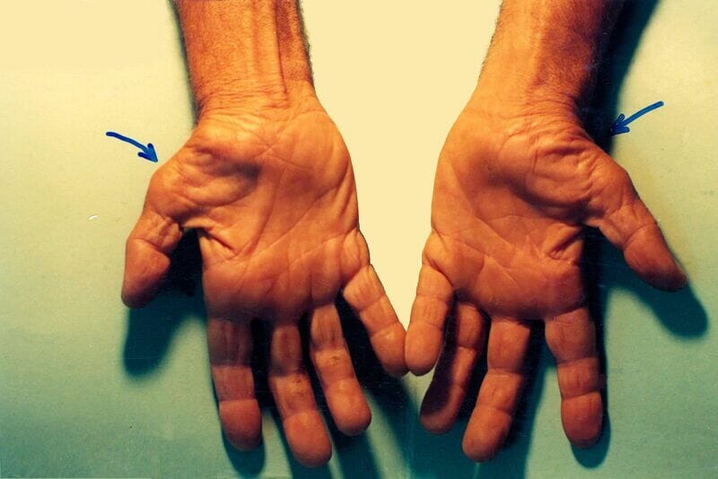
    • Thenar muscle atrophy Atrophy Decrease in the size of a cell, tissue, organ, or multiple organs, associated with a variety of pathological conditions such as abnormal cellular changes, ischemia, malnutrition, or hormonal changes. Cellular Adaptation
  • Provocative factors:
    • Sleep Sleep A readily reversible suspension of sensorimotor interaction with the environment, usually associated with recumbency and immobility. Physiology of Sleep:
      • Wrist-flexion posturing
      • Relative immobility/flat positioning (i.e., decreased perfusion)
    • Sustained hand Hand The hand constitutes the distal part of the upper limb and provides the fine, precise movements needed in activities of daily living. It consists of 5 metacarpal bones and 14 phalanges, as well as numerous muscles innervated by the median and ulnar nerves. Hand: Anatomy or arm Arm The arm, or “upper arm” in common usage, is the region of the upper limb that extends from the shoulder to the elbow joint and connects inferiorly to the forearm through the cubital fossa. It is divided into 2 fascial compartments (anterior and posterior). Arm: Anatomy positions
    • Repetitive actions of the hand Hand The hand constitutes the distal part of the upper limb and provides the fine, precise movements needed in activities of daily living. It consists of 5 metacarpal bones and 14 phalanges, as well as numerous muscles innervated by the median and ulnar nerves. Hand: Anatomy or wrist
  • Alleviating factors:
    • Changes in hand Hand The hand constitutes the distal part of the upper limb and provides the fine, precise movements needed in activities of daily living. It consists of 5 metacarpal bones and 14 phalanges, as well as numerous muscles innervated by the median and ulnar nerves. Hand: Anatomy/wrist posture (wrist extension Extension Examination of the Upper Limbs)
    • Shaking the hand Hand The hand constitutes the distal part of the upper limb and provides the fine, precise movements needed in activities of daily living. It consists of 5 metacarpal bones and 14 phalanges, as well as numerous muscles innervated by the median and ulnar nerves. Hand: Anatomy (i.e., improved perfusion)

Diagnosis

Carpal tunnel syndrome is a clinical diagnosis. A combination of clinical findings with confirmatory electrodiagnostic testing is more accurate for diagnosis.
Examination

  • Objective sensory Sensory Neurons which conduct nerve impulses to the central nervous system. Nervous System: Histology and motor Motor Neurons which send impulses peripherally to activate muscles or secretory cells. Nervous System: Histology deficits may be present (absence does not rule out a diagnosis).
  • Sensory Sensory Neurons which conduct nerve impulses to the central nervous system. Nervous System: Histology deficits occur late in the course of CTS:
    • Affects the fingers innervated by the median nerve Median Nerve A major nerve of the upper extremity. In humans, the fibers of the median nerve originate in the lower cervical and upper thoracic spinal cord (usually C6 to T1), travel via the brachial plexus, and supply sensory and motor innervation to parts of the forearm and hand. Cubital Fossa: Anatomy
    • Spare the thenar eminence
  • Objective weakness occurs in advanced CTS:
  • Atrophy Atrophy Decrease in the size of a cell, tissue, organ, or multiple organs, associated with a variety of pathological conditions such as abnormal cellular changes, ischemia, malnutrition, or hormonal changes. Cellular Adaptation of the thenar eminence may be present.

Provocative maneuvers

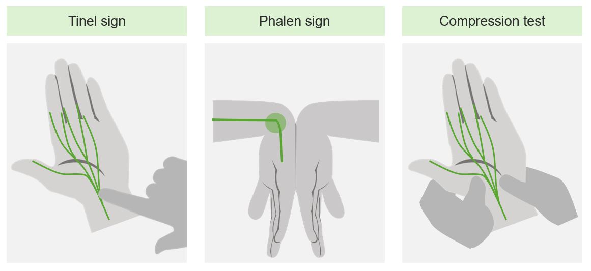
  • Phalen maneuver: 
  • Tinel test:
    • Firm percussion Percussion Act of striking a part with short, sharp blows as an aid in diagnosing the condition beneath the sound obtained. Pulmonary Examination over the course of the median nerve Median Nerve A major nerve of the upper extremity. In humans, the fibers of the median nerve originate in the lower cervical and upper thoracic spinal cord (usually C6 to T1), travel via the brachial plexus, and supply sensory and motor innervation to parts of the forearm and hand. Cubital Fossa: Anatomy proximal to or on top of the carpal tunnel
    • A positive test is defined as pain Pain An unpleasant sensation induced by noxious stimuli which are detected by nerve endings of nociceptive neurons. Pain: Types and Pathways and/or paresthesia in median innervated fingers on percussion Percussion Act of striking a part with short, sharp blows as an aid in diagnosing the condition beneath the sound obtained. Pulmonary Examination over the median nerve Median Nerve A major nerve of the upper extremity. In humans, the fibers of the median nerve originate in the lower cervical and upper thoracic spinal cord (usually C6 to T1), travel via the brachial plexus, and supply sensory and motor innervation to parts of the forearm and hand. Cubital Fossa: Anatomy.
  • Manual carpal compression Compression Blunt Chest Trauma:
    • Applying pressure over the transverse carpal ligament
    • Deemed positive if pain Pain An unpleasant sensation induced by noxious stimuli which are detected by nerve endings of nociceptive neurons. Pain: Types and Pathways and/or paresthesia noted in median innervated fingers within 30 seconds of compression Compression Blunt Chest Trauma
  • Hand Hand The hand constitutes the distal part of the upper limb and provides the fine, precise movements needed in activities of daily living. It consists of 5 metacarpal bones and 14 phalanges, as well as numerous muscles innervated by the median and ulnar nerves. Hand: Anatomy elevation test:
    • Raising hands above the head for 1 minute
    • Deemed positive if pain Pain An unpleasant sensation induced by noxious stimuli which are detected by nerve endings of nociceptive neurons. Pain: Types and Pathways and/or paresthesia noted in median innervated fingers within 30 seconds of elevation
Tests for carpal tunnel syndrome

Provocative maneuvers for clinical evaluation of carpal tunnel syndrome

Image by Lecturio.
Electrodiagnostic testing

  • Nerve conduction velocity:
    • Involves measurement of conduction velocity across the carpal tunnel and determination of the amplitude of sensory Sensory Neurons which conduct nerve impulses to the central nervous system. Nervous System: Histology and motor Motor Neurons which send impulses peripherally to activate muscles or secretory cells. Nervous System: Histology responses.
    • Nerve compression Nerve Compression Brachial Plexus Injuries causes damage to the myelin sheath manifesting as delayed distal latencies and slowed conduction velocities.
    • Sustained or severe compression Compression Blunt Chest Trauma leads to axonal loss resulting in the reduction of median-nerve action potential Action Potential Abrupt changes in the membrane potential that sweep along the cell membrane of excitable cells in response to excitation stimuli. Membrane Potential amplitude.
    • Gold standard for diagnosis; highly sensitive and specific
  • Electromyography Electromyography Recording of the changes in electric potential of muscle by means of surface or needle electrodes. Becker Muscular Dystrophy:
    • Useful to exclude other conditions
    • Useful to assess the severity of CTS if surgical decompression is being considered

Imaging

Imaging is useful if structural abnormality is suspected.

  • Neuromuscular ultrasound shows a significant increase in cross-sectional area of the median nerve Median Nerve A major nerve of the upper extremity. In humans, the fibers of the median nerve originate in the lower cervical and upper thoracic spinal cord (usually C6 to T1), travel via the brachial plexus, and supply sensory and motor innervation to parts of the forearm and hand. Cubital Fossa: Anatomy.
  • MRI is reserved for unusual cases to rule out mass Mass Three-dimensional lesion that occupies a space within the breast Imaging of the Breast lesions.

Grading Grading Methods which attempt to express in replicable terms the level of cell differentiation in neoplasms as increasing anaplasia correlates with the aggressiveness of the neoplasm. Grading, Staging, and Metastasis/ staging Staging Methods which attempt to express in replicable terms the extent of the neoplasm in the patient. Grading, Staging, and Metastasis

Clinical grading Grading Methods which attempt to express in replicable terms the level of cell differentiation in neoplasms as increasing anaplasia correlates with the aggressiveness of the neoplasm. Grading, Staging, and Metastasis of CTS severity:

  • Mild: 
    • Numbness, tingling Tingling Posterior Cord Syndrome, or discomfort in the median-nerve distribution 
    • No sensory Sensory Neurons which conduct nerve impulses to the central nervous system. Nervous System: Histology loss or weakness
    • No sleep Sleep A readily reversible suspension of sensorimotor interaction with the environment, usually associated with recumbency and immobility. Physiology of Sleep disruption
    • No difficulty with hand Hand The hand constitutes the distal part of the upper limb and provides the fine, precise movements needed in activities of daily living. It consists of 5 metacarpal bones and 14 phalanges, as well as numerous muscles innervated by the median and ulnar nerves. Hand: Anatomy function
    • No interference with activities of daily living
  • Moderate:
    • Sensory Sensory Neurons which conduct nerve impulses to the central nervous system. Nervous System: Histology loss with median distribution
    • Nocturnal symptoms occasionally disrupt sleep Sleep A readily reversible suspension of sensorimotor interaction with the environment, usually associated with recumbency and immobility. Physiology of Sleep.
    • Symptoms may interfere slightly with hand Hand The hand constitutes the distal part of the upper limb and provides the fine, precise movements needed in activities of daily living. It consists of 5 metacarpal bones and 14 phalanges, as well as numerous muscles innervated by the median and ulnar nerves. Hand: Anatomy function.
    • No interference with activities of daily living
  • Severe:
    • Weakness in the median distribution
    • Nocturnal symptoms routinely disrupt sleep Sleep A readily reversible suspension of sensorimotor interaction with the environment, usually associated with recumbency and immobility. Physiology of Sleep.
    • Symptoms are disabling and prevent carrying out ≥ 1 activities of daily living.

Electrodiagnostic grading Grading Methods which attempt to express in replicable terms the level of cell differentiation in neoplasms as increasing anaplasia correlates with the aggressiveness of the neoplasm. Grading, Staging, and Metastasis of CTS severity:

  • Mild:
  • Moderate:
    • Abnormal median sensory Sensory Neurons which conduct nerve impulses to the central nervous system. Nervous System: Histology latencies
    • Relative or absolute prolongation of median motor Motor Neurons which send impulses peripherally to activate muscles or secretory cells. Nervous System: Histology distal latency
    • No evidence of axonal loss
  • Severe: evidence of axonal loss

Management

Management is based on the acuity and severity of clinical symptoms and the degree of neurogenic injury as assessed by electrodiagnostic studies. 

Nonsurgical treatment options

For individuals with mild to moderate CTS, the rate of successful outcomes is 20%–93%. Options include:

  • Wrist splinting
  • Glucocorticoid injection
  • Oral glucocorticoids Glucocorticoids Glucocorticoids are a class within the corticosteroid family. Glucocorticoids are chemically and functionally similar to endogenous cortisol. There are a wide array of indications, which primarily benefit from the antiinflammatory and immunosuppressive effects of this class of drugs. Glucocorticoids
  • Perineural dextrose Dextrose Intravenous Fluids injection
  • Yoga
  • Carpal bone Bone Bone is a compact type of hardened connective tissue composed of bone cells, membranes, an extracellular mineralized matrix, and central bone marrow. The 2 primary types of bone are compact and spongy. Bones: Structure and Types mobilization ( physical therapy Physical Therapy Becker Muscular Dystrophy or occupational therapy Occupational Therapy Skilled treatment that helps individuals achieve independence in all facets of their lives. It assists in the development of skills needed for independent living. Fetal Alcohol Spectrum Disorder)
  • Therapeutic ultrasound therapy

Surgical decompression

  • Indications include:
    • Mild CTS unresponsive to conservative measures
    • Moderate to severe CTS associated with axonal loss or denervation
  • Open carpal tunnel release Release Release of a virus from the host cell following virus assembly and maturation. Egress can occur by host cell lysis, exocytosis, or budding through the plasma membrane. Virology technique
  • Endoscopic carpal tunnel release Release Release of a virus from the host cell following virus assembly and maturation. Egress can occur by host cell lysis, exocytosis, or budding through the plasma membrane. Virology technique
  • Ultrasound-guided minimally invasive carpal tunnel release Release Release of a virus from the host cell following virus assembly and maturation. Egress can occur by host cell lysis, exocytosis, or budding through the plasma membrane. Virology technique

Prognosis Prognosis A prediction of the probable outcome of a disease based on a individual’s condition and the usual course of the disease as seen in similar situations. Non-Hodgkin Lymphomas

  • Symptoms in untreated individuals with minimal or mild compression Compression Blunt Chest Trauma tend to worsen over 10–15 months.
  • Factors that predict progression:
  • Treatment with splinting or surgical decompression may lead to complete or marked improvement at 1 year in 70%–90% of individuals.

Differential Diagnosis

  • Cervical radiculopathy Cervical Radiculopathy Spinal Disk Herniation: C6 or C7 nerve-root involvement may present with arm Arm The arm, or “upper arm” in common usage, is the region of the upper limb that extends from the shoulder to the elbow joint and connects inferiorly to the forearm through the cubital fossa. It is divided into 2 fascial compartments (anterior and posterior). Arm: Anatomy pain Pain An unpleasant sensation induced by noxious stimuli which are detected by nerve endings of nociceptive neurons. Pain: Types and Pathways and paresthesia similar to that of CTS. Typically, there is also the presence of neck Neck The part of a human or animal body connecting the head to the rest of the body. Peritonsillar Abscess pain Pain An unpleasant sensation induced by noxious stimuli which are detected by nerve endings of nociceptive neurons. Pain: Types and Pathways exacerbated with neck Neck The part of a human or animal body connecting the head to the rest of the body. Peritonsillar Abscess movement; radiation Radiation Emission or propagation of acoustic waves (sound), electromagnetic energy waves (such as light; radio waves; gamma rays; or x-rays), or a stream of subatomic particles (such as electrons; neutrons; protons; or alpha particles). Osteosarcoma of pain Pain An unpleasant sensation induced by noxious stimuli which are detected by nerve endings of nociceptive neurons. Pain: Types and Pathways from the neck Neck The part of a human or animal body connecting the head to the rest of the body. Peritonsillar Abscess to the shoulder and arm Arm The arm, or “upper arm” in common usage, is the region of the upper limb that extends from the shoulder to the elbow joint and connects inferiorly to the forearm through the cubital fossa. It is divided into 2 fascial compartments (anterior and posterior). Arm: Anatomy; reduced reflexes; weakness of proximal arm Arm The arm, or “upper arm” in common usage, is the region of the upper limb that extends from the shoulder to the elbow joint and connects inferiorly to the forearm through the cubital fossa. It is divided into 2 fascial compartments (anterior and posterior). Arm: Anatomy muscles involving elbow flexion Flexion Examination of the Upper Limbs, extension Extension Examination of the Upper Limbs, and arm Arm The arm, or “upper arm” in common usage, is the region of the upper limb that extends from the shoulder to the elbow joint and connects inferiorly to the forearm through the cubital fossa. It is divided into 2 fascial compartments (anterior and posterior). Arm: Anatomy pronation Pronation Applies to movements of the forearm in turning the palm backward or downward. When referring to the foot, a combination of eversion and abduction movements in the tarsal and metatarsal joints (turning the foot up and in toward the midline of the body). Examination of the Upper Limbs; and sensory Sensory Neurons which conduct nerve impulses to the central nervous system. Nervous System: Histology loss in the palm or forearm Forearm The forearm is the region of the upper limb between the elbow and the wrist. The term “forearm” is used in anatomy to distinguish this area from the arm, a term that is commonly used to describe the entire upper limb. The forearm consists of 2 long bones (the radius and the ulna), the interosseous membrane, and multiple arteries, nerves, and muscles. Forearm: Anatomy outside the region of sensory Sensory Neurons which conduct nerve impulses to the central nervous system. Nervous System: Histology abnormalities caused by CTS. 
  • Brachial plexopathy Plexopathy Neuropathy is a nerve pathology presenting with sensory, motor, or autonomic impairment secondary to dysfunction of the affected nerve. The peripheral nerves are derived from several plexuses, with the brachial and lumbosacral plexuses supplying the major innervation to the extremities. Mononeuropathy (affecting a single nerve) and plexopathy (affecting the plexus) can occur from trauma, compression, and systemic diseases. Mononeuropathy and Plexopathy: manifests with predominant involvement of an individual nerve of the brachial plexus Brachial Plexus The large network of nerve fibers which distributes the innervation of the upper extremity. The brachial plexus extends from the neck into the axilla. In humans, the nerves of the plexus usually originate from the lower cervical and the first thoracic spinal cord segments (c5-c8 and T1), but variations are not uncommon. Peripheral Nerve Injuries in the Cervicothoracic Region, such as the axillary, long thoracic, anterior interosseous, radial, median, or musculocutaneous nerve Musculocutaneous Nerve A major nerve of the upper extremity. The fibers of the musculocutaneous nerve originate in the lower cervical spinal cord (usually C5 to C7), travel via the lateral cord of the brachial plexus, and supply sensory and motor innervation to the upper arm, elbow, and forearm. Axilla and Brachial Plexus: Anatomy. However, examination findings in brachial plexopathy Plexopathy Neuropathy is a nerve pathology presenting with sensory, motor, or autonomic impairment secondary to dysfunction of the affected nerve. The peripheral nerves are derived from several plexuses, with the brachial and lumbosacral plexuses supplying the major innervation to the extremities. Mononeuropathy (affecting a single nerve) and plexopathy (affecting the plexus) can occur from trauma, compression, and systemic diseases. Mononeuropathy and Plexopathy reveal weakness, sensory Sensory Neurons which conduct nerve impulses to the central nervous system. Nervous System: Histology loss, or diminished reflexes outside the distribution of the median nerve Median Nerve A major nerve of the upper extremity. In humans, the fibers of the median nerve originate in the lower cervical and upper thoracic spinal cord (usually C6 to T1), travel via the brachial plexus, and supply sensory and motor innervation to parts of the forearm and hand. Cubital Fossa: Anatomy and usually involve fibers from > 1 spinal nerve root.
  • Median neuropathy Neuropathy Leprosy: occurs in the proximal forearm Forearm The forearm is the region of the upper limb between the elbow and the wrist. The term “forearm” is used in anatomy to distinguish this area from the arm, a term that is commonly used to describe the entire upper limb. The forearm consists of 2 long bones (the radius and the ulna), the interosseous membrane, and multiple arteries, nerves, and muscles. Forearm: Anatomy where the nerve passes through the pronator teres Pronator teres Forearm: Anatomy muscle. The clinical presentation of median neuropathy Neuropathy Leprosy may include forearm Forearm The forearm is the region of the upper limb between the elbow and the wrist. The term “forearm” is used in anatomy to distinguish this area from the arm, a term that is commonly used to describe the entire upper limb. The forearm consists of 2 long bones (the radius and the ulna), the interosseous membrane, and multiple arteries, nerves, and muscles. Forearm: Anatomy pain Pain An unpleasant sensation induced by noxious stimuli which are detected by nerve endings of nociceptive neurons. Pain: Types and Pathways and sensory Sensory Neurons which conduct nerve impulses to the central nervous system. Nervous System: Histology loss involving the entire lateral palm. Clinical findings include sensory Sensory Neurons which conduct nerve impulses to the central nervous system. Nervous System: Histology loss over the thenar eminence (which is spared in CTS) and weakness of thumb flexion Flexion Examination of the Upper Limbs, wrist flexion Flexion Examination of the Upper Limbs, and arm Arm The arm, or “upper arm” in common usage, is the region of the upper limb that extends from the shoulder to the elbow joint and connects inferiorly to the forearm through the cubital fossa. It is divided into 2 fascial compartments (anterior and posterior). Arm: Anatomy pronation Pronation Applies to movements of the forearm in turning the palm backward or downward. When referring to the foot, a combination of eversion and abduction movements in the tarsal and metatarsal joints (turning the foot up and in toward the midline of the body). Examination of the Upper Limbs (which are median innervated muscles proximal to the carpal tunnel). Electromyography Electromyography Recording of the changes in electric potential of muscle by means of surface or needle electrodes. Becker Muscular Dystrophy or nerve conduction studies are required to localize the exact site of compression Compression Blunt Chest Trauma.
  • Motor Motor Neurons which send impulses peripherally to activate muscles or secretory cells. Nervous System: Histology neuron disease (e.g., amyotrophic lateral sclerosis Sclerosis A pathological process consisting of hardening or fibrosis of an anatomical structure, often a vessel or a nerve. Wilms Tumor [ ALS ALS Amyotrophic lateral sclerosis (ALS), also known as Lou Gehrig’s disease, is a sporadic or inherited neurodegenerative disease of upper motor neurons (UMNs) and lower motor neurons (LMNs). ALS is the most common progressive motor neuron disease in North America, primarily affecting men and individuals of Caucasian ethnicity. Amyotrophic Lateral Sclerosis]): presents with asymmetric hand Hand The hand constitutes the distal part of the upper limb and provides the fine, precise movements needed in activities of daily living. It consists of 5 metacarpal bones and 14 phalanges, as well as numerous muscles innervated by the median and ulnar nerves. Hand: Anatomy involvement that predominantly involves the thenar hand Hand The hand constitutes the distal part of the upper limb and provides the fine, precise movements needed in activities of daily living. It consists of 5 metacarpal bones and 14 phalanges, as well as numerous muscles innervated by the median and ulnar nerves. Hand: Anatomy intrinsic muscles. The absence of sensory Sensory Neurons which conduct nerve impulses to the central nervous system. Nervous System: Histology symptoms argues against CTS. ALS ALS Amyotrophic lateral sclerosis (ALS), also known as Lou Gehrig’s disease, is a sporadic or inherited neurodegenerative disease of upper motor neurons (UMNs) and lower motor neurons (LMNs). ALS is the most common progressive motor neuron disease in North America, primarily affecting men and individuals of Caucasian ethnicity. Amyotrophic Lateral Sclerosis never presents with the weakness of a single muscle.

References

  1. Kothari MJ. (2020). Carpal tunnel syndrome: etiology and epidemiology. Retrieved August 17, 2021, from https://www.uptodate.com/contents/carpal-tunnel-syndrome-etiology-and-epidemiology
  2. Kothari MJ. (2021). Carpal tunnel syndrome: Clinical manifestations and diagnosis. In Shefner J.M. et al. (Ed.), UpToDate. Retrieved August 18, 2021, from https://www.uptodate.com/contents/carpal-tunnel-syndrome-clinical-manifestations-and-diagnosis
  3. Kothari MJ. (2020). Carpal tunnel syndrome: Treatment and prognosis. Retrieved August 18, 2021, from https://www.uptodate.com/contents/carpal-tunnel-syndrome-treatment-and-prognosis
  4. Hunter AA, et al. (2021). Surgery for carpal tunnel syndrome. Retrieved August 18, 2021, from https://www.uptodate.com/contents/surgery-for-carpal-tunnel-syndrome
  5. Steinberg D. (2020). Carpal tunnel syndrome. MSD Manual Professional Version. Retrieved August 18, 2021, from https://www.msdmanuals.com/professional/musculoskeletal-and-connective-tissue-disorders/hand-disorders/carpal-tunnel-syndrome
  6. Ashworth NL. (2020). Carpal tunnel syndrome. Retrieved August 18, 2021, from https://emedicine.medscape.com/article/327330

Create your free account or log in to continue reading!

Sign up now and get free access to Lecturio with concept pages, medical videos, and questions for your medical education.

User Reviews

Details