Cardiovascular Examination

Examination of the cardiovascular system (CVS) is a critical component of a thorough physical examination. As with all components of a complete physical examination, the CVS examination consists of inspection Inspection Dermatologic Examination, palpation Palpation Application of fingers with light pressure to the surface of the body to determine consistency of parts beneath in physical diagnosis; includes palpation for determining the outlines of organs. Dermatologic Examination, and auscultation. The evaluation of the CVS focuses on the heart, but also includes an assessment of the arterial system throughout the body. A number of cardiovascular conditions can be diagnosed with a physical examination, including valvular heart disease, peripheral artery disease Peripheral artery disease Peripheral artery disease (PAD) is obstruction of the arterial lumen resulting in decreased blood flow to the distal limbs. The disease can be a result of atherosclerosis or thrombosis. Patients may be asymptomatic or have progressive claudication, skin discoloration, ischemic ulcers, or gangrene. Peripheral Artery Disease, and arrhythmia.

Last updated: Jul 6, 2023

Editorial responsibility: Stanley Oiseth, Lindsay Jones, Evelin Maza

Introduction

  • Equipment needed:
    • Stethoscope
    • Ruler (to measure jugular venous distention (JVD))
  • Positioning:
    • Ask the individual to lay at 30–45 degrees.
    • Expose the individual’s chest while maintaining modesty.
  • Environment:
    • Ensure good lighting, privacy, and hygiene. 
    • Drape individuals with a sterile Sterile Basic Procedures gown, towel, or sheet.
  • Initial steps:
    • Introduce yourself.
    • Explain the examination and why it is needed.
    • Review the heart rate Heart rate The number of times the heart ventricles contract per unit of time, usually per minute. Cardiac Physiology and blood pressure vital signs.

Inspection

Inspection Inspection Dermatologic Examination is the 1st part of the cardiovascular examination. Take note of pertinent positive and negative findings. Cardiovascular disease can be detected through a thorough inspection Inspection Dermatologic Examination of the entire body.

General appearance

  • General appearance/distress level of the individual:
    • Pertinent positives:
      • Well or no acute distress
      • Comfortable, normal affect
    • Pertinent negatives:
      • In distress (mild, moderate, or severe)
      • Disheveled
  • Level of consciousness:
    • Alert: normal response
    • Confused: disoriented to surroundings
    • Lethargic: drowsy (needs stimulation to initiate a response)
    • Obtunded: slowly responding (needs repeated stimulation to maintain attention Attention Focusing on certain aspects of current experience to the exclusion of others. It is the act of heeding or taking notice or concentrating. Psychiatric Assessment)
    • Stuporous: minimal response to stimulation
    • Comatose Comatose A profound state of unconsciousness associated with depressed cerebral activity from which the individual cannot be aroused. Coma generally occurs when there is dysfunction or injury involving both cerebral hemispheres or the brain stem reticular formation. Hyponatremia/unresponsive: no response to stimulation

Chest

  • Look for visible pulsations and thoracotomy Thoracotomy Surgical incision into the chest wall. Thoracic Surgery scars.
  • Chest deformities:
    • Pectus excavatum:
      • Sunken sternum Sternum A long, narrow, and flat bone commonly known as breastbone occurring in the midsection of the anterior thoracic segment or chest region, which stabilizes the rib cage and serves as the point of origin for several muscles that move the arms, head, and neck. Chest Wall: Anatomy and ribs Ribs A set of twelve curved bones which connect to the vertebral column posteriorly, and terminate anteriorly as costal cartilage. Together, they form a protective cage around the internal thoracic organs. Chest Wall: Anatomy
      • Sign of several connective tissue Connective tissue Connective tissues originate from embryonic mesenchyme and are present throughout the body except inside the brain and spinal cord. The main function of connective tissues is to provide structural support to organs. Connective tissues consist of cells and an extracellular matrix. Connective Tissue: Histology diseases (e.g., Marfan syndrome Marfan syndrome Marfan syndrome is a genetic condition with autosomal dominant inheritance. Marfan syndrome affects the elasticity of connective tissues throughout the body, most notably in the cardiovascular, ocular, and musculoskeletal systems. Marfan Syndrome)
    • Pectus carinatum:
      • “Pigeon chest”
      • Protrusion of the sternum Sternum A long, narrow, and flat bone commonly known as breastbone occurring in the midsection of the anterior thoracic segment or chest region, which stabilizes the rib cage and serves as the point of origin for several muscles that move the arms, head, and neck. Chest Wall: Anatomy and ribs Ribs A set of twelve curved bones which connect to the vertebral column posteriorly, and terminate anteriorly as costal cartilage. Together, they form a protective cage around the internal thoracic organs. Chest Wall: Anatomy
    • Signs of trauma:
      • Flail chest Flail chest Flail chest is a life-threatening traumatic injury that occurs when 3 or more contiguous ribs are fractured in 2 or more different locations. Patients present with chest pain, tachypnea, hypoxia, and paradoxical chest wall movement. Flail Chest: from fractures along the chest wall Chest wall The chest wall consists of skin, fat, muscles, bones, and cartilage. The bony structure of the chest wall is composed of the ribs, sternum, and thoracic vertebrae. The chest wall serves as armor for the vital intrathoracic organs and provides the stability necessary for the movement of the shoulders and arms. Chest Wall: Anatomy
      • Crepitus Crepitus Osteoarthritis: air in the subcutaneous tissue Subcutaneous tissue Loose connective tissue lying under the dermis, which binds skin loosely to subjacent tissues. It may contain a pad of adipocytes, which vary in number according to the area of the body and vary in size according to the nutritional state. Soft Tissue Abscess

Head

  • Eyes:
    • Conjunctival pallor:
      • Assess by gently pulling down the lower eyelid.
      • Suggests anemia Anemia Anemia is a condition in which individuals have low Hb levels, which can arise from various causes. Anemia is accompanied by a reduced number of RBCs and may manifest with fatigue, shortness of breath, pallor, and weakness. Subtypes are classified by the size of RBCs, chronicity, and etiology. Anemia: Overview and Types
    • Corneal arcus:
      • Yellowish-gray ring surrounding the iris
      • Indicates hypercholesterolemia Hypercholesterolemia A condition with abnormally high levels of cholesterol in the blood. It is defined as a cholesterol value exceeding the 95th percentile for the population. Lipid Disorders
    • Xanthelasma Xanthelasma Primary Biliary Cholangitis:
      • Yellow, raised lesions around the eyes
      • Indicates hypercholesterolemia Hypercholesterolemia A condition with abnormally high levels of cholesterol in the blood. It is defined as a cholesterol value exceeding the 95th percentile for the population. Lipid Disorders
  • Mouth:
    • Central cyanosis Central Cyanosis Tricuspid Valve Atresia (TVA):
      • Bluish discoloration of the lips Lips The lips are the soft and movable most external parts of the oral cavity. The blood supply of the lips originates from the external carotid artery, and the innervation is through cranial nerves. Lips and Tongue: Anatomy
      • Indicates low oxygen levels
    • High arched palate Palate The palate is the structure that forms the roof of the mouth and floor of the nasal cavity. This structure is divided into soft and hard palates. Palate: Anatomy:
      • Suggestive of Marfan syndrome Marfan syndrome Marfan syndrome is a genetic condition with autosomal dominant inheritance. Marfan syndrome affects the elasticity of connective tissues throughout the body, most notably in the cardiovascular, ocular, and musculoskeletal systems. Marfan Syndrome
      • Associated with ↑ risk of aortic aneurysm Aortic aneurysm An abnormal balloon- or sac-like dilatation in the wall of aorta. Thoracic Aortic Aneurysms/dissection
    • Dental hygiene: important if considering sources of infective endocarditis Infective endocarditis Infective endocarditis (IE) is caused by infection or inflammation of the inner lining of the heart (endocardium), most commonly affecting the heart valves. Endocarditis

Neck Neck The part of a human or animal body connecting the head to the rest of the body. Peritonsillar Abscess

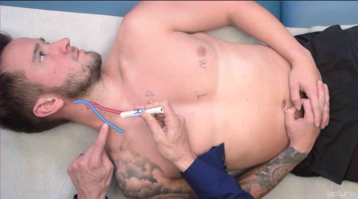
Evaluate for JVD:

Evaluation of jugular venous distension

Evaluation of jugular venous distension (JVD) with the individual supine at 30–45 degrees

Image by Lecturio. License: CC BY-NC-SA 4.0

Extremities

  • Evaluate the fingers and nails for abnormalities:
    • Clubbing of the fingers suggests chronic hypoxia Hypoxia Sub-optimal oxygen levels in the ambient air of living organisms. Ischemic Cell Damage.
    • Cyanosis Cyanosis A bluish or purplish discoloration of the skin and mucous membranes due to an increase in the amount of deoxygenated hemoglobin in the blood or a structural defect in the hemoglobin molecule. Pulmonary Examination of nails suggests hypoxia Hypoxia Sub-optimal oxygen levels in the ambient air of living organisms. Ischemic Cell Damage.
    • Splinter hemorrhages Splinter hemorrhages Endocarditis in the nails suggest septic emboli.
    • Osler nodes Osler nodes Endocarditis:
      • Tender, red nodules on finger pulps
      • Indicates infective endocarditis Infective endocarditis Infective endocarditis (IE) is caused by infection or inflammation of the inner lining of the heart (endocardium), most commonly affecting the heart valves. Endocarditis
    • Janeway lesions Janeway lesions Endocarditis:
      • Nontender, maculopapular Maculopapular Dermatologic Examination, erythematous pulp lesions of the palm
      • Indicates bacterial endocarditis Bacterial endocarditis Inflammation of the endocardium caused by bacteria that entered the bloodstream. The strains of bacteria vary with predisposing factors, such as congenital heart defects; heart valve diseases; heart valve prosthesis implantation; or intravenous drug use. Endocarditis
    • Capillary refill time:
    • Tar stain:
      • From smoking Smoking Willful or deliberate act of inhaling and exhaling smoke from burning substances or agents held by hand. Interstitial Lung Diseases
      • Indicates risk factors for cardiovascular disease
  • Cool peripheries may suggest poor cardiac output Cardiac output The volume of blood passing through the heart per unit of time. It is usually expressed as liters (volume) per minute so as not to be confused with stroke volume (volume per beat). Cardiac Mechanics/ hypovolemia Hypovolemia Sepsis in Children.
  • Sweaty/clammy skin Skin The skin, also referred to as the integumentary system, is the largest organ of the body. The skin is primarily composed of the epidermis (outer layer) and dermis (deep layer). The epidermis is primarily composed of keratinocytes that undergo rapid turnover, while the dermis contains dense layers of connective tissue. Skin: Structure and Functions can be associated with acute coronary syndrome.
  • Peripheral edema Peripheral edema Peripheral edema is the swelling of the lower extremities, namely, legs, feet, and ankles. Edema is a sign of heart failure Heart Failure A heterogeneous condition in which the heart is unable to pump out sufficient blood to meet the metabolic need of the body. Heart failure can be caused by structural defects, functional abnormalities (ventricular dysfunction), or a sudden overload beyond its capacity. Chronic heart failure is more common than acute heart failure which results from sudden insult to cardiac function, such as myocardial infarction. Total Anomalous Pulmonary Venous Return (TAPVR).

Palpation

The cardiovascular examination includes palpation Palpation Application of fingers with light pressure to the surface of the body to determine consistency of parts beneath in physical diagnosis; includes palpation for determining the outlines of organs. Dermatologic Examination of the pulses and the chest wall Chest wall The chest wall consists of skin, fat, muscles, bones, and cartilage. The bony structure of the chest wall is composed of the ribs, sternum, and thoracic vertebrae. The chest wall serves as armor for the vital intrathoracic organs and provides the stability necessary for the movement of the shoulders and arms. Chest Wall: Anatomy (precordium).

Pulses

Note the rate, rhythm, strength, character, and compare with contralateral pulses.
Precordium

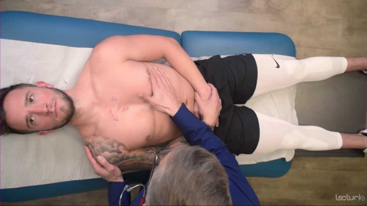
  • Place the right hand Hand The hand constitutes the distal part of the upper limb and provides the fine, precise movements needed in activities of daily living. It consists of 5 metacarpal bones and 14 phalanges, as well as numerous muscles innervated by the median and ulnar nerves. Hand: Anatomy across the individual’s left chest so it covers the area over the heart:
    • Rest the heel of the right hand Hand The hand constitutes the distal part of the upper limb and provides the fine, precise movements needed in activities of daily living. It consists of 5 metacarpal bones and 14 phalanges, as well as numerous muscles innervated by the median and ulnar nerves. Hand: Anatomy along the sternal border.
    • Lay the extended fingers below the left nipple Nipple The conic organs which usually give outlet to milk from the mammary glands. Examination of the Breast.
  • Identify the point of maximum impulse (where the heart beats the hardest):
    • A normal functioning ventricle will generate an impulse 2–3 cm in size. 
    • Best felt in the midclavicular line at the 5th intercostal space
    • A regular Regular Insulin, single, nonsustained tapping should be felt.
  • Check for thrills or heave/thrust:
Palpation of the precordium, with the heel of the hand over the sternal border and fingers below the left nipple

Palpation of the precordium: the heel of the hand over the sternal border with fingers below the left nipple

Image by Lecturio. License: CC BY-NC-SA 4.0

Auscultation

Auscultation is best performed on bare skin Skin The skin, also referred to as the integumentary system, is the largest organ of the body. The skin is primarily composed of the epidermis (outer layer) and dermis (deep layer). The epidermis is primarily composed of keratinocytes that undergo rapid turnover, while the dermis contains dense layers of connective tissue. Skin: Structure and Functions. Maintain the individual’s modesty while performing examination maneuvers on the chest.
Technique

  • Requires use of both the bell and the diaphragm Diaphragm The diaphragm is a large, dome-shaped muscle that separates the thoracic cavity from the abdominal cavity. The diaphragm consists of muscle fibers and a large central tendon, which is divided into right and left parts. As the primary muscle of inspiration, the diaphragm contributes 75% of the total inspiratory muscle force. Diaphragm: Anatomy of the stethoscope
  • The bell auscultates low-pitched sounds:
    • Bruits (nonlaminar flow Flow Blood flows through the heart, arteries, capillaries, and veins in a closed, continuous circuit. Flow is the movement of volume per unit of time. Flow is affected by the pressure gradient and the resistance fluid encounters between 2 points. Vascular resistance is the opposition to flow, which is caused primarily by blood friction against vessel walls. Vascular Resistance, Flow, and Mean Arterial Pressure associated with stenosis Stenosis Hypoplastic Left Heart Syndrome (HLHS))
    • Some heart murmurs Heart murmurs Heart sounds caused by vibrations resulting from the flow of blood through the heart. Heart murmurs can be examined by heart auscultation, and analyzed by their intensity (6 grades), duration, timing (systolic, diastolic, or continuous), location, transmission, and quality (musical, vibratory, blowing, etc). Heart Sounds
  • The diaphragm Diaphragm The diaphragm is a large, dome-shaped muscle that separates the thoracic cavity from the abdominal cavity. The diaphragm consists of muscle fibers and a large central tendon, which is divided into right and left parts. As the primary muscle of inspiration, the diaphragm contributes 75% of the total inspiratory muscle force. Diaphragm: Anatomy is used for all other sounds.
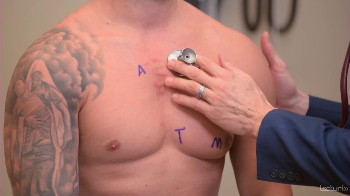
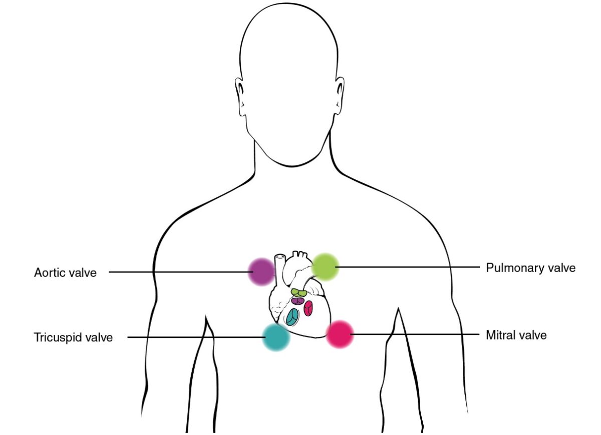
  • There are 4 points of auscultation of the precordium.
  • Mnemonic: APT-M:
    • A ( aortic valve Aortic valve The valve between the left ventricle and the ascending aorta which prevents backflow into the left ventricle. Heart: Anatomy): right 2nd intercostal space
    • P (pulmonic valve): left 2nd intercostal space 
    • T ( tricuspid valve Tricuspid valve The valve consisting of three cusps situated between the right atrium and right ventricle of the heart. Heart: Anatomy): left 4th intercostal space
    • M ( mitral valve Mitral valve The valve between the left atrium and left ventricle of the heart. Heart: Anatomy): lateral aspect of the 5th intercostal space

Heart sounds Heart sounds Heart sounds are brief, transient sounds produced by valve opening and closure and by movement of blood in the heart. They are divided into systolic and diastolic sounds. In most cases, only the first (S1) and second (S2) heart sounds are heard. These are high-frequency sounds and arise from aortic and pulmonary valve closure (S1), as well as mitral and tricuspid valve closure (S2). Heart Sounds

  • S1 S1 Heart Sounds (lub) is the 1st heart sound and marks the beginning of systole Systole Period of contraction of the heart, especially of the heart ventricles. Cardiac Cycle and the end of diastole Diastole Post-systolic relaxation of the heart, especially the heart ventricles. Cardiac Cycle:
    • Sound of the closure of the mitral and tricuspid valves
    • Loudest at the apex
    • Precedes the carotid pulse
  • S2 S2 Heart Sounds (dub) is the 2nd heart sound and marks the end of systole Systole Period of contraction of the heart, especially of the heart ventricles. Cardiac Cycle and the beginning of diastole Diastole Post-systolic relaxation of the heart, especially the heart ventricles. Cardiac Cycle:
    • Sound of the closure of the aortic and pulmonic valves
    • Loudest at the base
    • After the carotid pulse
  • Extra heart sounds Heart sounds Heart sounds are brief, transient sounds produced by valve opening and closure and by movement of blood in the heart. They are divided into systolic and diastolic sounds. In most cases, only the first (S1) and second (S2) heart sounds are heard. These are high-frequency sounds and arise from aortic and pulmonary valve closure (S1), as well as mitral and tricuspid valve closure (S2). Heart Sounds:
    • S3 S3 Heart Sounds is the 3rd heart sound (ventricular gallop):
      • Occurs just after S2 S2 Heart Sounds when the mitral valve Mitral valve The valve between the left atrium and left ventricle of the heart. Heart: Anatomy opens
      • Produced by a large amount of blood striking a compliant left ventricle (LV)
      • S3 S3 Heart Sounds is often heard in the setting of heart failure Heart Failure A heterogeneous condition in which the heart is unable to pump out sufficient blood to meet the metabolic need of the body. Heart failure can be caused by structural defects, functional abnormalities (ventricular dysfunction), or a sudden overload beyond its capacity. Chronic heart failure is more common than acute heart failure which results from sudden insult to cardiac function, such as myocardial infarction. Total Anomalous Pulmonary Venous Return (TAPVR) with reduced ejection fraction Ejection fraction Cardiac Cycle.
    • S4 S4 Heart Sounds is the 4th heart sound (atrial gallop):
      • Occurs just before S1 S1 Heart Sounds
      • Sound of the atria contracting to force blood into a noncompliant LV 
      • S4 S4 Heart Sounds is often heard in the setting of acute cardiovascular ischemia Ischemia A hypoperfusion of the blood through an organ or tissue caused by a pathologic constriction or obstruction of its blood vessels, or an absence of blood circulation. Ischemic Cell Damage.
    • Rubs:
      • The sounds produced with inflammation Inflammation Inflammation is a complex set of responses to infection and injury involving leukocytes as the principal cellular mediators in the body’s defense against pathogenic organisms. Inflammation is also seen as a response to tissue injury in the process of wound healing. The 5 cardinal signs of inflammation are pain, heat, redness, swelling, and loss of function. Inflammation of the parietal Parietal One of a pair of irregularly shaped quadrilateral bones situated between the frontal bone and occipital bone, which together form the sides of the cranium. Skull: Anatomy and visceral pericardium Visceral pericardium Heart: Anatomy.
      • A creaky/scratchy noise
  • Systolic murmurs Systolic Murmurs Heart murmurs which are systolic in timing. They occur between the first and the second heart sounds, between the closure of mitral valve and tricuspid valve and the closure of semilunar aortic and pulmonary valves. Systolic murmurs include ejection murmurs and regurgitant murmurs. Heart Sounds:
  • Diastolic murmurs Diastolic Murmurs Heart Sounds:

Audio:

In this audio clip, the normal S1 and S2 heart sounds are heard (normal S1, S2, sitting, listening with bell of the stethoscope). S1 corresponds to the closure of the atrioventricular valves, marking the beginning of systole. S2 corresponds to the closure of the semilunar valves, marking the beginning of diastole.
In this audio clip, the S3 gallop is heard. S3 occurs after S2, during the rapid filling phase of ventricular diastole.
In this audio clip, the S4 gallop is heard. S4 occurs before S1, during the atrial filling phase. S4 is heard in conditions where there is stiffness or low compliance in the ventricle.
This audio clip is an example of severe aortic stenosis. This is a harsh, crescendo-decrescendo murmur occurring between S1 and S2. Due to the severity of the aortic stenosis, the S2 heart sound is inaudible.
This audio clip is an example of a mitral stenosis murmur. The low-pitched, rumbling murmur starts after the opening snap of the mitral valve (after S2), and finishes with a short crescendo up to S1.

Special Maneuvers

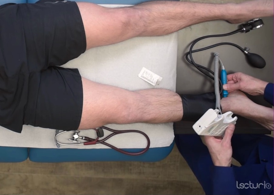
Ankle-brachial index

  • The ankle-brachial index is measured to screen for peripheral artery disease Peripheral artery disease Peripheral artery disease (PAD) is obstruction of the arterial lumen resulting in decreased blood flow to the distal limbs. The disease can be a result of atherosclerosis or thrombosis. Patients may be asymptomatic or have progressive claudication, skin discoloration, ischemic ulcers, or gangrene. Peripheral Artery Disease.
  • Measure systolic blood pressure in bilateral ankles and arms.
  • Divide each ankle pressure by the highest systolic pressure; the ratio determines the presence or absence of peripheral artery disease Peripheral artery disease Peripheral artery disease (PAD) is obstruction of the arterial lumen resulting in decreased blood flow to the distal limbs. The disease can be a result of atherosclerosis or thrombosis. Patients may be asymptomatic or have progressive claudication, skin discoloration, ischemic ulcers, or gangrene. Peripheral Artery Disease.
Table: Ankle-brachial index interpretation
Ankle-brachial index ratio Interpretation
> 1.4 Vessel hardening
1.0–1.4 Normal
0.9–1.0 Acceptable
0.8–0.9 Mild arterial disease
0.5–0.8 Moderate arterial disease
< 0.5 Severe arterial disease
Measuring the systolic pressure at the ankle to calculate the ankle brachial index

Measuring the systolic pressure at the ankle to calculate the ankle-brachial index

Image by Lecturio. License: CC BY-NC-SA 4.0

Clinical Relevance

  • Peripheral artery disease Peripheral artery disease Peripheral artery disease (PAD) is obstruction of the arterial lumen resulting in decreased blood flow to the distal limbs. The disease can be a result of atherosclerosis or thrombosis. Patients may be asymptomatic or have progressive claudication, skin discoloration, ischemic ulcers, or gangrene. Peripheral Artery Disease: obstruction of the arterial lumen resulting in decreased blood flow Blood flow Blood flow refers to the movement of a certain volume of blood through the vasculature over a given unit of time (e.g., mL per minute). Vascular Resistance, Flow, and Mean Arterial Pressure to the distal limbs, which can be a result of atherosclerosis Atherosclerosis Atherosclerosis is a common form of arterial disease in which lipid deposition forms a plaque in the blood vessel walls. Atherosclerosis is an incurable disease, for which there are clearly defined risk factors that often can be reduced through a change in lifestyle and behavior of the patient. Atherosclerosis or thrombosis Thrombosis Formation and development of a thrombus or blood clot in the blood vessel. Epidemic Typhus. Individuals may be either asymptomatic or have progressive claudication, skin Skin The skin, also referred to as the integumentary system, is the largest organ of the body. The skin is primarily composed of the epidermis (outer layer) and dermis (deep layer). The epidermis is primarily composed of keratinocytes that undergo rapid turnover, while the dermis contains dense layers of connective tissue. Skin: Structure and Functions discoloration, ischemic ulcers, or gangrene Gangrene Death and putrefaction of tissue usually due to a loss of blood supply. Small Bowel Obstruction. The diagnosis can be made by assessing the ankle-brachial index. Treatment includes lifestyle modifications and antiplatelet therapy.
  • Heart failure Heart Failure A heterogeneous condition in which the heart is unable to pump out sufficient blood to meet the metabolic need of the body. Heart failure can be caused by structural defects, functional abnormalities (ventricular dysfunction), or a sudden overload beyond its capacity. Chronic heart failure is more common than acute heart failure which results from sudden insult to cardiac function, such as myocardial infarction. Total Anomalous Pulmonary Venous Return (TAPVR): refers to the inability of the heart to supply the body with normal cardiac output Cardiac output The volume of blood passing through the heart per unit of time. It is usually expressed as liters (volume) per minute so as not to be confused with stroke volume (volume per beat). Cardiac Mechanics to meet metabolic needs. Heart failure Heart Failure A heterogeneous condition in which the heart is unable to pump out sufficient blood to meet the metabolic need of the body. Heart failure can be caused by structural defects, functional abnormalities (ventricular dysfunction), or a sudden overload beyond its capacity. Chronic heart failure is more common than acute heart failure which results from sudden insult to cardiac function, such as myocardial infarction. Total Anomalous Pulmonary Venous Return (TAPVR) is often a result of chronic hypertension Hypertension Hypertension, or high blood pressure, is a common disease that manifests as elevated systemic arterial pressures. Hypertension is most often asymptomatic and is found incidentally as part of a routine physical examination or during triage for an unrelated medical encounter. Hypertension or ischemic heart disease Ischemic heart disease Coronary heart disease (CHD), or ischemic heart disease, describes a situation in which an inadequate supply of blood to the myocardium exists due to a stenosis of the coronary arteries, typically from atherosclerosis. Coronary Heart Disease. The diagnosis can be suggested by physical exam findings of peripheral edema Peripheral edema Peripheral edema is the swelling of the lower extremities, namely, legs, feet, and ankles. Edema, increased JVD, and crackles in the lungs Lungs Lungs are the main organs of the respiratory system. Lungs are paired viscera located in the thoracic cavity and are composed of spongy tissue. The primary function of the lungs is to oxygenate blood and eliminate CO2. Lungs: Anatomy. Treatment includes lifestyle modifications and antihypertensive medications (most commonly ACE inhibitors ACE inhibitors Truncus Arteriosus and beta blockers).
  • Acute coronary syndrome: refers to acute ischemic heart disease Ischemic heart disease Coronary heart disease (CHD), or ischemic heart disease, describes a situation in which an inadequate supply of blood to the myocardium exists due to a stenosis of the coronary arteries, typically from atherosclerosis. Coronary Heart Disease resulting in unstable angina Unstable angina Precordial pain at rest, which may precede a myocardial infarction. Stable and Unstable Angina or myocardial infarction Myocardial infarction MI is ischemia and death of an area of myocardial tissue due to insufficient blood flow and oxygenation, usually from thrombus formation on a ruptured atherosclerotic plaque in the epicardial arteries. Clinical presentation is most commonly with chest pain, but women and patients with diabetes may have atypical symptoms. Myocardial Infarction. Individuals present with acute onset of chest pain Pain An unpleasant sensation induced by noxious stimuli which are detected by nerve endings of nociceptive neurons. Pain: Types and Pathways not relieved by rest. The diagnosis can be made by obtaining an electrocardiogram Electrocardiogram An electrocardiogram (ECG) is a graphic representation of the electrical activity of the heart plotted against time. Adhesive electrodes are affixed to the skin surface allowing measurement of cardiac impulses from many angles. The ECG provides 3-dimensional information about the conduction system of the heart, the myocardium, and other cardiac structures. Electrocardiogram (ECG) and measuring serum levels of cardiac enzymes Enzymes Enzymes are complex protein biocatalysts that accelerate chemical reactions without being consumed by them. Due to the body’s constant metabolic needs, the absence of enzymes would make life unsustainable, as reactions would occur too slowly without these molecules. Basics of Enzymes (e.g., troponin). Acute coronary syndrome is a medical emergency and rapid treatment should be geared towards revascularization Revascularization Thromboangiitis Obliterans (Buerger’s Disease) of the occluded area. 
  • Pericarditis Pericarditis Pericarditis is an inflammation of the pericardium, often with fluid accumulation. It can be caused by infection (often viral), myocardial infarction, drugs, malignancies, metabolic disorders, autoimmune disorders, or trauma. Acute, subacute, and chronic forms exist. Pericarditis: refers to inflammation Inflammation Inflammation is a complex set of responses to infection and injury involving leukocytes as the principal cellular mediators in the body’s defense against pathogenic organisms. Inflammation is also seen as a response to tissue injury in the process of wound healing. The 5 cardinal signs of inflammation are pain, heat, redness, swelling, and loss of function. Inflammation of the pericardium Pericardium A conical fibroserous sac surrounding the heart and the roots of the great vessels (aorta; venae cavae; pulmonary artery). Pericardium consists of two sacs: the outer fibrous pericardium and the inner serous pericardium. The latter consists of an outer parietal layer facing the fibrous pericardium, and an inner visceral layer (epicardium) resting next to the heart, and a pericardial cavity between these two layers. Heart: Anatomy. Pericarditis Pericarditis Pericarditis is an inflammation of the pericardium, often with fluid accumulation. It can be caused by infection (often viral), myocardial infarction, drugs, malignancies, metabolic disorders, autoimmune disorders, or trauma. Acute, subacute, and chronic forms exist. Pericarditis is often an idiopathic Idiopathic Dermatomyositis condition but can develop after myocardial infarction Myocardial infarction MI is ischemia and death of an area of myocardial tissue due to insufficient blood flow and oxygenation, usually from thrombus formation on a ruptured atherosclerotic plaque in the epicardial arteries. Clinical presentation is most commonly with chest pain, but women and patients with diabetes may have atypical symptoms. Myocardial Infarction. Individuals present with pleuritic chest pain Pain An unpleasant sensation induced by noxious stimuli which are detected by nerve endings of nociceptive neurons. Pain: Types and Pathways and friction rub, which can be heard while auscultating the heart. The chest pain Pain An unpleasant sensation induced by noxious stimuli which are detected by nerve endings of nociceptive neurons. Pain: Types and Pathways classically improves when the individual leans forward. Antiinflammatory medications can improve the severity of chest pain Pain An unpleasant sensation induced by noxious stimuli which are detected by nerve endings of nociceptive neurons. Pain: Types and Pathways.
  • Cardiac tamponade Tamponade Pericardial effusion, usually of rapid onset, exceeding ventricular filling pressures and causing collapse of the heart with a markedly reduced cardiac output. Pericarditis: occurs when fluid accumulates around the heart, ultimately compressing the heart and reducing cardiac output Cardiac output The volume of blood passing through the heart per unit of time. It is usually expressed as liters (volume) per minute so as not to be confused with stroke volume (volume per beat). Cardiac Mechanics. Individuals classically present with Beck triad: hypotension Hypotension Hypotension is defined as low blood pressure, specifically < 90/60 mm Hg, and is most commonly a physiologic response. Hypotension may be mild, serious, or life threatening, depending on the cause. Hypotension, muffled heart sounds Muffled Heart Sounds Pericardial Effusion and Cardiac Tamponade, and increased JVD. Cardiac tamponade Tamponade Pericardial effusion, usually of rapid onset, exceeding ventricular filling pressures and causing collapse of the heart with a markedly reduced cardiac output. Pericarditis is a medical emergency and must be treated with pericardiocentesis Pericardiocentesis Puncture and aspiration of fluid from the pericardium. Cardiac Surgery or pericardiotomy to remove the accumulated fluid.

References

  1. Walker HK, Hall WD, Hurst JW. (1990). Clinical Methods: The History, Physical, and Laboratory Examinations. 3rd edition. Butterworths.
  2. Mangione S, Nieman LZ. (1997). Cardiac auscultatory skills of internal medicine and family practice trainees: A comparison of diagnostic proficiency. JAMA. 278: 717–22.
  3. Silverman ME, Wooley CF. (2008). Samuel A. Levine and the History of Grading Systolic Murmurs. Am J Cardiol. 102 (8): p.1107–10.
  4. Mangione S, Nieman LZ, Gracely E, Kaye D. (1993). The teaching and practice of cardiac auscultation during internal medicine and cardiology training: nationwide survey. Ann Intern Med 119: 47–54.
  5. Nardone, Lucan LM, Palac DM. (1998). Physical examination: A revered skill under scrutiny. Southern Medical Journal 81: 770–73.

Create your free account or log in to continue reading!

Sign up now and get free access to Lecturio with concept pages, medical videos, and questions for your medical education.

User Reviews

Details