Cardiopulmonary Resuscitation (CPR)

Cardiopulmonary resuscitation Resuscitation The restoration to life or consciousness of one apparently dead. . Neonatal Respiratory Distress Syndrome ( CPR CPR The artificial substitution of heart and lung action as indicated for heart arrest resulting from electric shock, drowning, respiratory arrest, or other causes. The two major components of cardiopulmonary resuscitation are artificial ventilation and closed-chest cardiac massage. Cardiac Arrest) is an emergency procedure used in patients Patients Individuals participating in the health care system for the purpose of receiving therapeutic, diagnostic, or preventive procedures. Clinician–Patient Relationship with cardiac arrest Cardiac arrest Cardiac arrest is the sudden, complete cessation of cardiac output with hemodynamic collapse. Patients present as pulseless, unresponsive, and apneic. Rhythms associated with cardiac arrest are ventricular fibrillation/tachycardia, asystole, or pulseless electrical activity. Cardiac Arrest. Cardiopulmonary resuscitation Resuscitation The restoration to life or consciousness of one apparently dead. . Neonatal Respiratory Distress Syndrome combines the use of chest compressions, artificial ventilation Ventilation The total volume of gas inspired or expired per unit of time, usually measured in liters per minute. Ventilation: Mechanics of Breathing, and, when available, an automatic external defibrillator Defibrillator Cardiac electrical stimulators that apply brief high-voltage electroshocks to the heart. These stimulators are used to restore normal rhythm and contractile function in hearts of patients who are experiencing ventricular fibrillation or ventricular tachycardia that is not accompanied by a palpable pulse. Some defibrillators may also be used to correct certain noncritical dysrhythmias (called synchronized defibrillation or cardioversion), using relatively low-level discharges synchronized to the patient's ECG waveform. Cardiac Arrest (AED) to maintain circulatory flow Flow Blood flows through the heart, arteries, capillaries, and veins in a closed, continuous circuit. Flow is the movement of volume per unit of time. Flow is affected by the pressure gradient and the resistance fluid encounters between 2 points. Vascular resistance is the opposition to flow, which is caused primarily by blood friction against vessel walls. Vascular Resistance, Flow, and Mean Arterial Pressure and oxygenation to vital structures. Cardiopulmonary resuscitation Resuscitation The restoration to life or consciousness of one apparently dead. . Neonatal Respiratory Distress Syndrome is an integral part of basic life support Basic Life Support Airway Management ( BLS BLS Airway Management) and advanced cardiovascular life support (ACLS). High-quality CPR CPR The artificial substitution of heart and lung action as indicated for heart arrest resulting from electric shock, drowning, respiratory arrest, or other causes. The two major components of cardiopulmonary resuscitation are artificial ventilation and closed-chest cardiac massage. Cardiac Arrest improves the likelihood of survival. Some patients Patients Individuals participating in the health care system for the purpose of receiving therapeutic, diagnostic, or preventive procedures. Clinician–Patient Relationship in critical situations request a do not resuscitate (DNR) order, which instructs health care providers not to do CPR CPR The artificial substitution of heart and lung action as indicated for heart arrest resulting from electric shock, drowning, respiratory arrest, or other causes. The two major components of cardiopulmonary resuscitation are artificial ventilation and closed-chest cardiac massage. Cardiac Arrest if a patient suffers cardiac arrest Cardiac arrest Cardiac arrest is the sudden, complete cessation of cardiac output with hemodynamic collapse. Patients present as pulseless, unresponsive, and apneic. Rhythms associated with cardiac arrest are ventricular fibrillation/tachycardia, asystole, or pulseless electrical activity. Cardiac Arrest.

Last updated: Jan 17, 2023

Editorial responsibility: Stanley Oiseth, Lindsay Jones, Evelin Maza

Overview

Introduction

  • Annual incidence Incidence The number of new cases of a given disease during a given period in a specified population. It also is used for the rate at which new events occur in a defined population. It is differentiated from prevalence, which refers to all cases in the population at a given time. Measures of Disease Frequency of sudden cardiac arrest Cardiac arrest Cardiac arrest is the sudden, complete cessation of cardiac output with hemodynamic collapse. Patients present as pulseless, unresponsive, and apneic. Rhythms associated with cardiac arrest are ventricular fibrillation/tachycardia, asystole, or pulseless electrical activity. Cardiac Arrest in the United States: 180,000–450,000 +
  • Cardiopulmonary resuscitation Resuscitation The restoration to life or consciousness of one apparently dead. . Neonatal Respiratory Distress Syndrome ( CPR CPR The artificial substitution of heart and lung action as indicated for heart arrest resulting from electric shock, drowning, respiratory arrest, or other causes. The two major components of cardiopulmonary resuscitation are artificial ventilation and closed-chest cardiac massage. Cardiac Arrest) is attempted in approximately ⅔ of cases.
  • Early initiation of high-quality CPR CPR The artificial substitution of heart and lung action as indicated for heart arrest resulting from electric shock, drowning, respiratory arrest, or other causes. The two major components of cardiopulmonary resuscitation are artificial ventilation and closed-chest cardiac massage. Cardiac Arrest has been shown to dramatically increase survival rate.

Cardiopulmonary resuscitation Resuscitation The restoration to life or consciousness of one apparently dead. . Neonatal Respiratory Distress Syndrome sequence overview

Cardiopulmonary resuscitation Resuscitation The restoration to life or consciousness of one apparently dead. . Neonatal Respiratory Distress Syndrome consists of chest compressions and rescue breaths carried out sequentially. 
  • Identification Identification Defense Mechanisms of cardiac arrest Cardiac arrest Cardiac arrest is the sudden, complete cessation of cardiac output with hemodynamic collapse. Patients present as pulseless, unresponsive, and apneic. Rhythms associated with cardiac arrest are ventricular fibrillation/tachycardia, asystole, or pulseless electrical activity. Cardiac Arrest:
    • Patient seen losing consciousness or found unconscious Unconscious Those forces and content of the mind which are not ordinarily available to conscious awareness or to immediate recall. Psychotherapy 
    • Assess scene for safety.
    • Feel pulse and assess breathing simultaneously:
  • Prepare for CPR CPR The artificial substitution of heart and lung action as indicated for heart arrest resulting from electric shock, drowning, respiratory arrest, or other causes. The two major components of cardiopulmonary resuscitation are artificial ventilation and closed-chest cardiac massage. Cardiac Arrest:
    • Activation of emergency response system:
      • Outside hospital, call 911.
      • In hospital, call cardiac arrest Cardiac arrest Cardiac arrest is the sudden, complete cessation of cardiac output with hemodynamic collapse. Patients present as pulseless, unresponsive, and apneic. Rhythms associated with cardiac arrest are ventricular fibrillation/tachycardia, asystole, or pulseless electrical activity. Cardiac Arrest code.
    • Obtain automatic external defibrillator Defibrillator Cardiac electrical stimulators that apply brief high-voltage electroshocks to the heart. These stimulators are used to restore normal rhythm and contractile function in hearts of patients who are experiencing ventricular fibrillation or ventricular tachycardia that is not accompanied by a palpable pulse. Some defibrillators may also be used to correct certain noncritical dysrhythmias (called synchronized defibrillation or cardioversion), using relatively low-level discharges synchronized to the patient’s ECG waveform. Cardiac Arrest (AED), if available.
  • If pulse is present, but no breathing:
    • Provide rescue breath (1 every 6 seconds).
    • Recheck pulse every 2 minutes.
  • If no pulse present:
    • Start with chest compressions before giving rescue breath.
    • If trained in CPR CPR The artificial substitution of heart and lung action as indicated for heart arrest resulting from electric shock, drowning, respiratory arrest, or other causes. The two major components of cardiopulmonary resuscitation are artificial ventilation and closed-chest cardiac massage. Cardiac Arrest, initiate CPR CPR The artificial substitution of heart and lung action as indicated for heart arrest resulting from electric shock, drowning, respiratory arrest, or other causes. The two major components of cardiopulmonary resuscitation are artificial ventilation and closed-chest cardiac massage. Cardiac Arrest at rate of 30 chest compressions followed by 2 rescue breaths (30:2).
    • If sole lay rescuer, compression-only CPR CPR The artificial substitution of heart and lung action as indicated for heart arrest resulting from electric shock, drowning, respiratory arrest, or other causes. The two major components of cardiopulmonary resuscitation are artificial ventilation and closed-chest cardiac massage. Cardiac Arrest ( CPR CPR The artificial substitution of heart and lung action as indicated for heart arrest resulting from electric shock, drowning, respiratory arrest, or other causes. The two major components of cardiopulmonary resuscitation are artificial ventilation and closed-chest cardiac massage. Cardiac Arrest without mouth-to-mouth breaths)
    • Apply AED as soon as available.
    • Continue CPR CPR The artificial substitution of heart and lung action as indicated for heart arrest resulting from electric shock, drowning, respiratory arrest, or other causes. The two major components of cardiopulmonary resuscitation are artificial ventilation and closed-chest cardiac massage. Cardiac Arrest and deliver shocks as directed by AED.

Related videos

Rescue Breaths

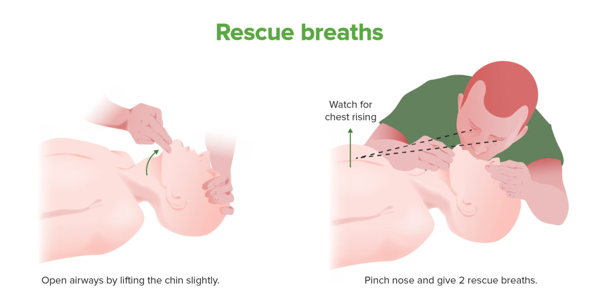
Rescue breath technique

  • If possible, use face shield or other form of personal protective equipment Personal protective equipment Specialized clothing or equipment worn for protection against health hazards. Personal protective equipment may include masks; respiratory protective devices; head protective devices; eye protective devices; ear protective devices; protective clothing; and protective footwear. Leptospira/Leptospirosis (PPE).
  • Clear mouth of objects before rescue breaths. 
  • Tilt head back slightly.
  • Lift chin Chin The anatomical frontal portion of the mandible, also known as the mentum, that contains the line of fusion of the two separate halves of the mandible (symphysis menti). This line of fusion divides inferiorly to enclose a triangular area called the mental protuberance. On each side, inferior to the second premolar tooth, is the mental foramen for the passage of blood vessels and a nerve. Melasma
  • Pinch nose Nose The nose is the human body’s primary organ of smell and functions as part of the upper respiratory system. The nose may be best known for inhaling oxygen and exhaling carbon dioxide, but it also contributes to other important functions, such as tasting. The anatomy of the nose can be divided into the external nose and the nasal cavity. Nose Anatomy (External & Internal).
  • Place your mouth fully over theirs. 
  • Blow hard enough to get chest rise (retilt head if chest does not rise).
Rescue breath technique

Rescue breath technique
Rescue breaths should be provided to apneic patients during CPR. Chest compressions should take precedence, especially in children, over rescue breaths.

Image by Lecturio.

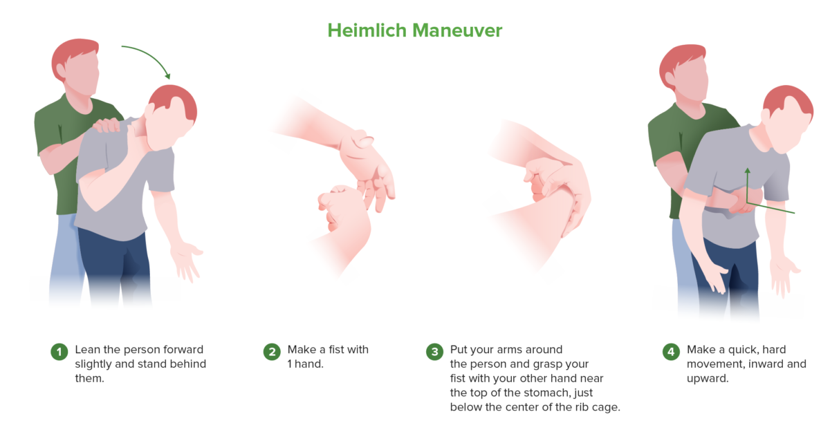
Heimlich maneuver Heimlich maneuver An emergency treatment commonly used to clear food and other foreign objects causing airway obstruction. Foreign Body Aspiration

If patient’s chest does not rise, foreign object may be obstructing airway Airway ABCDE Assessment:
  • With patient supine → open airway Airway ABCDE Assessment using jaw Jaw The jaw is made up of the mandible, which comprises the lower jaw, and the maxilla, which comprises the upper jaw. The mandible articulates with the temporal bone via the temporomandibular joint (TMJ). The 4 muscles of mastication produce the movements of the TMJ to ensure the efficient chewing of food. Jaw and Temporomandibular Joint: Anatomy lift →  if foreign body Foreign Body Foreign Body Aspiration is visible, remove object with finger sweep (do not perform blind finger sweep).
  • Attempt rescue breaths → if unsuccessful, retilt head and reattempt rescue breaths.
  • If no chest rise → kneel next to or straddle hips of patient → use 2 hands on top of each other and place heel of bottom hand Hand The hand constitutes the distal part of the upper limb and provides the fine, precise movements needed in activities of daily living. It consists of 5 metacarpal bones and 14 phalanges, as well as numerous muscles innervated by the median and ulnar nerves. Hand: Anatomy below xiphoid process Xiphoid process Chest Wall: Anatomy → press both hands into abdomen with quick upward thrust 5 times.
  • Open airway Airway ABCDE Assessment with jaw Jaw The jaw is made up of the mandible, which comprises the lower jaw, and the maxilla, which comprises the upper jaw. The mandible articulates with the temporal bone via the temporomandibular joint (TMJ). The 4 muscles of mastication produce the movements of the TMJ to ensure the efficient chewing of food. Jaw and Temporomandibular Joint: Anatomy lift → if foreign body Foreign Body Foreign Body Aspiration is visible, remove it → if not, repeat Heimlich maneuver Heimlich maneuver An emergency treatment commonly used to clear food and other foreign objects causing airway obstruction. Foreign Body Aspiration until ventilation Ventilation The total volume of gas inspired or expired per unit of time, usually measured in liters per minute. Ventilation: Mechanics of Breathing is successful.
Heimlich maneuver

Heimlich maneuver
In a choking patient or an unconscious patient on whom rescue breaths are not providing adequate ventilation, airway obstruction with a foreign object should be considered. The Heimlich maneuver works by producing positive pressure in the lungs, forcefully expelling any foreign body in the upper airway.
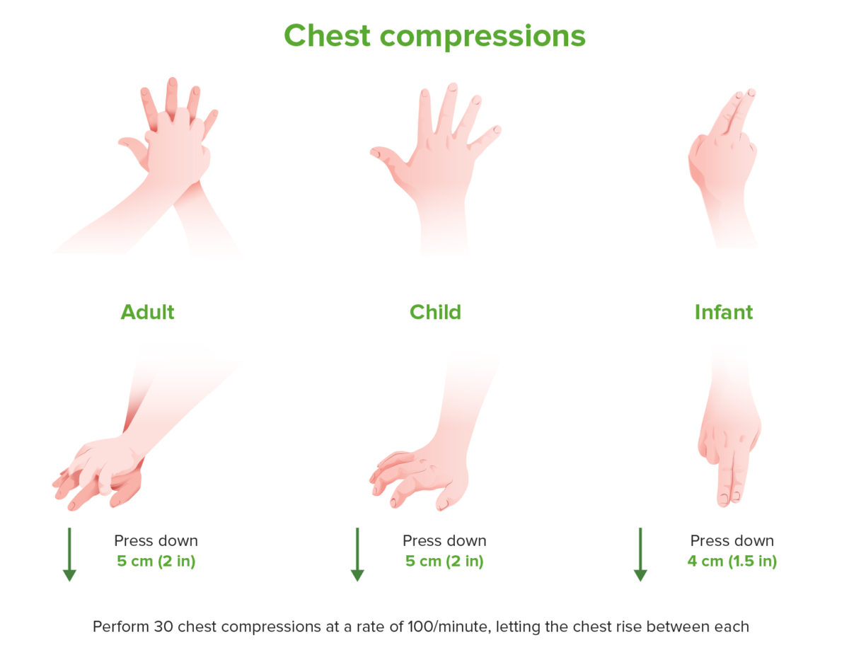
Image by Lecturio.

Chest Compressions

Positioning

  • Patient must be supine.
  • Patient must be lying on a firm surface.

Technique

  • In adults, use 2-hand chest compression Chest Compression Crush Syndrome technique:
    • 1 hand Hand The hand constitutes the distal part of the upper limb and provides the fine, precise movements needed in activities of daily living. It consists of 5 metacarpal bones and 14 phalanges, as well as numerous muscles innervated by the median and ulnar nerves. Hand: Anatomy on top of the other 
    • Place heel of bottom hand Hand The hand constitutes the distal part of the upper limb and provides the fine, precise movements needed in activities of daily living. It consists of 5 metacarpal bones and 14 phalanges, as well as numerous muscles innervated by the median and ulnar nerves. Hand: Anatomy over lower portion of the sternum Sternum A long, narrow, and flat bone commonly known as breastbone occurring in the midsection of the anterior thoracic segment or chest region, which stabilizes the rib cage and serves as the point of origin for several muscles that move the arms, head, and neck. Chest Wall: Anatomy, slightly below nipples.
  • Push “hard and fast”:
    • Appropriate rate: 100–120 compressions/min
    • Adequate depth: 5.0–7.6 cm (2–3 in) 
    • Allow full chest recoil Recoil Vessels can stretch and return to their original shape after receiving the stroke volume of blood ejected by the left ventricle during systole. Arteries: Histology.
Chest compressions

Hand placement in CPR for adults, children and infants
Hand positioning and depth of chest compression vary among adults, children and infants, while rate remains consistent.

Image by Lecturio.

Limit Limit A value (e.g., pressure or time) that should not be exceeded and which is specified by the operator to protect the lung Invasive Mechanical Ventilation pauses during CPR CPR The artificial substitution of heart and lung action as indicated for heart arrest resulting from electric shock, drowning, respiratory arrest, or other causes. The two major components of cardiopulmonary resuscitation are artificial ventilation and closed-chest cardiac massage. Cardiac Arrest

  • Pause only to give rescue breaths.
  • If possible, have other providers prepare to take turns performing compressions (change every 2 minutes).
  • Avoid hyperventilation Hyperventilation A pulmonary ventilation rate faster than is metabolically necessary for the exchange of gases. It is the result of an increased frequency of breathing, an increased tidal volume, or a combination of both. It causes an excess intake of oxygen and the blowing off of carbon dioxide. Respiratory Alkalosis: ventilation Ventilation The total volume of gas inspired or expired per unit of time, usually measured in liters per minute. Ventilation: Mechanics of Breathing rate around 6/min

Defibrillation

Early defibrillation Defibrillation Ventricular Fibrillation (V-fib) has been shown to significantly improve morbidity Morbidity The proportion of patients with a particular disease during a given year per given unit of population. Measures of Health Status and mortality Mortality All deaths reported in a given population. Measures of Health Status in ventricular fibrillation Ventricular fibrillation Ventricular fibrillation (VF or V-fib) is a type of ventricular tachyarrhythmia (> 300/min) often preceded by ventricular tachycardia. In this arrhythmia, the ventricle beats rapidly and sporadically. The ventricular contraction is uncoordinated, leading to a decrease in cardiac output and immediate hemodynamic collapse. Ventricular Fibrillation (V-fib) ( VF VF Ventricular fibrillation (VF or V-fib) is a type of ventricular tachyarrhythmia (> 300/min) often preceded by ventricular tachycardia. In this arrhythmia, the ventricle beats rapidly and sporadically. The ventricular contraction is uncoordinated, leading to a decrease in cardiac output and immediate hemodynamic collapse. Ventricular Fibrillation (V-fib))/ventricular tachycardia Tachycardia Abnormally rapid heartbeat, usually with a heart rate above 100 beats per minute for adults. Tachycardia accompanied by disturbance in the cardiac depolarization (cardiac arrhythmia) is called tachyarrhythmia. Sepsis in Children ( VT VT Ventricular tachycardia is any heart rhythm faster than 100 beats/min, with 3 or more irregular beats in a row, arising distal to the bundle of his. Ventricular tachycardia is the most common form of wide-complex tachycardia, and it is associated with a high mortality rate. Ventricular Tachycardia) cardiac arrest Cardiac arrest Cardiac arrest is the sudden, complete cessation of cardiac output with hemodynamic collapse. Patients present as pulseless, unresponsive, and apneic. Rhythms associated with cardiac arrest are ventricular fibrillation/tachycardia, asystole, or pulseless electrical activity. Cardiac Arrest.
Timing

  • Chest compressions should be initiated before placing defibrillator Defibrillator Cardiac electrical stimulators that apply brief high-voltage electroshocks to the heart. These stimulators are used to restore normal rhythm and contractile function in hearts of patients who are experiencing ventricular fibrillation or ventricular tachycardia that is not accompanied by a palpable pulse. Some defibrillators may also be used to correct certain noncritical dysrhythmias (called synchronized defibrillation or cardioversion), using relatively low-level discharges synchronized to the patient’s ECG waveform. Cardiac Arrest.
  • Rhythm analysis/ shock Shock Shock is a life-threatening condition associated with impaired circulation that results in tissue hypoxia. The different types of shock are based on the underlying cause: distributive (↑ cardiac output (CO), ↓ systemic vascular resistance (SVR)), cardiogenic (↓ CO, ↑ SVR), hypovolemic (↓ CO, ↑ SVR), obstructive (↓ CO), and mixed. Types of Shock should be performed as soon as possible without interrupting high-quality chest compression Chest Compression Crush Syndrome.
  • Ideal timing is to pause between switching providers; done every 2 minutes.

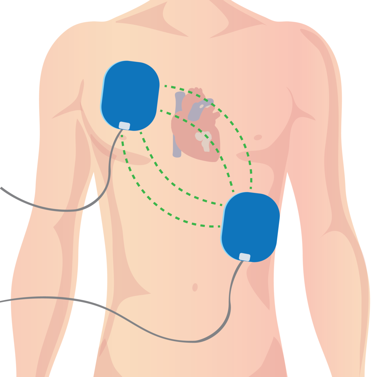
Positioning

  • Different models vary slightly, so follow instructions provided.
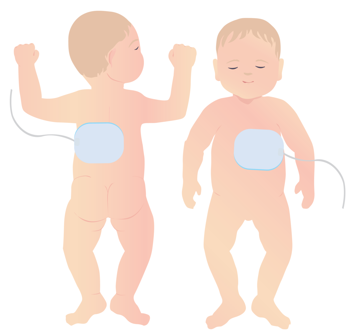
  • Usually, 1 pad is placed under the right collarbone and the other on the left side of the chest.

Analysis and shock Shock Shock is a life-threatening condition associated with impaired circulation that results in tissue hypoxia. The different types of shock are based on the underlying cause: distributive (↑ cardiac output (CO), ↓ systemic vascular resistance (SVR)), cardiogenic (↓ CO, ↑ SVR), hypovolemic (↓ CO, ↑ SVR), obstructive (↓ CO), and mixed. Types of Shock

  • Providers should not touch the patient during analysis/ shock Shock Shock is a life-threatening condition associated with impaired circulation that results in tissue hypoxia. The different types of shock are based on the underlying cause: distributive (↑ cardiac output (CO), ↓ systemic vascular resistance (SVR)), cardiogenic (↓ CO, ↑ SVR), hypovolemic (↓ CO, ↑ SVR), obstructive (↓ CO), and mixed. Types of Shock.
  • AED automatically analyzes patient rhythm and suggests shock Shock Shock is a life-threatening condition associated with impaired circulation that results in tissue hypoxia. The different types of shock are based on the underlying cause: distributive (↑ cardiac output (CO), ↓ systemic vascular resistance (SVR)), cardiogenic (↓ CO, ↑ SVR), hypovolemic (↓ CO, ↑ SVR), obstructive (↓ CO), and mixed. Types of Shock delivery and voltage.
  • Repeat analysis every 2-minute cycle.

Related videos

Differences in Children

Rescue breaths

  • Order of CPR CPR The artificial substitution of heart and lung action as indicated for heart arrest resulting from electric shock, drowning, respiratory arrest, or other causes. The two major components of cardiopulmonary resuscitation are artificial ventilation and closed-chest cardiac massage. Cardiac Arrest:
    • Cardiac arrest Cardiac arrest Cardiac arrest is the sudden, complete cessation of cardiac output with hemodynamic collapse. Patients present as pulseless, unresponsive, and apneic. Rhythms associated with cardiac arrest are ventricular fibrillation/tachycardia, asystole, or pulseless electrical activity. Cardiac Arrest in children is often due to hypoxia Hypoxia Sub-optimal oxygen levels in the ambient air of living organisms. Ischemic Cell Damage.
    • Perform 2 rescue breaths before starting compressions.
  • Differences in technique:
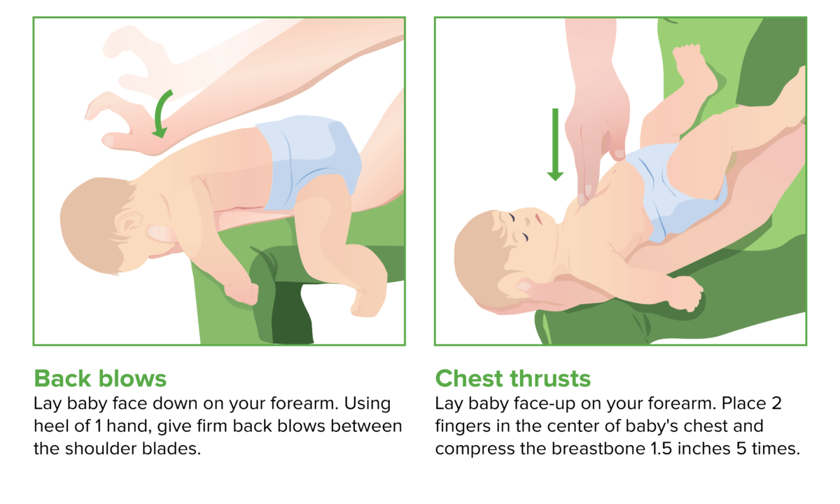
    • If choking suspected, do not use Heimlich maneuver Heimlich maneuver An emergency treatment commonly used to clear food and other foreign objects causing airway obstruction. Foreign Body Aspiration:
      • Use back blows in infants.
      • Use chest thrusts in small children.
    • Positioning of mouth:
      • For infants, place your mouth over their nose Nose The nose is the human body’s primary organ of smell and functions as part of the upper respiratory system. The nose may be best known for inhaling oxygen and exhaling carbon dioxide, but it also contributes to other important functions, such as tasting. The anatomy of the nose can be divided into the external nose and the nasal cavity. Nose Anatomy (External & Internal) and mouth. 
      • For children, pinch their nose Nose The nose is the human body’s primary organ of smell and functions as part of the upper respiratory system. The nose may be best known for inhaling oxygen and exhaling carbon dioxide, but it also contributes to other important functions, such as tasting. The anatomy of the nose can be divided into the external nose and the nasal cavity. Nose Anatomy (External & Internal) shut and place your mouth over theirs.
Back blows and chest thrusts

Back blows and chest thrusts (CPR in children)

Image by Lecturio.
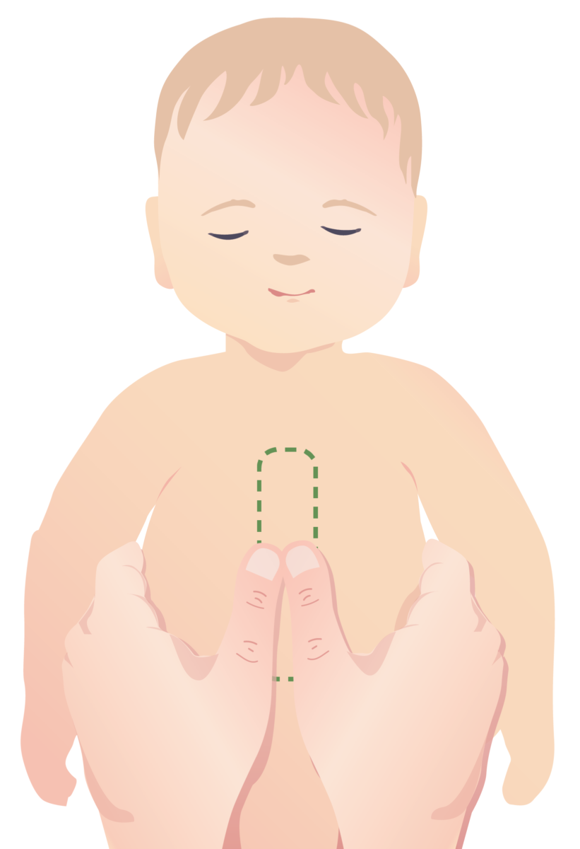
Chest compressions

  • In infants, use 2-finger technique or 2-thumb technique (when hands can fit around child).
  • In children, use 1-hand technique in center of chest.

Defibrillation Defibrillation Ventricular Fibrillation (V-fib)

  • Pediatric pads should be utilized:
    • Different size and shape
    • Different positioning
  • Voltage should be lowered, as specified by instructions in defibrillator Defibrillator Cardiac electrical stimulators that apply brief high-voltage electroshocks to the heart. These stimulators are used to restore normal rhythm and contractile function in hearts of patients who are experiencing ventricular fibrillation or ventricular tachycardia that is not accompanied by a palpable pulse. Some defibrillators may also be used to correct certain noncritical dysrhythmias (called synchronized defibrillation or cardioversion), using relatively low-level discharges synchronized to the patient’s ECG waveform. Cardiac Arrest.

Do Not Resuscitate (DNR)

A DNR order is a medical order, part of a patient’s advance directives Advance Directives The term advance directive (AD) refers to treatment preferences and/or the designation of a surrogate decision-maker in the event that a person becomes unable to make medical decisions on their own behalf. Advance directives represent the ethical principle of autonomy and may take the form of a living will, health care proxy, durable power of attorney for health care (DPAHC), and/or a physician’s order for life-sustaining treatment (POLST). Advance Directives.

Do not resuscitate orders prevent healthcare providers from performing the following procedures in case a patient’s breathing stops or heart rate Heart rate The number of times the heart ventricles contract per unit of time, usually per minute. Cardiac Physiology stops:

  • CPR CPR The artificial substitution of heart and lung action as indicated for heart arrest resulting from electric shock, drowning, respiratory arrest, or other causes. The two major components of cardiopulmonary resuscitation are artificial ventilation and closed-chest cardiac massage. Cardiac Arrest (chest compressions, artificial breathing)
  • Electrical cardioversion Cardioversion Atrial Fibrillation
  • Administering antiarrhythmic medications or cardiac resuscitation Resuscitation The restoration to life or consciousness of one apparently dead. . Neonatal Respiratory Distress Syndrome medications (e.g., epinephrine Epinephrine The active sympathomimetic hormone from the adrenal medulla. It stimulates both the alpha- and beta- adrenergic systems, causes systemic vasoconstriction and gastrointestinal relaxation, stimulates the heart, and dilates bronchi and cerebral vessels. Sympathomimetic Drugs)

Advance directives Advance Directives The term advance directive (AD) refers to treatment preferences and/or the designation of a surrogate decision-maker in the event that a person becomes unable to make medical decisions on their own behalf. Advance directives represent the ethical principle of autonomy and may take the form of a living will, health care proxy, durable power of attorney for health care (DPAHC), and/or a physician’s order for life-sustaining treatment (POLST). Advance Directives:

  • Patient’s ability to decide and reflect on their wishes about future medical care Medical care Conflict of Interest
  • Can be provided by family members (surrogate decision-maker)
  • Often includes more than a DNR order (e.g., vegetative state directives)

Being under a DNR order does not prohibit patients Patients Individuals participating in the health care system for the purpose of receiving therapeutic, diagnostic, or preventive procedures. Clinician–Patient Relationship from receiving appropriate medical care Medical care Conflict of Interest (surgeries, biopsies, dialysis Dialysis Renal replacement therapy refers to dialysis and/or kidney transplantation. Dialysis is a procedure by which toxins and excess water are removed from the circulation. Hemodialysis and peritoneal dialysis (PD) are the two types of dialysis, and their primary difference is the location of the filtration process (external to the body in hemodialysis versus inside the body for PD). Peritoneal Dialysis and Hemodialysis, blood samples, move to intensive care unit ( ICU ICU Hospital units providing continuous surveillance and care to acutely ill patients. West Nile Virus), etc ETC The electron transport chain (ETC) sends electrons through a series of proteins, which generate an electrochemical proton gradient that produces energy in the form of adenosine triphosphate (ATP). Electron Transport Chain (ETC).).

References

  1. Pozner, C. (2020). Basic life support (BLS) in adults. UpToDate. Retrieved December 10, 2020, from https://www.uptodate.com/contents/basic-life-support-bls-in-adults?search=CPR&source=search_result&selectedTitle=1~96&usage_type=default&display_rank=1
  2. Ralston, M. (2020). Pediatric basic life support (BLS) for health care providers. UpToDate. Retrieved December 10, 2020, from https://www.uptodate.com/contents/pediatric-basic-life-support-for-health-care-providers?search=CPR&usage_type=default&source=search_result&selectedTitle=3~96&display_rank=3#H16
  3. Silveira, M. (2020). Advance care planning and advance directives. UpToDate. Retrieved on December 10, 2020, from https://www.uptodate.com/contents/advance-care-planning-and-advance-directives?search=do-not-resuscitate&source=search_result&selectedTitle=1~71&usage_type=default&display_rank=1
  4. Brown III, C. (2020). The decision to intubate. UpToDate. Retrieved December 10, 2020, from https://www.uptodate.com/contents/the-decision-to-intubate?search=do-not-resuscitate&source=search_result&selectedTitle=3~71&usage_type=default&display_rank=3
  5. Tintinalli JE, et al. (2016). Resuscitation of children. In: Tintinalli’s Emergency Medicine: A Comprehensive Study Guide. 8th ed. New York, N.Y.: The McGraw Hill Companies.
  6. Kleinman ME, et al. (2015). Part 5: Adult basic life support and cardiopulmonary resuscitation quality — 2015 American Heart Association guidelines update for cardiopulmonary resuscitation and emergency cardiovascular care. Circulation.
  7. Kong MH et al. (2011). Systematic review of the incidence of sudden cardiac death in the United States. J Am Coll Cardiol.
  8. McNally B et al. (2005–2010). Centers for Disease Control and Prevention. Out-of-hospital cardiac arrest surveillance — Cardiac Arrest Registry to Enhance Survival (CARES), United States.
  9. Berg RA et al. (2010). Part 5: Adult basic life support: 2010 American Heart Association Guidelines for Cardiopulmonary Resuscitation and Emergency Cardiovascular Care. Circulation.

Create your free account or log in to continue reading!

Sign up now and get free access to Lecturio with concept pages, medical videos, and questions for your medical education.

User Reviews

Details