Cancer Immunotherapy

Cancer immunotherapy is a rapidly advancing medical therapy that takes advantage of the immune system Immune system The body's defense mechanism against foreign organisms or substances and deviant native cells. It includes the humoral immune response and the cell-mediated response and consists of a complex of interrelated cellular, molecular, and genetic components. Primary Lymphatic Organs to contain or eliminate cancer cells. Currently, immunotherapies have been incorporated into treatment regimens for different types of cancer. Various therapeutic approaches exist, including using cytokines Cytokines Non-antibody proteins secreted by inflammatory leukocytes and some non-leukocytic cells, that act as intercellular mediators. They differ from classical hormones in that they are produced by a number of tissue or cell types rather than by specialized glands. They generally act locally in a paracrine or autocrine rather than endocrine manner. Adaptive Immune Response, vaccines, oncolytic viruses Viruses Minute infectious agents whose genomes are composed of DNA or RNA, but not both. They are characterized by a lack of independent metabolism and the inability to replicate outside living host cells. Virology, T-cell manipulation or cellular adoptive immunotherapy, or antibodies Antibodies Immunoglobulins (Igs), also known as antibodies, are glycoprotein molecules produced by plasma cells that act in immune responses by recognizing and binding particular antigens. The various Ig classes are IgG (the most abundant), IgM, IgE, IgD, and IgA, which differ in their biologic features, structure, target specificity, and distribution. Immunoglobulins: Types and Functions to immune checkpoint molecules. These therapies provide new options for advanced cancers, including melanoma Melanoma Melanoma is a malignant tumor arising from melanocytes, the melanin-producing cells of the epidermis. These tumors are most common in fair-skinned individuals with a history of excessive sun exposure and sunburns. Melanoma, renal cell carcinoma Renal cell carcinoma Renal cell carcinoma (RCC) is a tumor that arises from the lining of the renal tubular system within the renal cortex. Renal cell carcinoma is responsible for 80%-85% of all primary renal neoplasms. Most RCCs arise sporadically, but smoking, hypertension, and obesity are linked to its development. Renal Cell Carcinoma, prostate Prostate The prostate is a gland in the male reproductive system. The gland surrounds the bladder neck and a portion of the urethra. The prostate is an exocrine gland that produces a weakly acidic secretion, which accounts for roughly 20% of the seminal fluid. adenocarcinoma, lung cancer Lung cancer Lung cancer is the malignant transformation of lung tissue and the leading cause of cancer-related deaths. The majority of cases are associated with long-term smoking. The disease is generally classified histologically as either small cell lung cancer or non-small cell lung cancer. Symptoms include cough, dyspnea, weight loss, and chest discomfort. Lung Cancer, urothelial carcinoma, Hodgkin lymphoma Lymphoma A general term for various neoplastic diseases of the lymphoid tissue. Imaging of the Mediastinum, and refractory B-cell ALL. With the immune system Immune system The body's defense mechanism against foreign organisms or substances and deviant native cells. It includes the humoral immune response and the cell-mediated response and consists of a complex of interrelated cellular, molecular, and genetic components. Primary Lymphatic Organs involved, these agents carry serious and potentially fulminant adverse effects and toxicities.

Last updated: May 17, 2024

Editorial responsibility: Stanley Oiseth, Lindsay Jones, Evelin Maza

Tumor Immunology

Immune system Immune system The body’s defense mechanism against foreign organisms or substances and deviant native cells. It includes the humoral immune response and the cell-mediated response and consists of a complex of interrelated cellular, molecular, and genetic components. Primary Lymphatic Organs

The immune system Immune system The body’s defense mechanism against foreign organisms or substances and deviant native cells. It includes the humoral immune response and the cell-mediated response and consists of a complex of interrelated cellular, molecular, and genetic components. Primary Lymphatic Organs provides defense (immunity) against invading pathogens ranging from viruses Viruses Minute infectious agents whose genomes are composed of DNA or RNA, but not both. They are characterized by a lack of independent metabolism and the inability to replicate outside living host cells. Virology to parasites, and components are interconnected by blood and the lymphatic circulation Circulation The movement of the blood as it is pumped through the cardiovascular system. ABCDE Assessment.

The 2 lines of defense Lines of Defense Inflammation (that overlap):

  • Innate immunity Innate immunity The capacity of a normal organism to remain unaffected by microorganisms and their toxins. It results from the presence of naturally occurring anti-infective agents, constitutional factors such as body temperature and immediate acting immune cells such as natural killer cells. Innate Immunity: Phagocytes and Antigen Presentation (which is nonspecific) involves:
    • Dendritic cells Dendritic cells Specialized cells of the hematopoietic system that have branch-like extensions. They are found throughout the lymphatic system, and in non-lymphoid tissues such as skin and the epithelia of the intestinal, respiratory, and reproductive tracts. They trap and process antigens, and present them to T-cells, thereby stimulating cell-mediated immunity. They are different from the non-hematopoietic follicular dendritic cells, which have a similar morphology and immune system function, but with respect to humoral immunity (antibody production). Skin: Structure and Functions 
    • Natural killer (NK) cells 
    • Macrophages Macrophages The relatively long-lived phagocytic cell of mammalian tissues that are derived from blood monocytes. Main types are peritoneal macrophages; alveolar macrophages; histiocytes; kupffer cells of the liver; and osteoclasts. They may further differentiate within chronic inflammatory lesions to epithelioid cells or may fuse to form foreign body giant cells or langhans giant cells. Innate Immunity: Phagocytes and Antigen Presentation
    • Neutrophils Neutrophils Granular leukocytes having a nucleus with three to five lobes connected by slender threads of chromatin, and cytoplasm containing fine inconspicuous granules and stainable by neutral dyes. Innate Immunity: Phagocytes and Antigen Presentation
    • Eosinophils Eosinophils Granular leukocytes with a nucleus that usually has two lobes connected by a slender thread of chromatin, and cytoplasm containing coarse, round granules that are uniform in size and stainable by eosin. Innate Immunity: Phagocytes and Antigen Presentation 
    • Basophils Basophils Granular leukocytes characterized by a relatively pale-staining, lobate nucleus and cytoplasm containing coarse dark-staining granules of variable size and stainable by basic dyes. Innate Immunity: Phagocytes and Antigen Presentation
    • Mast cells Mast cells Granulated cells that are found in almost all tissues, most abundantly in the skin and the gastrointestinal tract. Like the basophils, mast cells contain large amounts of histamine and heparin. Unlike basophils, mast cells normally remain in the tissues and do not circulate in the blood. Mast cells, derived from the bone marrow stem cells, are regulated by the stem cell factor. Innate Immunity: Phagocytes and Antigen Presentation 
  • Adaptive immunity (based on specific antigen Antigen Substances that are recognized by the immune system and induce an immune reaction. Vaccination recognition):
    • Cell-mediated immunity Cell-mediated immunity Manifestations of the immune response which are mediated by antigen-sensitized T-lymphocytes via lymphokines or direct cytotoxicity. This takes place in the absence of circulating antibody or where antibody plays a subordinate role. Squamous Cell Carcinoma (SCC): adaptive response in the cells/tissues involving the T cells T cells Lymphocytes responsible for cell-mediated immunity. Two types have been identified – cytotoxic (t-lymphocytes, cytotoxic) and helper T-lymphocytes (t-lymphocytes, helper-inducer). They are formed when lymphocytes circulate through the thymus gland and differentiate to thymocytes. When exposed to an antigen, they divide rapidly and produce large numbers of new T cells sensitized to that antigen. T cells: Types and Functions (which include CD4+ helper T cells T cells Lymphocytes responsible for cell-mediated immunity. Two types have been identified – cytotoxic (t-lymphocytes, cytotoxic) and helper T-lymphocytes (t-lymphocytes, helper-inducer). They are formed when lymphocytes circulate through the thymus gland and differentiate to thymocytes. When exposed to an antigen, they divide rapidly and produce large numbers of new T cells sensitized to that antigen. T cells: Types and Functions and CD8+ cytotoxic Cytotoxic Parvovirus B19 T cells T cells Lymphocytes responsible for cell-mediated immunity. Two types have been identified – cytotoxic (t-lymphocytes, cytotoxic) and helper T-lymphocytes (t-lymphocytes, helper-inducer). They are formed when lymphocytes circulate through the thymus gland and differentiate to thymocytes. When exposed to an antigen, they divide rapidly and produce large numbers of new T cells sensitized to that antigen. T cells: Types and Functions)
    • Humoral immunity: adaptive response in the fluids (“ humor Humor Defense Mechanisms”) involving B cells B cells Lymphoid cells concerned with humoral immunity. They are short-lived cells resembling bursa-derived lymphocytes of birds in their production of immunoglobulin upon appropriate stimulation. B cells: Types and Functions and immunoglobulins Immunoglobulins Immunoglobulins (Igs), also known as antibodies, are glycoprotein molecules produced by plasma cells that act in immune responses by recognizing and binding particular antigens. The various Ig classes are IgG (the most abundant), IgM, IgE, IgD, and IgA, which differ in their biologic features, structure, target specificity, and distribution. Immunoglobulins: Types and Functions

Tumor Tumor Inflammation recognition

Over 20,000 DNA-damaging events occur each day, which undergo repair via specific pathways, thus causing no lasting effect.
  • Unrepaired cells have the potential to have malignant changes.
  • More than 11,000 genomic mutations can occur, and many new tumor Tumor Inflammation antigens may be expressed.
    • Tumor-associated antigens (TAAs): proteins Proteins Linear polypeptides that are synthesized on ribosomes and may be further modified, crosslinked, cleaved, or assembled into complex proteins with several subunits. The specific sequence of amino acids determines the shape the polypeptide will take, during protein folding, and the function of the protein. Energy Homeostasis that can be expressed in or on tumor Tumor Inflammation or normal cells, to which the immune system Immune system The body’s defense mechanism against foreign organisms or substances and deviant native cells. It includes the humoral immune response and the cell-mediated response and consists of a complex of interrelated cellular, molecular, and genetic components. Primary Lymphatic Organs is tolerant. These antigens can include:
      • Products of mutated proto-oncogenes and tumor Tumor Inflammation suppressor genes Genes A category of nucleic acid sequences that function as units of heredity and which code for the basic instructions for the development, reproduction, and maintenance of organisms. DNA Types and Structure  (e.g., p53 mutations)
      • Overexpressed oncogenes Oncogenes Genes whose gain-of-function alterations lead to neoplastic cell transformation. They include, for example, genes for activators or stimulators of cell proliferation such as growth factors, growth factor receptors, protein kinases, signal transducers, nuclear phosphoproteins, and transcription factors. A prefix of ‘v-‘ before oncogene symbols indicates oncogenes captured and transmitted by retroviruses; the prefix ‘c-‘ before the gene symbol of an oncogene indicates it is the cellular homolog (proto-oncogenes) of a v-oncogene. Carcinogenesis (e.g., HER2 HER2 A cell surface protein-tyrosine kinase receptor that is overexpressed in a variety of adenocarcinomas. It has extensive homology to and heterodimerizes with the EGF receptor, the ERBB-3 receptor, and the ERBB-4 receptor. Activation of the erbB-2 receptor occurs through heterodimer formation with a ligand-bound erbB receptor family member. Targeted and Other Nontraditional Antineoplastic Therapy/ Neu Neu A cell surface protein-tyrosine kinase receptor that is overexpressed in a variety of adenocarcinomas. It has extensive homology to and heterodimerizes with the EGF receptor, the ERBB-3 receptor, and the ERBB-4 receptor. Activation of the erbB-2 receptor occurs through heterodimer formation with a ligand-bound erbB receptor family member. Targeted and Other Nontraditional Antineoplastic Therapy)
      • Overexpressed or aberrantly expressed nononcogenic proteins Proteins Linear polypeptides that are synthesized on ribosomes and may be further modified, crosslinked, cleaved, or assembled into complex proteins with several subunits. The specific sequence of amino acids determines the shape the polypeptide will take, during protein folding, and the function of the protein. Energy Homeostasis (e.g., gp100 in melanoma Melanoma Melanoma is a malignant tumor arising from melanocytes, the melanin-producing cells of the epidermis. These tumors are most common in fair-skinned individuals with a history of excessive sun exposure and sunburns. Melanoma)
      • Tumor Tumor Inflammation antigens produced by oncogenic viruses Viruses Minute infectious agents whose genomes are composed of DNA or RNA, but not both. They are characterized by a lack of independent metabolism and the inability to replicate outside living host cells. Virology 
      • Oncofetal antigens (substances produced by tumors and by fetal tissues)
      • Altered glycolipids Glycolipids Lipid attached to carbohydrate, outward-facing. The Cell: Cell Membrane and glycoproteins Glycoproteins Conjugated protein-carbohydrate compounds including mucins, mucoid, and amyloid glycoproteins. Basics of Carbohydrates
      • Cell type–specific differentiation antigens (e.g., prostate-specific antigen Prostate-specific antigen A glycoprotein that is a kallikrein-like serine proteinase and an esterase, produced by epithelial cells of both normal and malignant prostate tissue. It is an important marker for the diagnosis of prostate cancer. Prostate Cancer)
    • Tumor-specific antigens (TSAs): restricted to cancer cells (not found in healthy cells)
  • T lymphocytes T lymphocytes Lymphocytes responsible for cell-mediated immunity. Two types have been identified – cytotoxic (t-lymphocytes, cytotoxic) and helper T-lymphocytes (t-lymphocytes, helper-inducer). They are formed when lymphocytes circulate through the thymus gland and differentiate to thymocytes. When exposed to an antigen, they divide rapidly and produce large numbers of new T cells sensitized to that antigen. T cells: Types and Functions are capable of distinguishing self- from non–self-antigens (immunologic surveillance Surveillance Developmental Milestones and Normal Growth).
    • This response requires additional costimulatory signals:
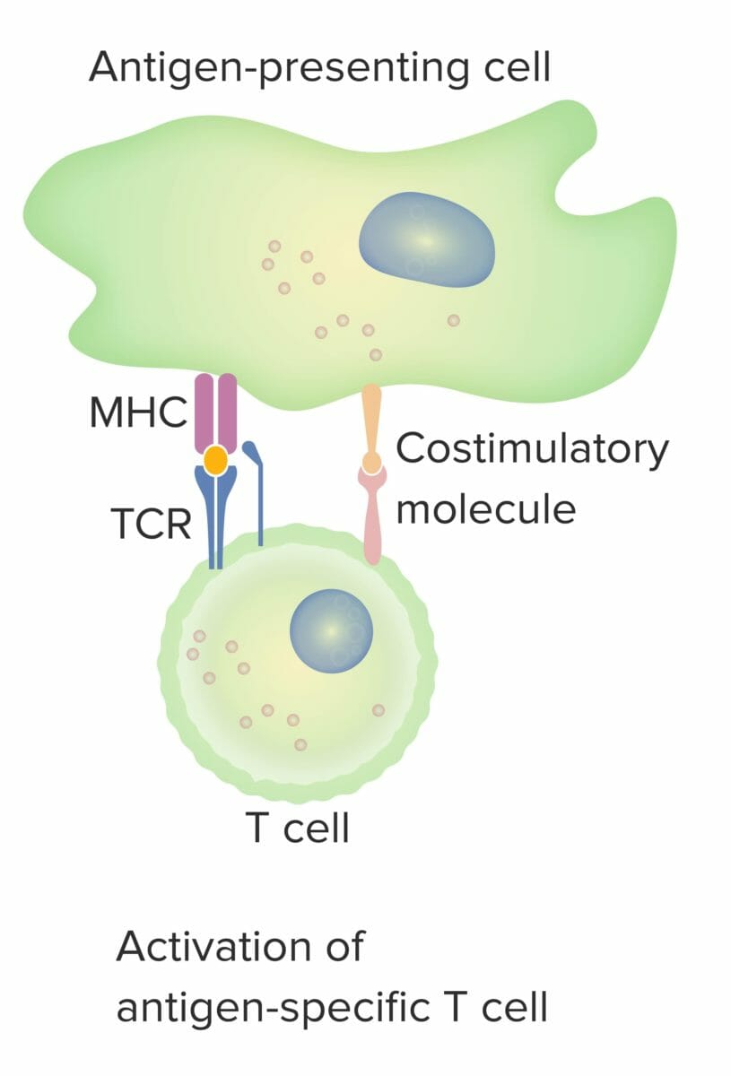
      • (1) T-cell receptor Receptor Receptors are proteins located either on the surface of or within a cell that can bind to signaling molecules known as ligands (e.g., hormones) and cause some type of response within the cell. Receptors ( TCR TCR Molecules on the surface of T-lymphocytes that recognize and combine with antigens. The receptors are non-covalently associated with a complex of several polypeptides collectively called CD3 antigens. Recognition of foreign antigen and the major histocompatibility complex is accomplished by a single heterodimeric antigen-receptor structure, composed of either alpha-beta or gamma-delta chains. Adaptive Cell-mediated Immunity) recognizes its cognate antigen Antigen Substances that are recognized by the immune system and induce an immune reaction. Vaccination, as presented by the antigen-presenting cell (e.g., dendritic cell).
      • (2) Costimulation Costimulation Adaptive Cell-mediated Immunity: best characterized by B7 protein (CD80/86) in the antigen-presenting cell interacting with the CD28 CD28 Costimulatory t-lymphocyte receptors that have specificity for CD80 antigen and CD86 antigen. Activation of this receptor results in increased t-cell proliferation, cytokine production and promotion of t-cell survival. T cells: Types and Functions of the T cell 
    • This CD28 CD28 Costimulatory t-lymphocyte receptors that have specificity for CD80 antigen and CD86 antigen. Activation of this receptor results in increased t-cell proliferation, cytokine production and promotion of t-cell survival. T cells: Types and Functions receptor Receptor Receptors are proteins located either on the surface of or within a cell that can bind to signaling molecules known as ligands (e.g., hormones) and cause some type of response within the cell. Receptors–B7 ligand combination, or “immunologic synapse Synapse The junction between 2 neurons is called a synapse. The synapse allows a neuron to pass an electrical or chemical signal to another neuron or target effector cell. Synapses and Neurotransmission,” stimulates the T cells T cells Lymphocytes responsible for cell-mediated immunity. Two types have been identified – cytotoxic (t-lymphocytes, cytotoxic) and helper T-lymphocytes (t-lymphocytes, helper-inducer). They are formed when lymphocytes circulate through the thymus gland and differentiate to thymocytes. When exposed to an antigen, they divide rapidly and produce large numbers of new T cells sensitized to that antigen. T cells: Types and Functions.
    • Other receptor Receptor Receptors are proteins located either on the surface of or within a cell that can bind to signaling molecules known as ligands (e.g., hormones) and cause some type of response within the cell. Receptors–ligand combinations are possible between activated T cells Activated T cells Adaptive Cell-mediated Immunity and other cells.
  • Regulation of response:
    • Stimulation of response is through the actions of agonist molecules (e.g., OX40).
    • Regulatory T cells Regulatory T cells Autoimmunity (Tregs) dampen the T-cell response.
    • Inhibition of the costimulatory signal (and thus, reduction of T-cell activity) is through immune checkpoint ( IC IC Inhaled Anesthetics) molecules, such as:
      • Programmed cell death-1 ( PD-1 PD-1 An inhibitory t-lymphocyte receptor that has specificity for CD274 antigen and programmed cell death 1 ligand 2 protein. Signaling by the receptor limits T cell proliferation and interferon gamma synthesis. The receptor also may play an essential role in the regulatory pathway that induces peripheral tolerance. T cells: Types and Functions)
      • Cytotoxic Cytotoxic Parvovirus B19 T-lymphocyte-associated protein 4 (CTLA-4)
    • Removal of this inhibition (checkpoint blockade immunotherapy) enhances immune response against the tumor Tumor Inflammation.
The 2-signal model of t-cell dependence on costimulation

The 2-signal model of T-cell dependence on costimulation:
When both signal 1 (T-cell receptor (TCR) binding the cognate antigen presented by the MHC molecule in the antigen-presenting cell) and signal 2 (costimulatory molecule interaction between the antigen-presenting cell and the T cell) are present, the mature T cell is fully activated.
The orange spot indicates proper binding between antigen and TCR.

Image by Lecturio.

Cancer immunoediting

Immunoediting is a process consisting of immunologic tumor Tumor Inflammation suppression Suppression Defense Mechanisms but can lead to tumor Tumor Inflammation progression. Immunoediting occurs in 3 main phases: 
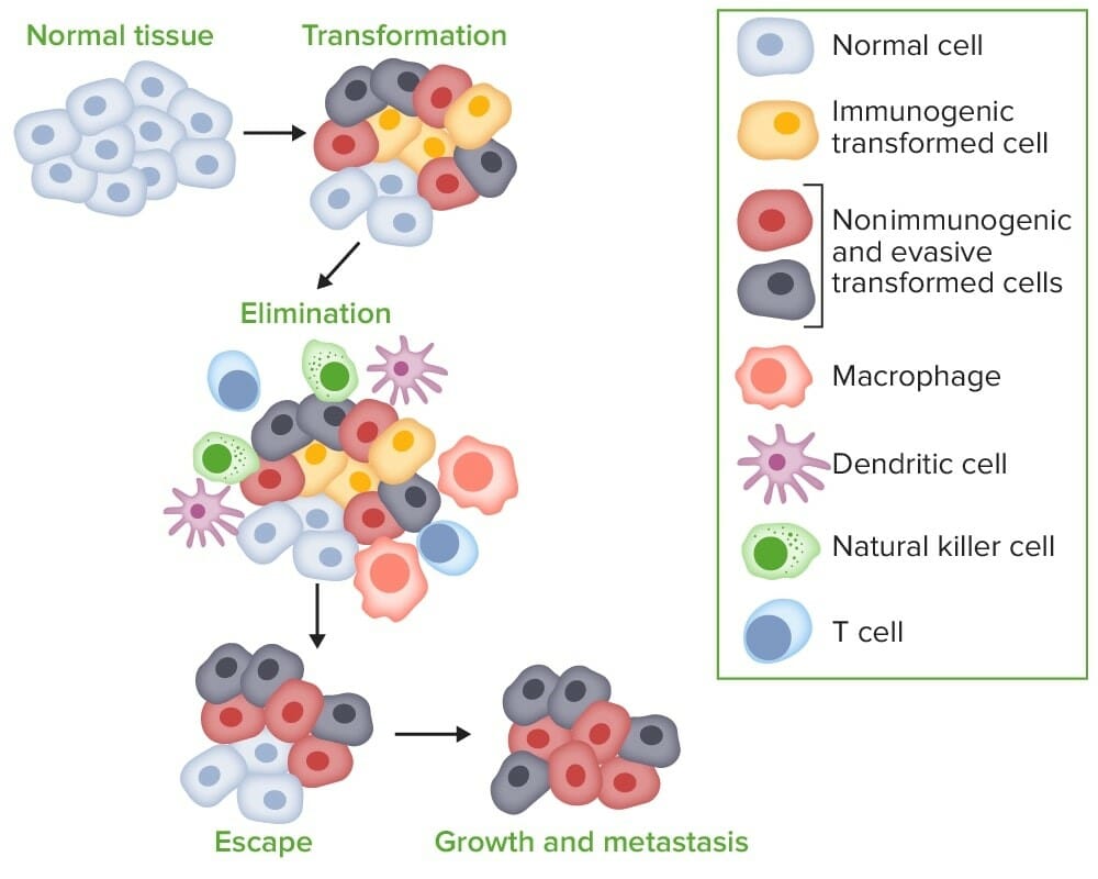
  • Elimination:
    • The initial damage and destruction of tumor Tumor Inflammation cells by innate and adaptive immunity
    • Completion of the phase means no cancer growth.
  • Equilibrium:
    • Occurs when tumor Tumor Inflammation cells survive the initial elimination attempt 
    • These cells are not able to progress, being maintained in a state of dormancy by the adaptive immune system Immune system The body’s defense mechanism against foreign organisms or substances and deviant native cells. It includes the humoral immune response and the cell-mediated response and consists of a complex of interrelated cellular, molecular, and genetic components. Primary Lymphatic Organs.
    • In this phase, tumor Tumor Inflammation immunogenicity is edited, where T cells T cells Lymphocytes responsible for cell-mediated immunity. Two types have been identified – cytotoxic (t-lymphocytes, cytotoxic) and helper T-lymphocytes (t-lymphocytes, helper-inducer). They are formed when lymphocytes circulate through the thymus gland and differentiate to thymocytes. When exposed to an antigen, they divide rapidly and produce large numbers of new T cells sensitized to that antigen. T cells: Types and Functions keep selectively attacking highly immunogenic tumor Tumor Inflammation cells.
    • This attack leaves other cells with less immunogenicity to potentially develop resistance Resistance Physiologically, the opposition to flow of air caused by the forces of friction. As a part of pulmonary function testing, it is the ratio of driving pressure to the rate of air flow. Ventilation: Mechanics of Breathing to the immune response.
  • Escape:
    • With constant immune mechanisms holding unstable tumor Tumor Inflammation cells in equilibrium, tumor-cell variants may emerge.
    • These cancer cells may express fewer antigens on their surfaces or lose their MHC class I expression.
    • Variants may also protect themselves from T-cell attack via expression of IC IC Inhaled Anesthetics molecules on their surfaces, like normal cells. 
    • Creation of an immunosuppressive state in the microenvironment is another way to grow without immunologic interference.
Carcinogenesis by immune evasion

Carcinogenesis by immune evasion:
When tumor cells transform from normal cells, the innate and adaptive immune systems detect and eliminate the transformed cells even before disease is clinically apparent (elimination). The process enters equilibrium, as tumor-cell variants may not be completely eliminated. However, the immune system attempts to control tumor-cell outgrowth by exerting selective pressure on highly immunogenic tumor cells. Tumor-cell immunogenicity is edited, leaving cells with reduced immunogenicity to grow and evade immunosurveillance, leading to progression of the cells into the escape phase, where the less immunogenic cells grow progressively and become clinically apparent cancer.

Image by Lecturio.

Cancer immunotherapy

Cancer immunotherapy stimulates the immune system Immune system The body’s defense mechanism against foreign organisms or substances and deviant native cells. It includes the humoral immune response and the cell-mediated response and consists of a complex of interrelated cellular, molecular, and genetic components. Primary Lymphatic Organs to respond to a malignancy Malignancy Hemothorax, activating different aspects of the immune system Immune system The body’s defense mechanism against foreign organisms or substances and deviant native cells. It includes the humoral immune response and the cell-mediated response and consists of a complex of interrelated cellular, molecular, and genetic components. Primary Lymphatic Organs to attack cancer cells. 

  • The immune system Immune system The body’s defense mechanism against foreign organisms or substances and deviant native cells. It includes the humoral immune response and the cell-mediated response and consists of a complex of interrelated cellular, molecular, and genetic components. Primary Lymphatic Organs is manipulated through:
    • Strengthening the host immune responses against tumors
    • Supplying modified immune system Immune system The body’s defense mechanism against foreign organisms or substances and deviant native cells. It includes the humoral immune response and the cell-mediated response and consists of a complex of interrelated cellular, molecular, and genetic components. Primary Lymphatic Organs components
    • Counteracting signals produced by cancer cells suppressing immune responses
  • Goals of immunotherapy:
    • Use the specificity and long-term memory Memory Complex mental function having four distinct phases: (1) memorizing or learning, (2) retention, (3) recall, and (4) recognition. Clinically, it is usually subdivided into immediate, recent, and remote memory. Psychiatric Assessment of the adaptive immune response Adaptive immune response Immune responses against pathogens are divided into the innate and adaptive immune response systems. The adaptive immune response, also called the acquired immune system, consists of 2 main mechanisms: the humoral- and cellular-mediated immune responses. Adaptive Immune Response
    • Achieve durable tumor Tumor Inflammation regression Regression Corneal Abrasions, Erosion, and Ulcers.
    • Cure disease.
    • Improve overall survival.
  • Therapeutic options:
    • Some agents act on different components of the immune system Immune system The body’s defense mechanism against foreign organisms or substances and deviant native cells. It includes the humoral immune response and the cell-mediated response and consists of a complex of interrelated cellular, molecular, and genetic components. Primary Lymphatic Organs:
      • Cytokines Cytokines Non-antibody proteins secreted by inflammatory leukocytes and some non-leukocytic cells, that act as intercellular mediators. They differ from classical hormones in that they are produced by a number of tissue or cell types rather than by specialized glands. They generally act locally in a paracrine or autocrine rather than endocrine manner. Adaptive Immune Response
      • T cells T cells Lymphocytes responsible for cell-mediated immunity. Two types have been identified – cytotoxic (t-lymphocytes, cytotoxic) and helper T-lymphocytes (t-lymphocytes, helper-inducer). They are formed when lymphocytes circulate through the thymus gland and differentiate to thymocytes. When exposed to an antigen, they divide rapidly and produce large numbers of new T cells sensitized to that antigen. T cells: Types and Functions and other cell types
      • Checkpoint molecules
    • Other agents use the response produced by viruses Viruses Minute infectious agents whose genomes are composed of DNA or RNA, but not both. They are characterized by a lack of independent metabolism and the inability to replicate outside living host cells. Virology or bacteria Bacteria Bacteria are prokaryotic single-celled microorganisms that are metabolically active and divide by binary fission. Some of these organisms play a significant role in the pathogenesis of diseases. Bacteriology on the immune system Immune system The body’s defense mechanism against foreign organisms or substances and deviant native cells. It includes the humoral immune response and the cell-mediated response and consists of a complex of interrelated cellular, molecular, and genetic components. Primary Lymphatic Organs.

Therapeutics

Cytokines Cytokines Non-antibody proteins secreted by inflammatory leukocytes and some non-leukocytic cells, that act as intercellular mediators. They differ from classical hormones in that they are produced by a number of tissue or cell types rather than by specialized glands. They generally act locally in a paracrine or autocrine rather than endocrine manner. Adaptive Immune Response and vaccines

  • Interleukin-2 Interleukin-2 A soluble substance elaborated by antigen- or mitogen-stimulated T-lymphocytes which induces DNA synthesis in naive lymphocytes. Interleukins (IL-2)
    • Has pleiotropic effects on both cytotoxic Cytotoxic Parvovirus B19 T cells T cells Lymphocytes responsible for cell-mediated immunity. Two types have been identified – cytotoxic (t-lymphocytes, cytotoxic) and helper T-lymphocytes (t-lymphocytes, helper-inducer). They are formed when lymphocytes circulate through the thymus gland and differentiate to thymocytes. When exposed to an antigen, they divide rapidly and produce large numbers of new T cells sensitized to that antigen. T cells: Types and Functions and Tregs  
    • High doses:
      • Promote the activity of CD8+ effector T cells Effector T cells Adaptive Cell-mediated Immunity and natural killer (NK) cells
      • Promote differentiation of CD4+ cells into T helper type 1 Type 1 Spinal Muscular Atrophy ( Th1 Th1 A subset of helper-inducer T-lymphocytes which synthesize and secrete interleukin-2; interferon-gamma; and interleukin-12. Due to their ability to kill antigen-presenting cells and their lymphokine-mediated effector activity, th1 cells are associated with vigorous delayed-type hypersensitivity reactions. T cells: Types and Functions) and Th2 Th2 A subset of helper-inducer T-lymphocytes which synthesize and secrete the interleukins il-4; il-5; il-6; and il-10. These cytokines influence b-cell development and antibody production as well as augmenting humoral responses. T cells: Types and Functions subclasses
    • Low doses:
      • ↑ Expansion of Tregs
      • Inhibit the formation of Th17 Th17 A subset of helper-effector T-lymphocytes which synthesize and secrete interleukins il-17; il-17f; and il-22. These cytokines are involved in host defenses and tissue inflammation in autoimmune diseases. T cells: Types and Functions cells involved in autoimmunity Autoimmunity Autoimmunity is a pathologic immune response toward self-antigens, resulting from a combination of factors: immunologic, genetic, and environmental. The immune system is equipped with self-tolerance, allowing immune cells such as T cells and B cells to recognize self-antigens and to not mount a reaction against them. Defects in this mechanism, along with environmental triggers (such as infections) and genetic susceptibility factors (most notable of which are the HLA genes) can lead to autoimmune diseases. Autoimmunity 
    • Used for melanoma Melanoma Melanoma is a malignant tumor arising from melanocytes, the melanin-producing cells of the epidermis. These tumors are most common in fair-skinned individuals with a history of excessive sun exposure and sunburns. Melanoma and renal cell carcinoma Renal cell carcinoma Renal cell carcinoma (RCC) is a tumor that arises from the lining of the renal tubular system within the renal cortex. Renal cell carcinoma is responsible for 80%-85% of all primary renal neoplasms. Most RCCs arise sporadically, but smoking, hypertension, and obesity are linked to its development. Renal Cell Carcinoma ( RCC RCC Renal cell carcinoma (RCC) is a tumor that arises from the lining of the renal tubular system within the renal cortex. Renal cell carcinoma is responsible for 80%-85% of all primary renal neoplasms. Most RCCs arise sporadically, but smoking, hypertension, and obesity are linked to its development. Renal Cell Carcinoma)
  • Interferon alfa-2b ( IFN IFN Interferon (IFN) is a cytokine with antiviral properties (it interferes with viral infections) and various roles in immunoregulation. The different types are type I IFN (IFN-ɑ and IFN-β), type II IFN (IFN-ɣ), and type III IFN (IFN-ƛ). Interferons⍺-2b):
    • Enhances Th1-mediated effects
    • Cytolytic and antiproliferative effects
    • Used as adjuvant Adjuvant Substances that augment, stimulate, activate, potentiate, or modulate the immune response at either the cellular or humoral level. The classical agents (freund’s adjuvant, bcg, corynebacterium parvum, et al.) contain bacterial antigens. Some are endogenous (e.g., histamine, interferon, transfer factor, tuftsin, interleukin-1). Their mode of action is either non-specific, resulting in increased immune responsiveness to a wide variety of antigens, or antigen-specific, i.e., affecting a restricted type of immune response to a narrow group of antigens. The therapeutic efficacy of many biological response modifiers is related to their antigen-specific immunoadjuvanticity. Vaccination treatment for high-risk melanoma Melanoma Melanoma is a malignant tumor arising from melanocytes, the melanin-producing cells of the epidermis. These tumors are most common in fair-skinned individuals with a history of excessive sun exposure and sunburns. Melanoma
  • Lenalidomide and pomalidomide:
  • Vaccines (therapeutic):
    • Sipuleucel-T (provenge):
      • Currently, approved vaccine-based therapy for advanced (castration-resistant hormone-refractory) prostate Prostate The prostate is a gland in the male reproductive system. The gland surrounds the bladder neck and a portion of the urethra. The prostate is an exocrine gland that produces a weakly acidic secretion, which accounts for roughly 20% of the seminal fluid. cancer
      • Autologous dendritic cell preparation targeting prostatic acid phosphatase (PAP)
      • Blood mononuclear cells are obtained via leukapheresis.
      • Antigen-presenting cells Antigen-presenting cells A heterogeneous group of immunocompetent cells that mediate the cellular immune response by processing and presenting antigens to the T-cells. Traditional antigen-presenting cells include macrophages; dendritic cells; langerhans cells; and B-lymphocytes. Follicular dendritic cells are not traditional antigen-presenting cells, but because they hold antigen on their cell surface in the form of immune complexes for b-cell recognition they are considered so by some authors. Adaptive Immune Response are isolated and then activated in vitro by PAP fused to granulocyte macrophage colony-stimulating factor Macrophage colony-stimulating factor A mononuclear phagocyte colony-stimulating factor (M-CSF) synthesized by mesenchymal cells. The compound stimulates the survival, proliferation, and differentiation of hematopoietic cells of the monocyte-macrophage series. M-CSF is a disulfide-bonded glycoprotein dimer with a mw of 70 kda. It binds to a specific high affinity receptor. White Myeloid Cells: Histology ( GM-CSF GM-CSF An acidic glycoprotein of mw 23 kda with internal disulfide bonds. The protein is produced in response to a number of inflammatory mediators by mesenchymal cells present in the hemopoietic environment and at peripheral sites of inflammation. GM-CSF is able to stimulate the production of neutrophilic granulocytes, macrophages, and mixed granulocyte-macrophage colonies from bone marrow cells and can stimulate the formation of eosinophil colonies from fetal liver progenitor cells. GM-CSF can also stimulate some functional activities in mature granulocytes and macrophages. White Myeloid Cells: Histology).
      • Reinfusion of the product produces T-cell activity against tumors expressing PAP.
    • Bacille Calmette–Guérin (BCG)
      • Derived from attenuated strain of Mycobacterium bovis Mycobacterium bovis The bovine variety of the tubercle Bacillus. It is called also Mycobacterium tuberculosis var. Bovis. Mycobacterium
      • Approved for the intravesical treatment of early-stage bladder Bladder A musculomembranous sac along the urinary tract. Urine flows from the kidneys into the bladder via the ureters, and is held there until urination. Pyelonephritis and Perinephric Abscess cancer
      • Activates an immune reaction against the tumor Tumor Inflammation

Oncolytic viruses Viruses Minute infectious agents whose genomes are composed of DNA or RNA, but not both. They are characterized by a lack of independent metabolism and the inability to replicate outside living host cells. Virology

  • Emerging class of cancer therapeutics using viruses Viruses Minute infectious agents whose genomes are composed of DNA or RNA, but not both. They are characterized by a lack of independent metabolism and the inability to replicate outside living host cells. Virology to infect and destroy tumor Tumor Inflammation cells
  • The viruses Viruses Minute infectious agents whose genomes are composed of DNA or RNA, but not both. They are characterized by a lack of independent metabolism and the inability to replicate outside living host cells. Virology are genetically modified to preferentially lyse cancer cells and spare normal cells.
  • Talimogene laherparepvec (T-VEC)
    • The 1st approved oncolytic immunotherapy 
    • Genetically engineered herpes simplex Herpes Simplex A group of acute infections caused by herpes simplex virus type 1 or type 2 that is characterized by the development of one or more small fluid-filled vesicles with a raised erythematous base on the skin or mucous membrane. It occurs as a primary infection or recurs due to a reactivation of a latent infection. Congenital TORCH Infections virus Virus Viruses are infectious, obligate intracellular parasites composed of a nucleic acid core surrounded by a protein capsid. Viruses can be either naked (non-enveloped) or enveloped. The classification of viruses is complex and based on many factors, including type and structure of the nucleoid and capsid, the presence of an envelope, the replication cycle, and the host range. Virology, modified to express GM-CSF GM-CSF An acidic glycoprotein of mw 23 kda with internal disulfide bonds. The protein is produced in response to a number of inflammatory mediators by mesenchymal cells present in the hemopoietic environment and at peripheral sites of inflammation. GM-CSF is able to stimulate the production of neutrophilic granulocytes, macrophages, and mixed granulocyte-macrophage colonies from bone marrow cells and can stimulate the formation of eosinophil colonies from fetal liver progenitor cells. GM-CSF can also stimulate some functional activities in mature granulocytes and macrophages. White Myeloid Cells: Histology
    • Used in unresectable melanoma Melanoma Melanoma is a malignant tumor arising from melanocytes, the melanin-producing cells of the epidermis. These tumors are most common in fair-skinned individuals with a history of excessive sun exposure and sunburns. Melanoma
    • Can be combined with ICs

Cellular adoptive immunotherapy

These therapies are developed based on the manipulation of the individual’s cells through in vitro expansion of tumor-specific T cells T cells Lymphocytes responsible for cell-mediated immunity. Two types have been identified – cytotoxic (t-lymphocytes, cytotoxic) and helper T-lymphocytes (t-lymphocytes, helper-inducer). They are formed when lymphocytes circulate through the thymus gland and differentiate to thymocytes. When exposed to an antigen, they divide rapidly and produce large numbers of new T cells sensitized to that antigen. T cells: Types and Functions, which are reinfused by autologous transplantation.

Techniques of manipulating T cells T cells Lymphocytes responsible for cell-mediated immunity. Two types have been identified – cytotoxic (t-lymphocytes, cytotoxic) and helper T-lymphocytes (t-lymphocytes, helper-inducer). They are formed when lymphocytes circulate through the thymus gland and differentiate to thymocytes. When exposed to an antigen, they divide rapidly and produce large numbers of new T cells sensitized to that antigen. T cells: Types and Functions (and other immune cell types):
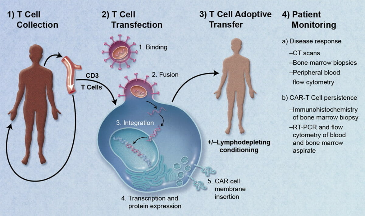
  • Chimeric antigen Antigen Substances that are recognized by the immune system and induce an immune reaction. Vaccination receptor Receptor Receptors are proteins located either on the surface of or within a cell that can bind to signaling molecules known as ligands (e.g., hormones) and cause some type of response within the cell. Receptors (CAR) T cells T cells Lymphocytes responsible for cell-mediated immunity. Two types have been identified – cytotoxic (t-lymphocytes, cytotoxic) and helper T-lymphocytes (t-lymphocytes, helper-inducer). They are formed when lymphocytes circulate through the thymus gland and differentiate to thymocytes. When exposed to an antigen, they divide rapidly and produce large numbers of new T cells sensitized to that antigen. T cells: Types and Functions:
    • The T cells T cells Lymphocytes responsible for cell-mediated immunity. Two types have been identified – cytotoxic (t-lymphocytes, cytotoxic) and helper T-lymphocytes (t-lymphocytes, helper-inducer). They are formed when lymphocytes circulate through the thymus gland and differentiate to thymocytes. When exposed to an antigen, they divide rapidly and produce large numbers of new T cells sensitized to that antigen. T cells: Types and Functions are equipped with CAR, a synthetic receptor Receptor Receptors are proteins located either on the surface of or within a cell that can bind to signaling molecules known as ligands (e.g., hormones) and cause some type of response within the cell. Receptors that binds to cancer cells.
    • These T cells T cells Lymphocytes responsible for cell-mediated immunity. Two types have been identified – cytotoxic (t-lymphocytes, cytotoxic) and helper T-lymphocytes (t-lymphocytes, helper-inducer). They are formed when lymphocytes circulate through the thymus gland and differentiate to thymocytes. When exposed to an antigen, they divide rapidly and produce large numbers of new T cells sensitized to that antigen. T cells: Types and Functions target the proteins Proteins Linear polypeptides that are synthesized on ribosomes and may be further modified, crosslinked, cleaved, or assembled into complex proteins with several subunits. The specific sequence of amino acids determines the shape the polypeptide will take, during protein folding, and the function of the protein. Energy Homeostasis of leukemic B cells B cells Lymphoid cells concerned with humoral immunity. They are short-lived cells resembling bursa-derived lymphocytes of birds in their production of immunoglobulin upon appropriate stimulation. B cells: Types and Functions.
    • When malignant B cells B cells Lymphoid cells concerned with humoral immunity. They are short-lived cells resembling bursa-derived lymphocytes of birds in their production of immunoglobulin upon appropriate stimulation. B cells: Types and Functions are recognized, cells are destroyed via release of cytokines Cytokines Non-antibody proteins secreted by inflammatory leukocytes and some non-leukocytic cells, that act as intercellular mediators. They differ from classical hormones in that they are produced by a number of tissue or cell types rather than by specialized glands. They generally act locally in a paracrine or autocrine rather than endocrine manner. Adaptive Immune Response, perforin Perforin A calcium-dependent pore-forming protein synthesized in cytolytic lymphocytes and sequestered in secretory granules. Upon immunological reaction between a cytolytic lymphocyte and a target cell, perforin is released at the plasma membrane and polymerizes into transmembrane tubules (forming pores) which lead to death of a target cell. Lymphocytes: Histology, and granzymes Granzymes A family of serine endopeptidases found in the secretory granules of leukocytes such as cytotoxic T-lymphocytes and natural killer cells. When secreted into the intercellular space granzymes act to eliminate transformed and virus-infected host cells. Lymphocytes: Histology.
    • Adverse effects of therapy include:
      • Marked cytokine release syndrome (cytokine storm) 
      • Neurotoxicity
  • Tumor-infiltrating lymphocytes Lymphocytes Lymphocytes are heterogeneous WBCs involved in immune response. Lymphocytes develop from the bone marrow, starting from hematopoietic stem cells (HSCs) and progressing to common lymphoid progenitors (CLPs). B and T lymphocytes and natural killer (NK) cells arise from the lineage. Lymphocytes: Histology (TILs):
    • Cell population of TILs is obtained directly from the resected tumor Tumor Inflammation.
    • Expansion of cells is facilitated by coculture with IL-2.
    • Prior to infusion, the affected individual receives chemotherapy Chemotherapy Osteosarcoma or radiation Radiation Emission or propagation of acoustic waves (sound), electromagnetic energy waves (such as light; radio waves; gamma rays; or x-rays), or a stream of subatomic particles (such as electrons; neutrons; protons; or alpha particles). Osteosarcoma to reduce Tregs.
    • When reintroduced, the TILs (mostly CD8+ cells) recognize and attack the tumor Tumor Inflammation antigens.
  • Immune-mobilizing monoclonal TCRs against cancer (ImmTACs):
    • Engineered TCR TCR Molecules on the surface of T-lymphocytes that recognize and combine with antigens. The receptors are non-covalently associated with a complex of several polypeptides collectively called CD3 antigens. Recognition of foreign antigen and the major histocompatibility complex is accomplished by a single heterodimeric antigen-receptor structure, composed of either alpha-beta or gamma-delta chains. Adaptive Cell-mediated Immunity therapy
    • In some cases, T cells T cells Lymphocytes responsible for cell-mediated immunity. Two types have been identified – cytotoxic (t-lymphocytes, cytotoxic) and helper T-lymphocytes (t-lymphocytes, helper-inducer). They are formed when lymphocytes circulate through the thymus gland and differentiate to thymocytes. When exposed to an antigen, they divide rapidly and produce large numbers of new T cells sensitized to that antigen. T cells: Types and Functions may not recognize the tumor Tumor Inflammation cells or be sufficiently activated or expanded.
    • These T cells T cells Lymphocytes responsible for cell-mediated immunity. Two types have been identified – cytotoxic (t-lymphocytes, cytotoxic) and helper T-lymphocytes (t-lymphocytes, helper-inducer). They are formed when lymphocytes circulate through the thymus gland and differentiate to thymocytes. When exposed to an antigen, they divide rapidly and produce large numbers of new T cells sensitized to that antigen. T cells: Types and Functions can be equipped with a new TCR TCR Molecules on the surface of T-lymphocytes that recognize and combine with antigens. The receptors are non-covalently associated with a complex of several polypeptides collectively called CD3 antigens. Recognition of foreign antigen and the major histocompatibility complex is accomplished by a single heterodimeric antigen-receptor structure, composed of either alpha-beta or gamma-delta chains. Adaptive Cell-mediated Immunity to target specific cancer antigens.
    • An example is the investigational drug, tebentafusp, a soluble TCR TCR Molecules on the surface of T-lymphocytes that recognize and combine with antigens. The receptors are non-covalently associated with a complex of several polypeptides collectively called CD3 antigens. Recognition of foreign antigen and the major histocompatibility complex is accomplished by a single heterodimeric antigen-receptor structure, composed of either alpha-beta or gamma-delta chains. Adaptive Cell-mediated Immunity that targets gp100, a uveal melanoma Melanoma Melanoma is a malignant tumor arising from melanocytes, the melanin-producing cells of the epidermis. These tumors are most common in fair-skinned individuals with a history of excessive sun exposure and sunburns. Melanoma antigen Antigen Substances that are recognized by the immune system and induce an immune reaction. Vaccination.
  • Bispecific T-cell engagers (BiTEs):
    • Bispecific antibodies Antibodies Immunoglobulins (Igs), also known as antibodies, are glycoprotein molecules produced by plasma cells that act in immune responses by recognizing and binding particular antigens. The various Ig classes are IgG (the most abundant), IgM, IgE, IgD, and IgA, which differ in their biologic features, structure, target specificity, and distribution. Immunoglobulins: Types and Functions (BsAbs) that recruit T cells T cells Lymphocytes responsible for cell-mediated immunity. Two types have been identified – cytotoxic (t-lymphocytes, cytotoxic) and helper T-lymphocytes (t-lymphocytes, helper-inducer). They are formed when lymphocytes circulate through the thymus gland and differentiate to thymocytes. When exposed to an antigen, they divide rapidly and produce large numbers of new T cells sensitized to that antigen. T cells: Types and Functions to the malignant cells
    • Target CD3 CD3 Complex of at least five membrane-bound polypeptides in mature T-lymphocytes that are non-covalently associated with one another and with the T-cell receptor. The CD3 complex includes the gamma, delta, epsilon, zeta, and eta chains (subunits). When antigen binds to the T-cell receptor, the CD3 complex transduces the activating signals to the cytoplasm of the T-cell. The CD3 gamma and delta chains (subunits) are separate from and not related to the gamma/delta chains of the T-cell receptor. T cells: Types and Functions and tumor-specific antigens, ultimately leading to cytotoxicity of T cells T cells Lymphocytes responsible for cell-mediated immunity. Two types have been identified – cytotoxic (t-lymphocytes, cytotoxic) and helper T-lymphocytes (t-lymphocytes, helper-inducer). They are formed when lymphocytes circulate through the thymus gland and differentiate to thymocytes. When exposed to an antigen, they divide rapidly and produce large numbers of new T cells sensitized to that antigen. T cells: Types and Functions against the tumor Tumor Inflammation 
    • Blinatumomab: approved BiTE
  • NK-cell therapy:
    • Unlike T cells T cells Lymphocytes responsible for cell-mediated immunity. Two types have been identified – cytotoxic (t-lymphocytes, cytotoxic) and helper T-lymphocytes (t-lymphocytes, helper-inducer). They are formed when lymphocytes circulate through the thymus gland and differentiate to thymocytes. When exposed to an antigen, they divide rapidly and produce large numbers of new T cells sensitized to that antigen. T cells: Types and Functions, NK cells NK cells A specialized subset of T-lymphocytes that exhibit features of innate immunity similar to that of natural killer cells. They are reactive to glycolipids presented in the context of the major histocompatibility complex (MHC) class I-like molecule, CD1D antigen. Lymphocytes: Histology are not tailored to a specific antigen Antigen Substances that are recognized by the immune system and induce an immune reaction. Vaccination.
    • NK cells NK cells A specialized subset of T-lymphocytes that exhibit features of innate immunity similar to that of natural killer cells. They are reactive to glycolipids presented in the context of the major histocompatibility complex (MHC) class I-like molecule, CD1D antigen. Lymphocytes: Histology can be enhanced with CARs to effectively target malignant cells.
Cellular adaptive immunotherapy

Cellular adaptive immunotherapy:
1) T cells are collected.
2) The T cells are engineered to express chimeric antigen receptors (CARs), followed by in vitro expansion.
3) These cells are then infused into the same individual. These cells go into circulation, recognizing and destroying malignant cells.
4) Subsequent monitoring of disease response follows.
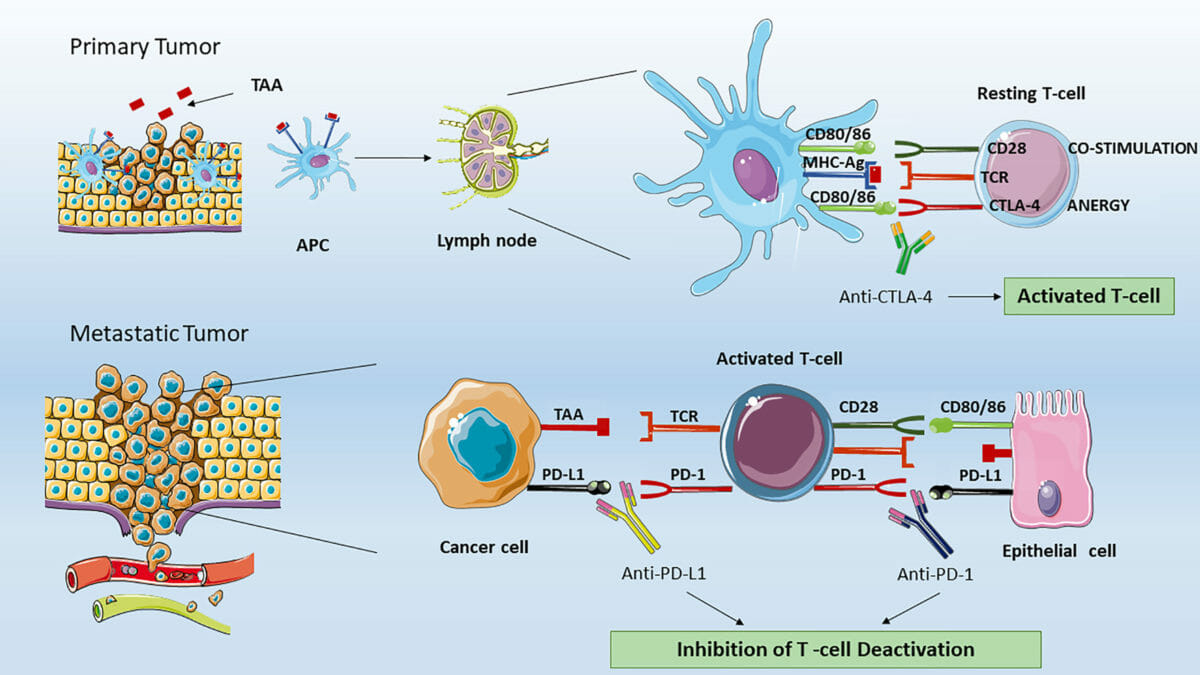
Image: “CAR-Engineered T-Cell Adoptive Transfer” by Caron A. Jacobson and Jerome Ritz. License: Public Domain

Checkpoint inhibitors

  • PD-1 PD-1 An inhibitory t-lymphocyte receptor that has specificity for CD274 antigen and programmed cell death 1 ligand 2 protein. Signaling by the receptor limits T cell proliferation and interferon gamma synthesis. The receptor also may play an essential role in the regulatory pathway that induces peripheral tolerance. T cells: Types and Functions:
    • A transmembrane protein (checkpoint molecule) in T cells T cells Lymphocytes responsible for cell-mediated immunity. Two types have been identified – cytotoxic (t-lymphocytes, cytotoxic) and helper T-lymphocytes (t-lymphocytes, helper-inducer). They are formed when lymphocytes circulate through the thymus gland and differentiate to thymocytes. When exposed to an antigen, they divide rapidly and produce large numbers of new T cells sensitized to that antigen. T cells: Types and Functions that binds:
      • Programmed cell death Cell death Injurious stimuli trigger the process of cellular adaptation, whereby cells respond to withstand the harmful changes in their environment. Overwhelmed adaptive mechanisms lead to cell injury. Mild stimuli produce reversible injury. If the stimulus is severe or persistent, injury becomes irreversible. Apoptosis is programmed cell death, a mechanism with both physiologic and pathologic effects. Cell Injury and Death ligand 1 (PD-L1): found in multiple tissues, including tumor Tumor Inflammation cells
      • PD-L2: found in hematopoietic cells
    • PD-1 PD-1 An inhibitory t-lymphocyte receptor that has specificity for CD274 antigen and programmed cell death 1 ligand 2 protein. Signaling by the receptor limits T cell proliferation and interferon gamma synthesis. The receptor also may play an essential role in the regulatory pathway that induces peripheral tolerance. T cells: Types and Functions–PD-L1/2 interaction:
      • Inhibits apoptosis Apoptosis A regulated cell death mechanism characterized by distinctive morphologic changes in the nucleus and cytoplasm, including the endonucleolytic cleavage of genomic DNA, at regularly spaced, internucleosomal sites, I.e., DNA fragmentation. It is genetically-programmed and serves as a balance to mitosis in regulating the size of animal tissues and in mediating pathologic processes associated with tumor growth. Ischemic Cell Damage of tumor Tumor Inflammation cells and promotes peripheral T-effector-cell exhaustion 
      • Increases conversion of T-effector cells to Tregs
      • Blocking this interaction produces therapeutic effect → ↑ T-cell activity against tumor Tumor Inflammation cells
  • Inhibitors of PD-1 PD-1 An inhibitory t-lymphocyte receptor that has specificity for CD274 antigen and programmed cell death 1 ligand 2 protein. Signaling by the receptor limits T cell proliferation and interferon gamma synthesis. The receptor also may play an essential role in the regulatory pathway that induces peripheral tolerance. T cells: Types and Functions–PD-L1 interaction:
    • Antibodies Antibodies Immunoglobulins (Igs), also known as antibodies, are glycoprotein molecules produced by plasma cells that act in immune responses by recognizing and binding particular antigens. The various Ig classes are IgG (the most abundant), IgM, IgE, IgD, and IgA, which differ in their biologic features, structure, target specificity, and distribution. Immunoglobulins: Types and Functions inhibiting PD-1 PD-1 An inhibitory t-lymphocyte receptor that has specificity for CD274 antigen and programmed cell death 1 ligand 2 protein. Signaling by the receptor limits T cell proliferation and interferon gamma synthesis. The receptor also may play an essential role in the regulatory pathway that induces peripheral tolerance. T cells: Types and Functions:
      • Pembrolizumab Pembrolizumab Squamous Cell Carcinoma (SCC)
      • Nivolumab Nivolumab A genetically engineered, fully humanized immunoglobulin g4 monoclonal antibody that binds to the pd-1 receptor, activating an immune response to tumor cells. It is used as monotherapy or in combination with ipilimumab for the treatment of advanced malignant melanoma. It is also used in the treatment of advanced or recurring non-small cell lung cancer; renal cell carcinoma; and Hodgkin’s lymphoma. Melanoma
      • Dostarlimab
    • Antibodies Antibodies Immunoglobulins (Igs), also known as antibodies, are glycoprotein molecules produced by plasma cells that act in immune responses by recognizing and binding particular antigens. The various Ig classes are IgG (the most abundant), IgM, IgE, IgD, and IgA, which differ in their biologic features, structure, target specificity, and distribution. Immunoglobulins: Types and Functions inhibiting PD-L1:
      • Atezolizumab (1st approved PD-L1 inhibitor)
      • Durvalumab
      • Avelumab
  • CTLA-4:
    • Checkpoint molecule with a major role in immune response down-regulation Down-Regulation A negative regulatory effect on physiological processes at the molecular, cellular, or systemic level. At the molecular level, the major regulatory sites include membrane receptors, genes (gene expression regulation), mRNAs, and proteins. Pharmacokinetics and Pharmacodynamics
    • Found on the surface of CD4+ and CD8+ T lymphocytes T lymphocytes Lymphocytes responsible for cell-mediated immunity. Two types have been identified – cytotoxic (t-lymphocytes, cytotoxic) and helper T-lymphocytes (t-lymphocytes, helper-inducer). They are formed when lymphocytes circulate through the thymus gland and differentiate to thymocytes. When exposed to an antigen, they divide rapidly and produce large numbers of new T cells sensitized to that antigen. T cells: Types and Functions, where it has high affinity for costimulatory receptors Receptors Receptors are proteins located either on the surface of or within a cell that can bind to signaling molecules known as ligands (e.g., hormones) and cause some type of response within the cell. Receptors CD80 and CD86 on antigen-presenting cells Antigen-presenting cells A heterogeneous group of immunocompetent cells that mediate the cellular immune response by processing and presenting antigens to the T-cells. Traditional antigen-presenting cells include macrophages; dendritic cells; langerhans cells; and B-lymphocytes. Follicular dendritic cells are not traditional antigen-presenting cells, but because they hold antigen on their cell surface in the form of immune complexes for b-cell recognition they are considered so by some authors. Adaptive Immune Response 
    • ↓ T-cell proliferation and IL-2 production
    • Inhibition leads to increased immune response and tumor Tumor Inflammation shrinkage.
  • Antibodies Antibodies Immunoglobulins (Igs), also known as antibodies, are glycoprotein molecules produced by plasma cells that act in immune responses by recognizing and binding particular antigens. The various Ig classes are IgG (the most abundant), IgM, IgE, IgD, and IgA, which differ in their biologic features, structure, target specificity, and distribution. Immunoglobulins: Types and Functions inhibiting CTLA–4: ipilimumab (1st checkpoint antibody approved for use)
  • Notable adverse effects of checkpoint inhibitors:
    • Cytokine release syndrome:
      • Similar to CAR T-cell therapy
      • Manifests with fever Fever Fever is defined as a measured body temperature of at least 38°C (100.4°F). Fever is caused by circulating endogenous and/or exogenous pyrogens that increase levels of prostaglandin E2 in the hypothalamus. Fever is commonly associated with chills, rigors, sweating, and flushing of the skin. Fever with or without multiple organ dysfunction
      • Corticosteroids Corticosteroids Chorioretinitis or other immunomodulators can improve immune-mediated toxic manifestations.
    • Fatigue Fatigue The state of weariness following a period of exertion, mental or physical, characterized by a decreased capacity for work and reduced efficiency to respond to stimuli. Fibromyalgia
    • Infusion-related reactions
    • Dermatologic toxicity Toxicity Dosage Calculation
    • Opportunistic infections Infections Invasion of the host organism by microorganisms or their toxins or by parasites that can cause pathological conditions or diseases. Chronic Granulomatous Disease
    • Colitis Colitis Inflammation of the colon section of the large intestine, usually with symptoms such as diarrhea (often with blood and mucus), abdominal pain, and fever. Pseudomembranous Colitis
    • Endocrine toxicities (e.g., hyperthyroidism Hyperthyroidism Hypersecretion of thyroid hormones from the thyroid gland. Elevated levels of thyroid hormones increase basal metabolic rate. Thyrotoxicosis and Hyperthyroidism or hypothyroidism Hypothyroidism Hypothyroidism is a condition characterized by a deficiency of thyroid hormones. Iodine deficiency is the most common cause worldwide, but Hashimoto’s disease (autoimmune thyroiditis) is the leading cause in non-iodine-deficient regions. Hypothyroidism, adrenal insufficiency Adrenal Insufficiency Conditions in which the production of adrenal corticosteroids falls below the requirement of the body. Adrenal insufficiency can be caused by defects in the adrenal glands, the pituitary gland, or the hypothalamus. Adrenal Insufficiency and Addison Disease, diabetes Diabetes Diabetes mellitus (DM) is a metabolic disease characterized by hyperglycemia and dysfunction of the regulation of glucose metabolism by insulin. Type 1 DM is diagnosed mostly in children and young adults as the result of autoimmune destruction of β cells in the pancreas and the resulting lack of insulin. Type 2 DM has a significant association with obesity and is characterized by insulin resistance. Diabetes Mellitus mellitus)
    • Neutropenia Neutropenia Neutrophils are an important component of the immune system and play a significant role in the eradication of infections. Low numbers of circulating neutrophils, referred to as neutropenia, predispose the body to recurrent infections or sepsis, though patients can also be asymptomatic. Neutropenia, thrombocytopenia Thrombocytopenia Thrombocytopenia occurs when the platelet count is < 150,000 per microliter. The normal range for platelets is usually 150,000-450,000/µL of whole blood. Thrombocytopenia can be a result of decreased production, increased destruction, or splenic sequestration of platelets. Patients are often asymptomatic until platelet counts are < 50,000/µL. Thrombocytopenia
Immune checkpoint inhibitors

Immune checkpoint inhibitors:
Top: Antigen-presenting cells process tumor-associated antigens (TAAs) and complex them to MHC molecules.
The antigen-presenting cells migrate to the lymph node (in T-cell–dominant areas) and present TAAs to the naive T cells.
Activation of the T cell requires 2 signals. The 1st is mediated by the binding of TAA to a T-cell receptor (TCR). The 2nd signal can be from the binding of T-cell CD28 to costimulatory CD80/CD86, which activates the T cell.
However, when the T-cell-CTLA-4 interacts with the same CD80/CD86 antigen-presenting–cell molecules, the effect is inhibitory (T-cell anergy or no T-cell activation occurs).
Therefore, CTLA-4 and CD28 compete for the binding to CD80/CD86 proteins. The anti-CTLA-4 blocking action of ipilimumab restores CD28 proactivator signaling, resulting in antitumor T-lymphocyte responses.
Bottom: In peripheral tissues, the activated T cell can be deactivated by the binding of T-cell programmed death cell-1 (PD-1) with programmed cell death ligand 1 (PD-L1) (or PD-L2) expressed on tumor cells. The anti-PD-1 or anti–PD-L1 blocking action by monoclonal antibodies (e.g., nivolumab, pembrolizumab, atezolizumab) restores effective antitumor T-lymphocyte activity.

Image: “Figure 1 Effect of ICIs on T lymphocytes” by Franzin, R. et al. License: CC BY 4.0

Comparison of Therapeutics

Comparison of agents that enhance the immune system Immune system The body’s defense mechanism against foreign organisms or substances and deviant native cells. It includes the humoral immune response and the cell-mediated response and consists of a complex of interrelated cellular, molecular, and genetic components. Primary Lymphatic Organs (use of cytokines Cytokines Non-antibody proteins secreted by inflammatory leukocytes and some non-leukocytic cells, that act as intercellular mediators. They differ from classical hormones in that they are produced by a number of tissue or cell types rather than by specialized glands. They generally act locally in a paracrine or autocrine rather than endocrine manner. Adaptive Immune Response, organisms to infect tumor Tumor Inflammation cells, and manipulation of immune cells)
Therapeutic class Agents Indications
Immunostimulatory cytokines Cytokines Non-antibody proteins secreted by inflammatory leukocytes and some non-leukocytic cells, that act as intercellular mediators. They differ from classical hormones in that they are produced by a number of tissue or cell types rather than by specialized glands. They generally act locally in a paracrine or autocrine rather than endocrine manner. Adaptive Immune Response IL–2
  • Melanoma Melanoma Melanoma is a malignant tumor arising from melanocytes, the melanin-producing cells of the epidermis. These tumors are most common in fair-skinned individuals with a history of excessive sun exposure and sunburns. Melanoma
  • RCC RCC Renal cell carcinoma (RCC) is a tumor that arises from the lining of the renal tubular system within the renal cortex. Renal cell carcinoma is responsible for 80%-85% of all primary renal neoplasms. Most RCCs arise sporadically, but smoking, hypertension, and obesity are linked to its development. Renal Cell Carcinoma
IFNα-2b
  • Melanoma Melanoma Melanoma is a malignant tumor arising from melanocytes, the melanin-producing cells of the epidermis. These tumors are most common in fair-skinned individuals with a history of excessive sun exposure and sunburns. Melanoma
  • AIDS-related Kaposi Kaposi A multicentric, malignant neoplastic vascular proliferation characterized by the development of bluish-red cutaneous nodules, usually on the lower extremities, most often on the toes or feet, and slowly increasing in size and number and spreading to more proximal areas. The tumors have endothelium-lined channels and vascular spaces admixed with variably sized aggregates of spindle-shaped cells, and often remain confined to the skin and subcutaneous tissue, but widespread visceral involvement may occur. Hhv-8 is the suspected cause. There is also a high incidence in AIDS patients. AIDS-defining Conditions sarcoma
  • Follicular lymphoma Lymphoma A general term for various neoplastic diseases of the lymphoid tissue. Imaging of the Mediastinum
  • Hairy-cell leukemia
  • Genital warts Warts Benign epidermal proliferations or tumors; some are viral in origin. Female Genitourinary Examination
Vaccines Sipuleucel-T Castration-resistant prostate Prostate The prostate is a gland in the male reproductive system. The gland surrounds the bladder neck and a portion of the urethra. The prostate is an exocrine gland that produces a weakly acidic secretion, which accounts for roughly 20% of the seminal fluid. adenocarcinoma
Bacille Calmette–Guérin (BCG) Bladder Bladder A musculomembranous sac along the urinary tract. Urine flows from the kidneys into the bladder via the ureters, and is held there until urination. Pyelonephritis and Perinephric Abscess cancer
Immunomodulatory agents
  • Lenalidomide
  • Pomalidomide
Multiple myeloma Multiple myeloma Multiple myeloma (MM) is a malignant condition of plasma cells (activated B lymphocytes) primarily seen in the elderly. Monoclonal proliferation of plasma cells results in cytokine-driven osteoclastic activity and excessive secretion of IgG antibodies. Multiple Myeloma
Oncolytic viruses Viruses Minute infectious agents whose genomes are composed of DNA or RNA, but not both. They are characterized by a lack of independent metabolism and the inability to replicate outside living host cells. Virology T-VEC Melanoma Melanoma Melanoma is a malignant tumor arising from melanocytes, the melanin-producing cells of the epidermis. These tumors are most common in fair-skinned individuals with a history of excessive sun exposure and sunburns. Melanoma
CAR T-cell therapy Agents are available under the REMS program of the FDA (for autologous use only).
  • ALL
  • Follicular lymphoma Lymphoma A general term for various neoplastic diseases of the lymphoid tissue. Imaging of the Mediastinum
  • Large B-cell lymphoma Lymphoma A general term for various neoplastic diseases of the lymphoid tissue. Imaging of the Mediastinum
  • Mantle-cell lymphoma Lymphoma A general term for various neoplastic diseases of the lymphoid tissue. Imaging of the Mediastinum
  • Multiple myeloma Multiple myeloma Multiple myeloma (MM) is a malignant condition of plasma cells (activated B lymphocytes) primarily seen in the elderly. Monoclonal proliferation of plasma cells results in cytokine-driven osteoclastic activity and excessive secretion of IgG antibodies. Multiple Myeloma
BiTE (CD3-directed therapy) Blinatumomab ALL
AIDS: acquired immunodeficiency syndrome
FDA: Food and Drug Administration
IL-2: interleukin-2
IFNα-2b: interferon-alfa 2b
RCC: renal cell cancer
REMS: Risk Evaluation and Mitigation Strategy
T-VEC: talimogene laherparepvec
Comparison of immune checkpoint inhibitors
Therapeutic class Agents Indications
Anti-PD-1 Pembrolizumab Pembrolizumab Squamous Cell Carcinoma (SCC)
  • Breast cancer Breast cancer Breast cancer is a disease characterized by malignant transformation of the epithelial cells of the breast. Breast cancer is the most common form of cancer and 2nd most common cause of cancer-related death among women. Breast Cancer
  • Cutaneous squamous-cell cancer
  • Cervical cancer Cervical cancer Cervical cancer, or invasive cervical carcinoma (ICC), is the 3rd most common cancer in women in the world, with > 50% of the cases being fatal. In the United States, ICC is the 13th most common cancer and the cause of < 3% of all cancer deaths due to the slow progression of precursor lesions and, more importantly, effective cancer screening. Cervical Cancer
  • Endometrial cancer Endometrial Cancer Endometrial carcinoma (EC) is the most common gynecologic malignancy in the developed world, and it has several histologic types. Endometrioid carcinoma (known as type 1 EC) typically develops from atypical endometrial hyperplasia, is hormonally responsive, and carries a favorable prognosis. Endometrial Hyperplasia and Endometrial Cancer
  • Esophageal cancer Esophageal cancer Esophageal cancer is 1 of the most common causes of cancer-related deaths worldwide. Nearly all esophageal cancers are either adenocarcinoma (commonly affecting the distal esophagus) or squamous cell carcinoma (affecting the proximal two-thirds of the esophagus). Esophageal Cancer
  • Gastric cancer Gastric cancer Gastric cancer is the 3rd-most common cause of cancer-related deaths worldwide. The majority of cases are from adenocarcinoma. The modifiable risk factors include Helicobacter pylori infection, smoking, and nitrate-rich diets. Gastric Cancer
  • Head and neck Neck The part of a human or animal body connecting the head to the rest of the body. Peritonsillar Abscess cancer
  • HCC HCC Hepatocellular carcinoma (HCC) typically arises in a chronically diseased or cirrhotic liver and is the most common primary liver cancer. Diagnosis may include ultrasound, CT, MRI, biopsy (if inconclusive imaging), and/or biomarkers. Hepatocellular Carcinoma (HCC) and Liver Metastases
  • Hodgkin lymphoma Lymphoma A general term for various neoplastic diseases of the lymphoid tissue. Imaging of the Mediastinum
  • Melanoma Melanoma Melanoma is a malignant tumor arising from melanocytes, the melanin-producing cells of the epidermis. These tumors are most common in fair-skinned individuals with a history of excessive sun exposure and sunburns. Melanoma
  • Merkel cell cancer
  • MSI-H or dMMR cancer
  • RCC RCC Renal cell carcinoma (RCC) is a tumor that arises from the lining of the renal tubular system within the renal cortex. Renal cell carcinoma is responsible for 80%-85% of all primary renal neoplasms. Most RCCs arise sporadically, but smoking, hypertension, and obesity are linked to its development. Renal Cell Carcinoma
  • Urothelial cancer
Nivolumab Nivolumab A genetically engineered, fully humanized immunoglobulin g4 monoclonal antibody that binds to the pd-1 receptor, activating an immune response to tumor cells. It is used as monotherapy or in combination with ipilimumab for the treatment of advanced malignant melanoma. It is also used in the treatment of advanced or recurring non-small cell lung cancer; renal cell carcinoma; and Hodgkin’s lymphoma. Melanoma
  • Colorectal cancer Colorectal cancer Colorectal cancer (CRC) is the 2nd leading cause of cancer-related deaths in the United States. Colorectal cancer is a heterogeneous disease that arises from genetic and epigenetic abnormalities, with influence from environmental factors. Colorectal Cancer
  • Melanoma Melanoma Melanoma is a malignant tumor arising from melanocytes, the melanin-producing cells of the epidermis. These tumors are most common in fair-skinned individuals with a history of excessive sun exposure and sunburns. Melanoma
  • Esophageal cancer Esophageal cancer Esophageal cancer is 1 of the most common causes of cancer-related deaths worldwide. Nearly all esophageal cancers are either adenocarcinoma (commonly affecting the distal esophagus) or squamous cell carcinoma (affecting the proximal two-thirds of the esophagus). Esophageal Cancer
  • Gastric cancer Gastric cancer Gastric cancer is the 3rd-most common cause of cancer-related deaths worldwide. The majority of cases are from adenocarcinoma. The modifiable risk factors include Helicobacter pylori infection, smoking, and nitrate-rich diets. Gastric Cancer
  • Head and neck Neck The part of a human or animal body connecting the head to the rest of the body. Peritonsillar Abscess cancer
  • Hodgkin lymphoma Lymphoma A general term for various neoplastic diseases of the lymphoid tissue. Imaging of the Mediastinum
  • HCC HCC Hepatocellular carcinoma (HCC) typically arises in a chronically diseased or cirrhotic liver and is the most common primary liver cancer. Diagnosis may include ultrasound, CT, MRI, biopsy (if inconclusive imaging), and/or biomarkers. Hepatocellular Carcinoma (HCC) and Liver Metastases
  • Mesothelioma Mesothelioma Malignant mesothelioma (usually referred to as simply mesothelioma) is the malignant growth of mesothelial cells, most commonly affecting the pleura. The majority of cases are associated with occupational exposure to asbestos that occurred > 20 years before clinical onset, which includes dyspnea, chest pain, coughing, fatigue, and weight loss. Malignant Mesothelioma
  • RCC RCC Renal cell carcinoma (RCC) is a tumor that arises from the lining of the renal tubular system within the renal cortex. Renal cell carcinoma is responsible for 80%-85% of all primary renal neoplasms. Most RCCs arise sporadically, but smoking, hypertension, and obesity are linked to its development. Renal Cell Carcinoma
  • Urothelial cancer
Dostarlimab
  • Endometrial cancer Endometrial Cancer Endometrial carcinoma (EC) is the most common gynecologic malignancy in the developed world, and it has several histologic types. Endometrioid carcinoma (known as type 1 EC) typically develops from atypical endometrial hyperplasia, is hormonally responsive, and carries a favorable prognosis. Endometrial Hyperplasia and Endometrial Cancer
  • dMMR cancer (solid tumors)
Anti-PD-L1 Atezolizumab
  • HCC HCC Hepatocellular carcinoma (HCC) typically arises in a chronically diseased or cirrhotic liver and is the most common primary liver cancer. Diagnosis may include ultrasound, CT, MRI, biopsy (if inconclusive imaging), and/or biomarkers. Hepatocellular Carcinoma (HCC) and Liver Metastases
  • Melanoma Melanoma Melanoma is a malignant tumor arising from melanocytes, the melanin-producing cells of the epidermis. These tumors are most common in fair-skinned individuals with a history of excessive sun exposure and sunburns. Melanoma
  • NSCLC
  • SCLC
  • Urothelial cancer
Avelumab
  • Merkel-cell cancer
  • RCC RCC Renal cell carcinoma (RCC) is a tumor that arises from the lining of the renal tubular system within the renal cortex. Renal cell carcinoma is responsible for 80%-85% of all primary renal neoplasms. Most RCCs arise sporadically, but smoking, hypertension, and obesity are linked to its development. Renal Cell Carcinoma
  • Urothelial cancer
Durvalumab
  • NSCLC
  • SCLC
Anti–CTLA-4 Ipilimumab
  • Colorectal carcinoma
  • HCC HCC Hepatocellular carcinoma (HCC) typically arises in a chronically diseased or cirrhotic liver and is the most common primary liver cancer. Diagnosis may include ultrasound, CT, MRI, biopsy (if inconclusive imaging), and/or biomarkers. Hepatocellular Carcinoma (HCC) and Liver Metastases
  • Malignant pleural mesothelioma Mesothelioma Malignant mesothelioma (usually referred to as simply mesothelioma) is the malignant growth of mesothelial cells, most commonly affecting the pleura. The majority of cases are associated with occupational exposure to asbestos that occurred > 20 years before clinical onset, which includes dyspnea, chest pain, coughing, fatigue, and weight loss. Malignant Mesothelioma
  • NSCLC
  • RCC RCC Renal cell carcinoma (RCC) is a tumor that arises from the lining of the renal tubular system within the renal cortex. Renal cell carcinoma is responsible for 80%-85% of all primary renal neoplasms. Most RCCs arise sporadically, but smoking, hypertension, and obesity are linked to its development. Renal Cell Carcinoma
dMMR: deficient mismatch repair
HCC: hepatocellular cancer
MSI-H: microsatellite instability—high
NSCLC: non–small cell lung cancer
RCC: renal cell cancer
SCLC: small cell lung cancer

References

  1. Chen, G., Bodogai, M., Tamehiro, N., Shen, C., Dou, J. (2018). Cancer immunotherapy: theory and application. Journal of Immunology Research 2018:7502161. https://doi.org/10.1155/2018/7502161
  2. Disis, M. (2014). Mechanism of action of immunotherapy. Seminars in Oncology 41(Suppl 5):S3-S13. 41. 10.1053/j.seminoncol.2014.09.004 
  3. Greenberg, P. (2021) Adoptive cell therapy: CAR T, TCR, TIL, NK. Cancer Research Institute. Retrieved January 8, 2022, from https://www.cancerresearch.org/en-us/immunotherapy/treatment-types/adoptive-cell-therapy#tcr
  4. Levinson, W., Chin-Hong P., Joyce E.A., Nussbaum J., Schwartz, B. (Eds.), (2022). Tumor immunity. Chapter 67 of Review of Medical Microbiology & Immunology: A Guide to Clinical Infectious Diseases, 17th ed. McGraw Hill. https://accessmedicine.mhmedical.com/content.aspx?bookid=3123&sectionid=262002060
  5. Oiseth S.J.,  Aziz M.S. (2017). Cancer immunotherapy: a brief review of the history, possibilities, and challenges ahead. Journal of Cancer Metastasis and Treatment. https://oaepublishstorage.blob.core.windows.net/82580177-5632-4585-bf24-c54aa1afe386/2275.pdf
  6. Postow, M. (2021) Toxicities associated with checkpoint inhibitor immunotherapy. UpToDate. Retrieved January 9, 2022, from https://www.uptodate.com/contents/toxicities-associated-with-checkpoint-inhibitor-immunotherapy
  7. Shoushtari,  A.N. (2021). Principles of cancer immunotherapy. UpToDate. Retrieved January 7, 2022, from https://www.uptodate.com/contents/principles-of-cancer-immunotherapy
  8. Tian, Z., Liu, M., Zhang, Y., et al. (2021). Bispecific T cell engagers: an emerging therapy for management of hematologic malignancies. Journal of Hematology and Oncology 14:75. https://doi.org/10.1186/s13045-021-01084-4
  9. Vesely, M.D., Schreiber, R.D. (2013). Cancer immunoediting: antigens, mechanisms, and implications to cancer immunotherapy. Annals of the New York Academy of Sciences 1284(1):1–5. https://doi.org/10.1111/nyas.12105

Create your free account or log in to continue reading!

Sign up now and get free access to Lecturio with concept pages, medical videos, and questions for your medical education.

User Reviews

Details