Advertisement

Advertisement

Advertisement

Advertisement

Imaging of the Breast

Female breasts Breasts The breasts are found on the anterior thoracic wall and consist of mammary glands surrounded by connective tissue. The mammary glands are modified apocrine sweat glands that produce milk, which serves as nutrition for infants. Breasts are rudimentary and usually nonfunctioning in men. Breasts: Anatomy, made of glandular, adipose, and connective tissue Connective tissue Connective tissues originate from embryonic mesenchyme and are present throughout the body except inside the brain and spinal cord. The main function of connective tissues is to provide structural support to organs. Connective tissues consist of cells and an extracellular matrix. Connective Tissue: Histology, are hormone-sensitive organs that undergo changes along with the menstrual cycle Menstrual cycle The menstrual cycle is the cyclic pattern of hormonal and tissular activity that prepares a suitable uterine environment for the fertilization and implantation of an ovum. The menstrual cycle involves both an endometrial and ovarian cycle that are dependent on one another for proper functioning. There are 2 phases of the ovarian cycle and 3 phases of the endometrial cycle. Menstrual Cycle and during pregnancy Pregnancy The status during which female mammals carry their developing young (embryos or fetuses) in utero before birth, beginning from fertilization to birth. Pregnancy: Diagnosis, Physiology, and Care. Breasts Breasts The breasts are found on the anterior thoracic wall and consist of mammary glands surrounded by connective tissue. The mammary glands are modified apocrine sweat glands that produce milk, which serves as nutrition for infants. Breasts are rudimentary and usually nonfunctioning in men. Breasts: Anatomy may be affected by various diseases, in which different imaging methods are important to arrive at the correct diagnosis and management. Mammography Mammography Radiographic examination of the breast. Breast Cancer Screening is used for breast cancer Breast cancer Breast cancer is a disease characterized by malignant transformation of the epithelial cells of the breast. Breast cancer is the most common form of cancer and 2nd most common cause of cancer-related death among women. Breast Cancer screening Screening Preoperative Care and diagnostic evaluation of various breast-related symptoms. Ultrasonography is rarely used for screening Screening Preoperative Care, but it is typically used for diagnostic workup and during procedures (e.g., breast biopsy Biopsy Removal and pathologic examination of specimens from the living body. Ewing Sarcoma). MRI of the breasts Breasts The breasts are found on the anterior thoracic wall and consist of mammary glands surrounded by connective tissue. The mammary glands are modified apocrine sweat glands that produce milk, which serves as nutrition for infants. Breasts are rudimentary and usually nonfunctioning in men. Breasts: Anatomy is used as a supplementary screening Screening Preoperative Care tool for those at high risk for developing breast cancer Breast cancer Breast cancer is a disease characterized by malignant transformation of the epithelial cells of the breast. Breast cancer is the most common form of cancer and 2nd most common cause of cancer-related death among women. Breast Cancer. Additionally, in individuals with breast implants, inconclusive mammographic and/or breast ultrasound Breast Ultrasound Fibrocystic Change findings, and diagnosed breast cancer Breast cancer Breast cancer is a disease characterized by malignant transformation of the epithelial cells of the breast. Breast cancer is the most common form of cancer and 2nd most common cause of cancer-related death among women. Breast Cancer needing evaluation pretreatment and posttreatment, MRI is an important breast radiologic tool.

Last updated: Feb 14, 2023

Editorial responsibility: Stanley Oiseth, Lindsay Jones, Evelin Maza

Advertisement

Advertisement

Advertisement

Advertisement

Advertisement

Advertisement

Advertisement

Advertisement

Mammography

Mammogram Mammogram Fibrocystic Change

  • Film-screen mammogram Mammogram Fibrocystic Change: uses X-rays X-rays X-rays are high-energy particles of electromagnetic radiation used in the medical field for the generation of anatomical images. X-rays are projected through the body of a patient and onto a film, and this technique is called conventional or projectional radiography. X-rays, radiographic film and intensifying screens to produce an image of the breasts Breasts The breasts are found on the anterior thoracic wall and consist of mammary glands surrounded by connective tissue. The mammary glands are modified apocrine sweat glands that produce milk, which serves as nutrition for infants. Breasts are rudimentary and usually nonfunctioning in men. Breasts: Anatomy
  • Digital mammogram Mammogram Fibrocystic Change: a digital detector takes the place of the film and screen

Indications

  • Screening Screening Preoperative Care:
    • All women ≥ 40 years of age are recommended to undergo mammography Mammography Radiographic examination of the breast. Breast Cancer Screening for early detection of breast cancer Breast cancer Breast cancer is a disease characterized by malignant transformation of the epithelial cells of the breast. Breast cancer is the most common form of cancer and 2nd most common cause of cancer-related death among women. Breast Cancer or precancerous Precancerous Pathological conditions that tend eventually to become malignant. Barrett Esophagus lesions.
    • Frequency recommendations vary with medical organizations.
    • High-risk individuals (e.g., calculated lifetime risk of breast cancer Breast cancer Breast cancer is a disease characterized by malignant transformation of the epithelial cells of the breast. Breast cancer is the most common form of cancer and 2nd most common cause of cancer-related death among women. Breast Cancer > 20%) by age 30, are recommended to have:
  • Diagnostic mammogram Mammogram Fibrocystic Change:
    • Evaluation of palpable mass in women > 30 years of age 
    • Abnormality found on routine screening Screening Preoperative Care mammogram Mammogram Fibrocystic Change
    • Evaluation of women presenting with breast pain Pain An unpleasant sensation induced by noxious stimuli which are detected by nerve endings of nociceptive neurons. Pain: Types and Pathways or tenderness, nipple Nipple The conic organs which usually give outlet to milk from the mammary glands. Examination of the Breast discharge, skin Skin The skin, also referred to as the integumentary system, is the largest organ of the body. The skin is primarily composed of the epidermis (outer layer) and dermis (deep layer). The epidermis is primarily composed of keratinocytes that undergo rapid turnover, while the dermis contains dense layers of connective tissue. Skin: Structure and Functions and nipple Nipple The conic organs which usually give outlet to milk from the mammary glands. Examination of the Breast changes

Contraindications Contraindications A condition or factor associated with a recipient that makes the use of a drug, procedure, or physical agent improper or inadvisable. Contraindications may be absolute (life threatening) or relative (higher risk of complications in which benefits may outweigh risks). Noninvasive Ventilation

There are no absolute contraindications Contraindications A condition or factor associated with a recipient that makes the use of a drug, procedure, or physical agent improper or inadvisable. Contraindications may be absolute (life threatening) or relative (higher risk of complications in which benefits may outweigh risks). Noninvasive Ventilation, but there are relative ones (owing to adverse effects of radiation Radiation Emission or propagation of acoustic waves (sound), electromagnetic energy waves (such as light; radio waves; gamma rays; or x-rays), or a stream of subatomic particles (such as electrons; neutrons; protons; or alpha particles). Osteosarcoma exposure).

  • Pregnancy Pregnancy The status during which female mammals carry their developing young (embryos or fetuses) in utero before birth, beginning from fertilization to birth. Pregnancy: Diagnosis, Physiology, and Care ( mammography Mammography Radiographic examination of the breast. Breast Cancer Screening can be postponed if individual is asymptomatic and has average risk for breast cancer Breast cancer Breast cancer is a disease characterized by malignant transformation of the epithelial cells of the breast. Breast cancer is the most common form of cancer and 2nd most common cause of cancer-related death among women. Breast Cancer)
  • Lactation Lactation The processes of milk secretion by the maternal mammary glands after parturition. The proliferation of the mammary glandular tissue, milk synthesis, and milk expulsion or let down are regulated by the interactions of several hormones including estradiol; progesterone; prolactin; and oxytocin. Breastfeeding:
    • Lobules Lobules Breasts: Anatomy attenuate X-rays X-rays X-rays are high-energy particles of electromagnetic radiation used in the medical field for the generation of anatomical images. X-rays are projected through the body of a patient and onto a film, and this technique is called conventional or projectional radiography. X-rays, thus increasing the density of the breasts Breasts The breasts are found on the anterior thoracic wall and consist of mammary glands surrounded by connective tissue. The mammary glands are modified apocrine sweat glands that produce milk, which serves as nutrition for infants. Breasts are rudimentary and usually nonfunctioning in men. Breasts: Anatomy
    • Visualization of lesions will be difficult.
  • Women < 30 years of age: increased sensitivity to radiation Radiation Emission or propagation of acoustic waves (sound), electromagnetic energy waves (such as light; radio waves; gamma rays; or x-rays), or a stream of subatomic particles (such as electrons; neutrons; protons; or alpha particles). Osteosarcoma

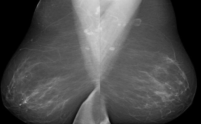
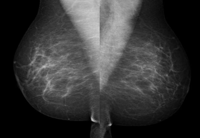
Mammogram Mammogram Fibrocystic Change views

Table: Mammogram Mammogram Fibrocystic Change views
Views Description
Standard views Mediolateral oblique (MLO) view Better view of the superior lateral quadrant of the breast Quadrant of The Breast Examination of the Breast and axilla Axilla The axilla is a pyramid-shaped space located between the upper thorax and the arm. The axilla has a base, an apex, and 4 walls (anterior, medial, lateral, posterior). The base of the pyramid is made up of the axillary skin. The apex is the axillary inlet, located between the 1st rib, superior border of the scapula, and clavicle. Axilla and Brachial Plexus: Anatomy
Craniocaudal view (CC)
  • Medial part of the breast: lower part of image
  • Lateral part of the breast: upper part of image
Specialized views Spot compression Compression Blunt Chest Trauma Better visualization of suspicious masses, calcifications, or asymmetric breast tissue
Magnification
XCCL (exaggerated CC lateral) view Modified CC view, focused on the lateral part of breast
XCCM (exaggerated CC medial) view Modified CC view, focused on the medial part of the breast

Normal findings

  • Normal breast is composed of:
    • Parenchyma (ducts and lobules Lobules Breasts: Anatomy)
    • Connective tissue Connective tissue Connective tissues originate from embryonic mesenchyme and are present throughout the body except inside the brain and spinal cord. The main function of connective tissues is to provide structural support to organs. Connective tissues consist of cells and an extracellular matrix. Connective Tissue: Histology
    • Fat
  • Breast findings:
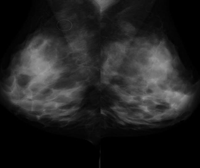
    • Brighter signal represents fibroglandular tissue.
    • Gray areas represent adipose tissue Adipose tissue Adipose tissue is a specialized type of connective tissue that has both structural and highly complex metabolic functions, including energy storage, glucose homeostasis, and a multitude of endocrine capabilities. There are three types of adipose tissue, white adipose tissue, brown adipose tissue, and beige or “brite” adipose tissue, which is a transitional form. Adipose Tissue: Histology.
  • Breast density: 
    • Findings can be:
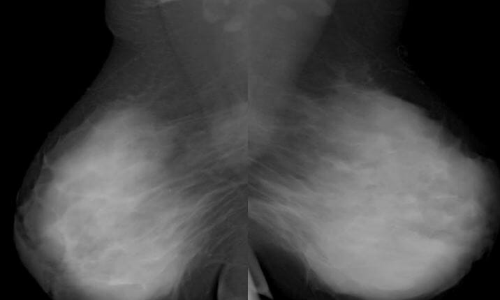
      • Extremely dense (which can obscure lesions or cancers)
      • Heterogeneously dense 
      • Scattered fibroglandular 
      • Almost entirely fatty
    • Young women typically have denser breast tissue.
    • Older women have a higher amount of fatty tissue.

Mammographic assessment categories

Table: Mammographic assessment categories
Category Assessment Follow-up
BI-RADS 0 Incomplete assessment Additional mammography Mammography Radiographic examination of the breast. Breast Cancer Screening views or ultrasound follow-up needed
BI-RADS 1 Negative Continue with routine screening Screening Preoperative Care
BI-RADS 2 Benign Benign Fibroadenoma findings Continue with routine screening Screening Preoperative Care
BI-RADS 3 Probably benign Benign Fibroadenoma findings Diagnostic mammography Mammography Radiographic examination of the breast. Breast Cancer Screening or ultrasound in 6 months
BI-RADS 4 Suspicious abnormality Biopsy Biopsy Removal and pathologic examination of specimens from the living body. Ewing Sarcoma should be considered.
BI-RADS 5 Highly suggestive of malignancy Malignancy Hemothorax Biopsy Biopsy Removal and pathologic examination of specimens from the living body. Ewing Sarcoma should be performed.
BI-RADS 6 Biopsy-proven malignancy Malignancy Hemothorax Management for breast cancer Breast cancer Breast cancer is a disease characterized by malignant transformation of the epithelial cells of the breast. Breast cancer is the most common form of cancer and 2nd most common cause of cancer-related death among women. Breast Cancer
BI-RADS: Breast Imaging Reporting and Data System

Breast Ultrasound

Indications

  • Evaluation of any palpable finding (ideal initial study for women who are < 30 years of age, pregnant, or lactating)
  • Further evaluation of abnormal masses found during mammography Mammography Radiographic examination of the breast. Breast Cancer Screening or MRI scans
  • Method for guidance for biopsy Biopsy Removal and pathologic examination of specimens from the living body. Ewing Sarcoma of breast tissue
  • Assessment of the axillary, supraclavicular, and infraclavicular lymph nodes Lymph Nodes They are oval or bean shaped bodies (1 – 30 mm in diameter) located along the lymphatic system. Lymphatic Drainage System: Anatomy in cases of suspicion of breast cancer Breast cancer Breast cancer is a disease characterized by malignant transformation of the epithelial cells of the breast. Breast cancer is the most common form of cancer and 2nd most common cause of cancer-related death among women. Breast Cancer
  • Can infrequently be used as an adjunct to screening Screening Preoperative Care in women with dense breast tissue 

Normal findings

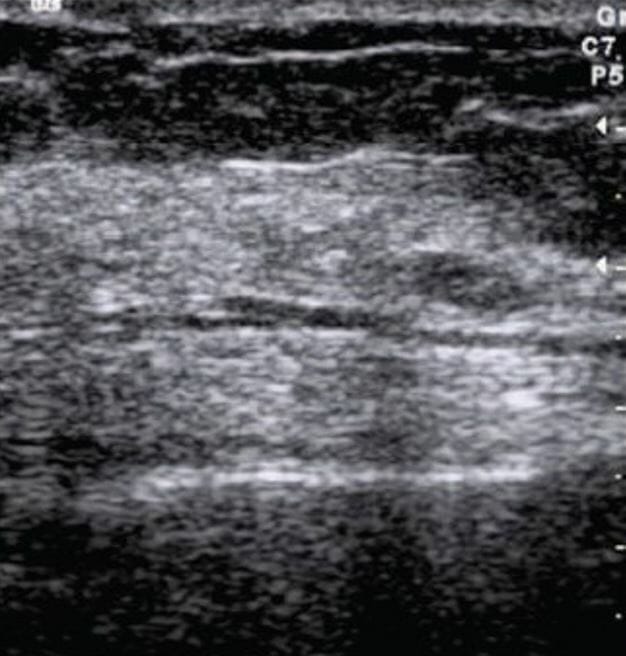
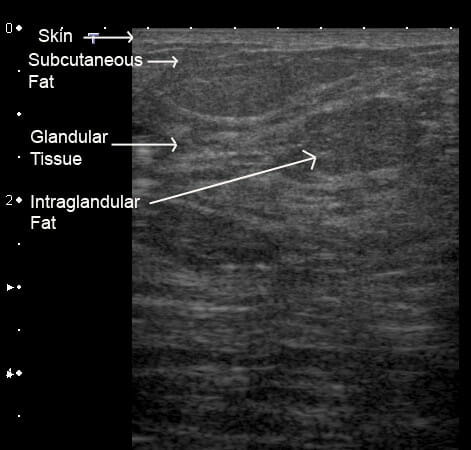
  • Young nonlactating breast:
    • Mainly composed of echogenic fibroglandular tissue, with little to no fat 
    • Greater amounts of adipose tissue Adipose tissue Adipose tissue is a specialized type of connective tissue that has both structural and highly complex metabolic functions, including energy storage, glucose homeostasis, and a multitude of endocrine capabilities. There are three types of adipose tissue, white adipose tissue, brown adipose tissue, and beige or “brite” adipose tissue, which is a transitional form. Adipose Tissue: Histology are deposited with increasing age/ parity Parity The number of offspring a female has borne. It is contrasted with gravidity, which refers to the number of pregnancies, regardless of outcome. Pregnancy: Diagnosis, Physiology, and Care.
  • Lactating breast: prominent fluid-filled ducts with echogenic epithelial lining

Breast MRI

MRI of the breasts Breasts The breasts are found on the anterior thoracic wall and consist of mammary glands surrounded by connective tissue. The mammary glands are modified apocrine sweat glands that produce milk, which serves as nutrition for infants. Breasts are rudimentary and usually nonfunctioning in men. Breasts: Anatomy

Radiologic technique that uses magnetic fields and radiofrequency pulses to produce highly detailed images of the breasts Breasts The breasts are found on the anterior thoracic wall and consist of mammary glands surrounded by connective tissue. The mammary glands are modified apocrine sweat glands that produce milk, which serves as nutrition for infants. Breasts are rudimentary and usually nonfunctioning in men. Breasts: Anatomy.

Indications

  • Screening Screening Preoperative Care:
    • Indicated as supplementary screening Screening Preoperative Care along with mammography Mammography Radiographic examination of the breast. Breast Cancer Screening in women with a high risk of breast cancer Breast cancer Breast cancer is a disease characterized by malignant transformation of the epithelial cells of the breast. Breast cancer is the most common form of cancer and 2nd most common cause of cancer-related death among women. Breast Cancer
      • BRCA1 or BRCA2 mutations
      • Strong family history Family History Adult Health Maintenance of breast and/or ovarian cancer Ovarian cancer Ovarian cancer is a malignant tumor arising from the ovarian tissue and is classified according to the type of tissue from which it originates. The 3 major types of ovarian cancer are epithelial ovarian carcinomas (EOCs), ovarian germ cell tumors (OGCTs), and sex cord-stromal tumors (SCSTs). Ovarian Cancer
      • History of radiation Radiation Emission or propagation of acoustic waves (sound), electromagnetic energy waves (such as light; radio waves; gamma rays; or x-rays), or a stream of subatomic particles (such as electrons; neutrons; protons; or alpha particles). Osteosarcoma therapy to the chest (e.g., for Hodgkin lymphoma Lymphoma A general term for various neoplastic diseases of the lymphoid tissue. Imaging of the Mediastinum)
    • Used as follow-up for an abnormal or inconclusive mammography Mammography Radiographic examination of the breast. Breast Cancer Screening result
  • Detection of occult breast cancer Breast cancer Breast cancer is a disease characterized by malignant transformation of the epithelial cells of the breast. Breast cancer is the most common form of cancer and 2nd most common cause of cancer-related death among women. Breast Cancer in an individual presenting with axillary lymph nodes Lymph Nodes They are oval or bean shaped bodies (1 – 30 mm in diameter) located along the lymphatic system. Lymphatic Drainage System: Anatomy diagnosed with metastatic disease
  • In diagnosed breast cancer Breast cancer Breast cancer is a disease characterized by malignant transformation of the epithelial cells of the breast. Breast cancer is the most common form of cancer and 2nd most common cause of cancer-related death among women. Breast Cancer, MRI aids AIDS Chronic HIV infection and depletion of CD4 cells eventually results in acquired immunodeficiency syndrome (AIDS), which can be diagnosed by the presence of certain opportunistic diseases called AIDS-defining conditions. These conditions include a wide spectrum of bacterial, viral, fungal, and parasitic infections as well as several malignancies and generalized conditions. HIV Infection and AIDS in preoperative evaluation and staging Staging Methods which attempt to express in replicable terms the extent of the neoplasm in the patient. Grading, Staging, and Metastasis, as well as in measurement of response to chemotherapy Chemotherapy Osteosarcoma.
  • Evaluation of breast implants
  • Evaluation of inconclusive mammograms or ultrasounds
  • In cases of dense breast tissue, which hinders other tests

Contraindications Contraindications A condition or factor associated with a recipient that makes the use of a drug, procedure, or physical agent improper or inadvisable. Contraindications may be absolute (life threatening) or relative (higher risk of complications in which benefits may outweigh risks). Noninvasive Ventilation

A breast MRI is contraindicated in MRI-incompatible hardware or anaphylaxis Anaphylaxis An acute hypersensitivity reaction due to exposure to a previously encountered antigen. The reaction may include rapidly progressing urticaria, respiratory distress, vascular collapse, systemic shock, and death. Type I Hypersensitivity Reaction to gadolinium Gadolinium An element of the rare earth family of metals. It has the atomic symbol gd, atomic number 64, and atomic weight 157. 25. Its oxide is used in the control rods of some nuclear reactors. Magnetic Resonance Imaging (MRI).

MRI findings

Findings are grouped into 3 categories:
  • Focus/foci: area of enhancement measuring < 5 mm MM Multiple myeloma (MM) is a malignant condition of plasma cells (activated B lymphocytes) primarily seen in the elderly. Monoclonal proliferation of plasma cells results in cytokine-driven osteoclastic activity and excessive secretion of IgG antibodies. Multiple Myeloma in diameter 
  • Mass:
    • A lesion that occupies a space within the breast
    • The shape, margins, and enhancement pattern of the mass are of special diagnostic value.
    • Benign Benign Fibroadenoma lesions often show progressive enhancement.
    • Among the findings seen in malignancy Malignancy Hemothorax:
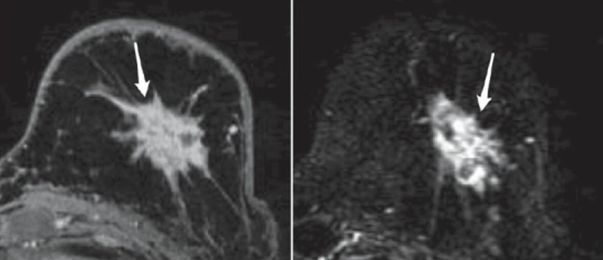
      • Masses with irregular or spiculated margins
      • Rapid uptake and washout of gadolinium Gadolinium An element of the rare earth family of metals. It has the atomic symbol gd, atomic number 64, and atomic weight 157. 25. Its oxide is used in the control rods of some nuclear reactors. Magnetic Resonance Imaging (MRI)
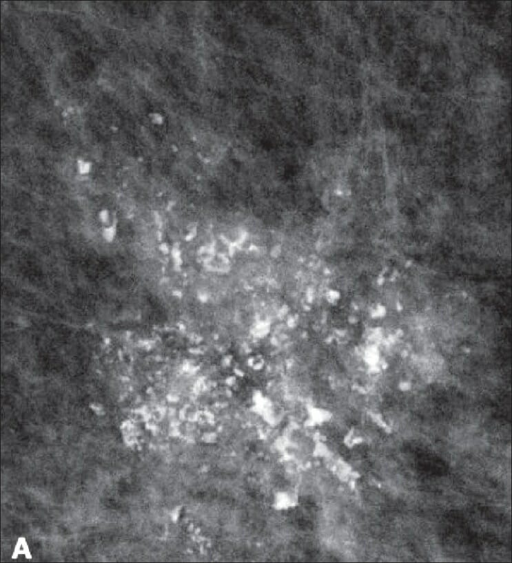
  • Non–mass-like enhancement:
    • Areas of enhancement without a detectable 3-dimensional mass
    • Distribution can be diffuse, regional, segmental, or linear.
    • Enhancement can vary: heterogeneous, homogeneous Homogeneous Imaging of the Spleen, clumped, or clustered

Abnormal Findings

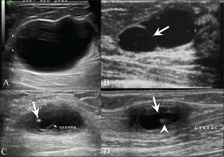
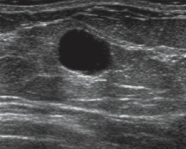
Breast cysts Cysts Any fluid-filled closed cavity or sac that is lined by an epithelium. Cysts can be of normal, abnormal, non-neoplastic, or neoplastic tissues. Fibrocystic Change

  • Ultrasonography findings:
    • Simple cyst:
      • Well-defined round/oval lesion 
      • No internal echo ( anechoic Anechoic A structure that produces no echo at all (looks completely black) Ultrasound (Sonography))
      • Thin, regular Regular Insulin walls
      • Posterior acoustic enhancement
    • Complex cyst:
      • Presence of internal echoes caused by accumulation of pus, blood, or milk
      • Absence of posterior wall enhancement
      • Chronic cysts Cysts Any fluid-filled closed cavity or sac that is lined by an epithelium. Cysts can be of normal, abnormal, non-neoplastic, or neoplastic tissues. Fibrocystic Change may be ill defined.
  • Mammography Mammography Radiographic examination of the breast. Breast Cancer Screening findings:
    • Oval or spherical shape
    • Smooth, well-defined borders

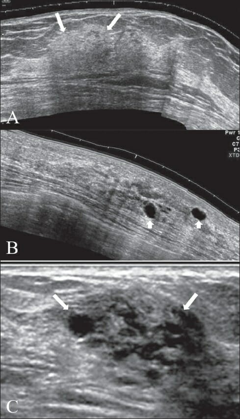
Fibrocystic Fibrocystic Fibrocystic Change changes

  • Ultrasonography findings (highly variable Variable Variables represent information about something that can change. The design of the measurement scales, or of the methods for obtaining information, will determine the data gathered and the characteristics of that data. As a result, a variable can be qualitative or quantitative, and may be further classified into subgroups. Types of Variables):
    • Focal areas of thickening (patchy increase in echogenicity)
    • Single cysts Cysts Any fluid-filled closed cavity or sac that is lined by an epithelium. Cysts can be of normal, abnormal, non-neoplastic, or neoplastic tissues. Fibrocystic Change or clusters of small cysts Cysts Any fluid-filled closed cavity or sac that is lined by an epithelium. Cysts can be of normal, abnormal, non-neoplastic, or neoplastic tissues. Fibrocystic Change
  • Mammography Mammography Radiographic examination of the breast. Breast Cancer Screening findings:
    • Heterogeneous and usually dense parenchyma 
    • Partially circumscribed masses 
    • Tea-cup, low-density round calcifications 

Fibroadenoma Fibroadenoma Fibroadenomas are the most common benign tumor of the female breast and the most common breast tumor in adolescent and young women. The tumors are well-circumscribed, mobile, and unencapsulated, with a rubbery or firm consistency. Fibroadenoma

  • Ultrasonography findings:
    • Well-defined spherical lesion
    • Smooth borders ( capsule Capsule An envelope of loose gel surrounding a bacterial cell which is associated with the virulence of pathogenic bacteria. Some capsules have a well-defined border, whereas others form a slime layer that trails off into the medium. Most capsules consist of relatively simple polysaccharides but there are some bacteria whose capsules are made of polypeptides. Bacteroides)
    • Uniform hypoechogenicity
    • May or may not present with internal echoes
    • Transverse diameter > anteroposterior diameter (insinuating that it does not invade through fascial planes)
  • Mammography Mammography Radiographic examination of the breast. Breast Cancer Screening findings:
    • Well-circumscribed oval or spherical mass
    • Smooth, regular Regular Insulin borders
    • Hypodense or isodense to glandular tissue
    • May present with lobulations
    • Popcorn-shaped calcifications (in involuting fibroadenomas)

Lipoma Lipoma A lipoma is a benign neoplasm of fat cells (adipocytes) and the most common soft tissue tumor in adults. The etiology is unknown, but obesity is a predisposing factor; genetics also play a role, with multiple lipomas occurring in various inherited disorders. Lipoma

  • Ultrasonography findings:
    • Well-defined echogenic lesion
    • Soft consistency Consistency Dermatologic Examination; can be deformed by compression Compression Blunt Chest Trauma with the transducer Transducer A device placed on the patient’s body to visualize a target Ultrasound (Sonography)
    • Lamellar appearance and thin capsule Capsule An envelope of loose gel surrounding a bacterial cell which is associated with the virulence of pathogenic bacteria. Some capsules have a well-defined border, whereas others form a slime layer that trails off into the medium. Most capsules consist of relatively simple polysaccharides but there are some bacteria whose capsules are made of polypeptides. Bacteroides
  • Mammography Mammography Radiographic examination of the breast. Breast Cancer Screening findings:
    • Radiolucent Radiolucent An object of low density that is permeable to X-rays (looks black) X-rays mass with no calcification
    • May have a thin, peripheral, fluid density capsule Capsule An envelope of loose gel surrounding a bacterial cell which is associated with the virulence of pathogenic bacteria. Some capsules have a well-defined border, whereas others form a slime layer that trails off into the medium. Most capsules consist of relatively simple polysaccharides but there are some bacteria whose capsules are made of polypeptides. Bacteroides
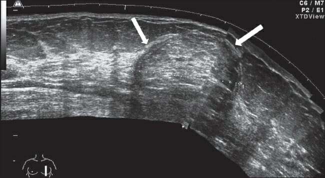
Ultrasound image of a well-defined subtly-echogenic mass

Ultrasound image of a well-defined subtly echogenic mass (lipoma) with a lamellar pattern and a well-defined, thin capsule (arrows)

Image: “Lipoma” by Gokhale, S. License: CC BY 2.0
Breast cancer Breast cancer Breast cancer is a disease characterized by malignant transformation of the epithelial cells of the breast. Breast cancer is the most common form of cancer and 2nd most common cause of cancer-related death among women. Breast Cancer

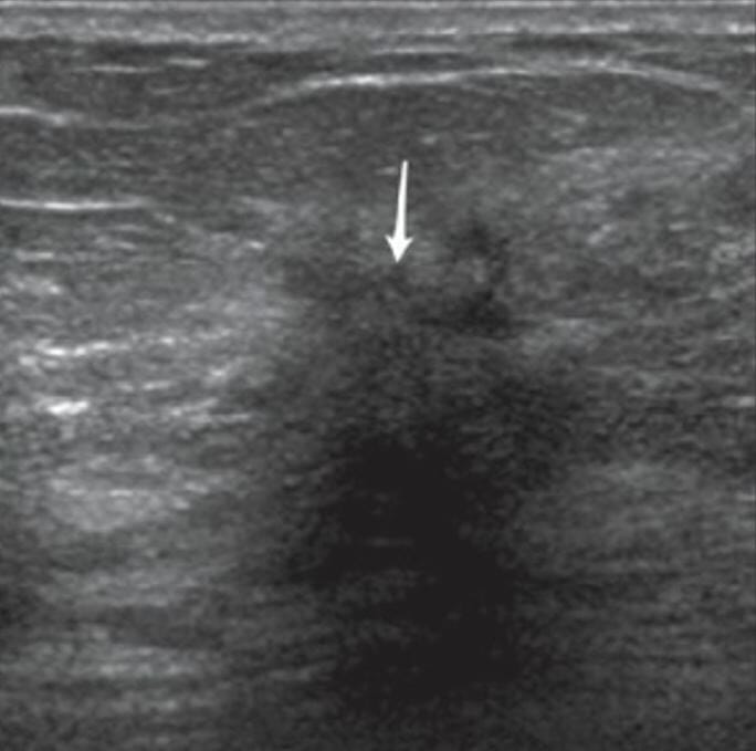
  • Ultrasonographic characteristics of malignant lesions:
    • Usually hypoechoic Hypoechoic A structure that produces a low-amplitude echo (darker grays) Ultrasound (Sonography) 
    • Ill-defined or irregular borders
    • Anteroposterior diameter > transverse diameter (insinuating that it does invade through fascial planes)
    • Spiculated margins
    • Posterior acoustic shadowing
    • Microcalcifications
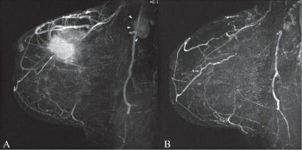
    • Significant vascularity seen on Doppler Doppler Ultrasonography applying the doppler effect, with frequency-shifted ultrasound reflections produced by moving targets (usually red blood cells) in the bloodstream along the ultrasound axis in direct proportion to the velocity of movement of the targets, to determine both direction and velocity of blood flow. Ultrasound (Sonography) imaging
  • Mammography Mammography Radiographic examination of the breast. Breast Cancer Screening:
    • Characteristics of benign Benign Fibroadenoma breast masses:
      • Well-defined, circumscribed mass
      • Radiolucent Radiolucent An object of low density that is permeable to X-rays (looks black) X-rays ring surrounding the lesion ( halo sign Halo sign Aspergillus/Aspergillosis)
      • Diffuse microcalcifications
      • Presence of fat in a mass
    • Characteristics of malignant lesions:
      • Hyperdense masses 
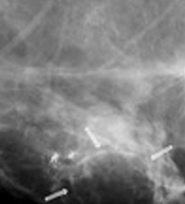
      • Fine linear branching microcalcifications
      • Irregular borders
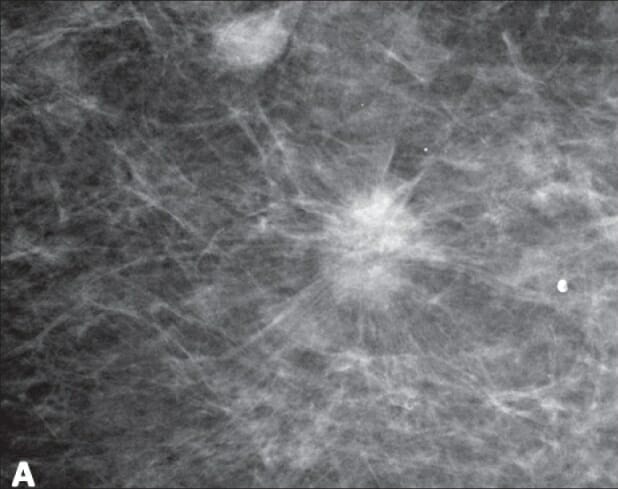
      • Spiculation
      • Architectural distortions
  • MRI findings:
    • Irregular borders
    • Spiculations
    • Segmental distribution and calcifications
    • Enhancing internal septations
    • Perilesional edema Edema Edema is a condition in which excess serous fluid accumulates in the body cavity or interstitial space of connective tissues. Edema is a symptom observed in several medical conditions. It can be categorized into 2 types, namely, peripheral (in the extremities) and internal (in an organ or body cavity). Edema: clustered ring enhancement Ring Enhancement Brain Abscess
    • Heterogeneously enhancing pattern

Fat necrosis Necrosis The death of cells in an organ or tissue due to disease, injury or failure of the blood supply. Ischemic Cell Damage

  • Ultrasonograpy findings:
    • Hypoechoic Hypoechoic A structure that produces a low-amplitude echo (darker grays) Ultrasound (Sonography) mass with well-defined margins
    • May or may not have mural nodule Nodule Chalazion(s)
    • Can produce significant shadowing secondary to coarse calcifications
  • Mammography Mammography Radiographic examination of the breast. Breast Cancer Screening findings:
    • May present as an ill-defined, irregular, spiculated mass (similar to breast cancer Breast cancer Breast cancer is a disease characterized by malignant transformation of the epithelial cells of the breast. Breast cancer is the most common form of cancer and 2nd most common cause of cancer-related death among women. Breast Cancer)
    • Oil cysts Cysts Any fluid-filled closed cavity or sac that is lined by an epithelium. Cysts can be of normal, abnormal, non-neoplastic, or neoplastic tissues. Fibrocystic Change
    • May appear with egg-shell calcifications

Breast hamartoma Hamartoma A focal malformation resembling a neoplasm, composed of an overgrowth of mature cells and tissues that normally occur in the affected area. Colorectal Cancer

  • Benign Benign Fibroadenoma slow-growing breast lesion (uncommon)
  • Contains fat and fibrous Fibrous Fibrocystic Change tissue and presents as painless masses
  • Ultrasonography findings:
    • Margins are difficult to detect because it resembles normal breast tissue.
    • May present with internal echoes that are a mix of hyperechoic Hyperechoic A structure that produces a high-amplitude echo (lighter grays and white) Ultrasound (Sonography) and hypoechoic Hypoechoic A structure that produces a low-amplitude echo (darker grays) Ultrasound (Sonography) components 
  • Mammography Mammography Radiographic examination of the breast. Breast Cancer Screening findings:
    • Well-circumscribed spherical or oval compressible mass with thin capsules
    • Heterogeneous: contains both fat and soft-tissue internal densities
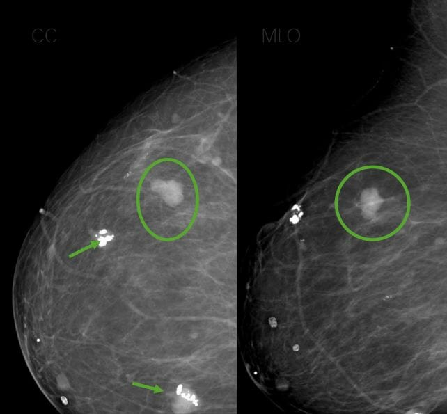
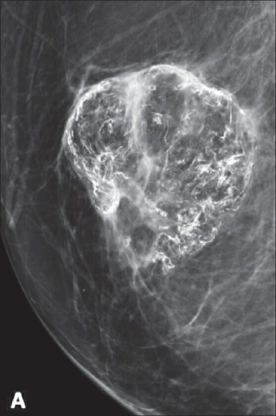
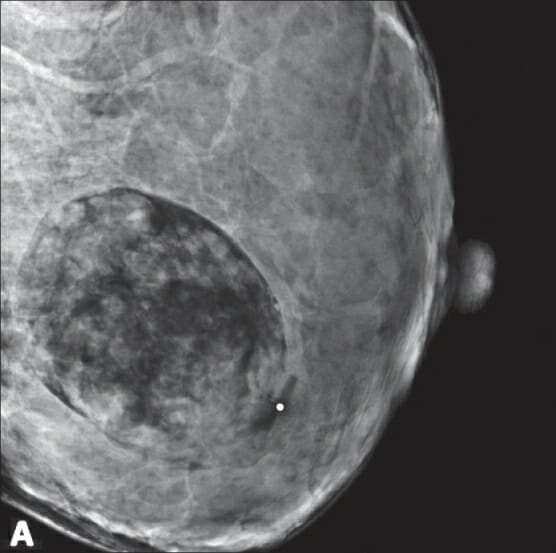
Mammogram showing a fat-containing oval mass, suggestive of hamartoma

Mammogram showing a fat-containing oval mass, suggestive of hamartoma

Image: “A 26-year-old pregnant female with a new palpable mass” by Ojeda-Fournier H, Nguyen JQ. License: CC BY 2.0
References

  1. Aydin, H. (2019). The MRI characteristics of non-mass enhancement lesions of the breast: associations with malignancy. Br J Radiol 92:20180464. https://doi.org/10.1259/bjr.20180464
  2. Farrokh, D., Hashemi, J., Ansaripour, E. (2011). Breast hamartoma: mammographic findings. Iran J Radiol 8:258–260. https://doi.org/10.5812/iranjradiol.4492
  3. Freimanis, R.I., Ayoub, J.S. (2011). Radiology of the breast. Chapter 5 of Chen, M.M., Pope, T.L., Ott, D.J. (Eds.), Basic Radiology, 2nd ed. McGraw Hill. https://accessmedicine.mhmedical.com/content.aspx?bookid=360&sectionid=39669012
  4. Reeves, R.A., Kaufman, T. (2021). Mammography. StatPearls. Retrieved November 23, 2021, from https://www.ncbi.nlm.nih.gov/books/NBK559310/
  5. Slanetz, P. (2021). MRI of the breasts and emerging technologies. UpToDate. Retrieved December 11, 2021, from https://www.uptodate.com/contents/mri-of-the-breast-and-emerging-technologies
  6. Tozaki, M., Igarashi, T., Fukuda, K. (2006). Breast MRI using the VIBE sequence: clustered ring enhancement in the differential diagnosis of lesions showing non-masslike enhancement. AJR Am J Roentgenol 187:313–321. https://pubmed.ncbi.nlm.nih.gov/16861532/

Create your free account or log in to continue reading!

Sign up now and get free access to Lecturio with concept pages, medical videos, and questions for your medical education.

User Reviews

Unwrap New Skills This Holiday 🎄 Save 30% on all plans now!

Details