Breast Cancer

Breast cancer is a disease characterized by malignant transformation Transformation Change brought about to an organism's genetic composition by unidirectional transfer (transfection; transduction, genetic; conjugation, genetic, etc.) and incorporation of foreign DNA into prokaryotic or eukaryotic cells by recombination of part or all of that DNA into the cell's genome. Bacteriology of the epithelial cells of the breast. Breast cancer is the most common form of cancer and 2nd most common cause of cancer-related death among women. Genetic factors, age, and hormonal and environmental influence contribute to the progression of the disease. The most common histologic type is infiltrating ductal carcinoma, which is > 75% of all breast cancers. Screening Screening Preoperative Care mammography Mammography Radiographic examination of the breast. Breast Cancer Screening is recommended for early disease detection. Diagnosis is by core needle biopsy Core Needle Biopsy Fibrocystic Change, with biologic factors determined by immunohistochemical testing. Surgery, systemic treatment ( chemotherapy Chemotherapy Osteosarcoma, biologic therapy, endocrine therapy), and radiation Radiation Emission or propagation of acoustic waves (sound), electromagnetic energy waves (such as light; radio waves; gamma rays; or x-rays), or a stream of subatomic particles (such as electrons; neutrons; protons; or alpha particles). Osteosarcoma therapy (RT) are part of the early-stage and locally advanced disease management. In metastatic breast cancer, systemic treatment is utilized with palliative measures.

Last updated: May 17, 2024

Editorial responsibility: Stanley Oiseth, Lindsay Jones, Evelin Maza

Overview

Epidemiology

  • Breast cancer is the most common cancer in women.
  • Accounts for 29% of all malignant diseases among women in the United States
  • Incidence Incidence The number of new cases of a given disease during a given period in a specified population. It also is used for the rate at which new events occur in a defined population. It is differentiated from prevalence, which refers to all cases in the population at a given time. Measures of Disease Frequency: 125 cases per 100,000 women
  • Risk increases with age, with 90% of cases occurring in women > 40 years of age
  • Male breast cancer accounts for < 1% of total cases.
  • An important cause of death in women:
    • United States: the 2nd leading cause of cancer-related deaths
    • Developing countries: the leading cause of cancer-related deaths
  • Early detection and improved treatments have reduced death rates.

Risk factors

Unmodifiable factors that increase the risk:

  • Family history Family History Adult Health Maintenance:
    • Breast cancer in 1st- or 2nd-degree relatives (mother, grandmother, sister)
    • Ashkenazi Jewish descent
  • Hormonal influences: long hormone exposure due to early menarche Menarche The first menstrual cycle marked by the initiation of menstruation. Menstrual Cycle and/or late menopause Menopause Menopause is a physiologic process in women characterized by the permanent cessation of menstruation that occurs after the loss of ovarian activity. Menopause can only be diagnosed retrospectively, after 12 months without menstrual bleeding. Menopause
  • Genetic mutations Genetic Mutations Carcinogenesis (examples):
    • BRCA1 (on chromosome Chromosome In a prokaryotic cell or in the nucleus of a eukaryotic cell, a structure consisting of or containing DNA which carries the genetic information essential to the cell. Basic Terms of Genetics 17q)
    • BRCA2 (on chromosome Chromosome In a prokaryotic cell or in the nucleus of a eukaryotic cell, a structure consisting of or containing DNA which carries the genetic information essential to the cell. Basic Terms of Genetics 13q)
    • p53 (on chromosome Chromosome In a prokaryotic cell or in the nucleus of a eukaryotic cell, a structure consisting of or containing DNA which carries the genetic information essential to the cell. Basic Terms of Genetics 17)
  • Increasing age
  • Breast cancer on the contralateral side

Modifiable risk factors:

  • Lifestyle factors that increase the risk:
    • High-fat diet
    • Obesity Obesity Obesity is a condition associated with excess body weight, specifically with the deposition of excessive adipose tissue. Obesity is considered a global epidemic. Major influences come from the western diet and sedentary lifestyles, but the exact mechanisms likely include a mixture of genetic and environmental factors. Obesity (especially after menopause Menopause Menopause is a physiologic process in women characterized by the permanent cessation of menstruation that occurs after the loss of ovarian activity. Menopause can only be diagnosed retrospectively, after 12 months without menstrual bleeding. Menopause)
    • Heavy alcohol use
    • Tobacco
  • Hormonal influences that increase the risk:
    • Higher age at 1st delivery (> 30 years of age)
    • Nulliparity
    • Hormone replacement therapy Hormone Replacement Therapy Hormone replacement therapy (HRT) is used to treat symptoms associated with female menopause and in combination to suppress ovulation. Risks and side effects include uterine bleeding, predisposition to cancer, breast tenderness, hyperpigmentation, migraine headaches, hypertension, bloating, and mood changes. Noncontraceptive Estrogen and Progestins after menopause Menopause Menopause is a physiologic process in women characterized by the permanent cessation of menstruation that occurs after the loss of ovarian activity. Menopause can only be diagnosed retrospectively, after 12 months without menstrual bleeding. Menopause (> 5 years)
  • Hormonal influences that decrease risk: breastfeeding Breastfeeding Breastfeeding is often the primary source of nutrition for the newborn. During pregnancy, hormonal stimulation causes the number and size of mammary glands in the breast to significantly increase. After delivery, prolactin stimulates milk production, while oxytocin stimulates milk expulsion through the lactiferous ducts, where it is sucked out through the nipple by the infant. Breastfeeding for at least 6 months

Mnemonics:

BReast- CA CA Condylomata acuminata are a clinical manifestation of genital HPV infection. Condylomata acuminata are described as raised, pearly, flesh-colored, papular, cauliflower-like lesions seen in the anogenital region that may cause itching, pain, or bleeding. Condylomata Acuminata (Genital Warts)ncer 1 and 2” = Mutated genes Genes A category of nucleic acid sequences that function as units of heredity and which code for the basic instructions for the development, reproduction, and maintenance of organisms. DNA Types and Structure are the BRCA1 and BRCA2 genes Genes A category of nucleic acid sequences that function as units of heredity and which code for the basic instructions for the development, reproduction, and maintenance of organisms. DNA Types and Structure.

Histologic classification

Non-invasive:

  • Ductal carcinoma in situ (DCIS)
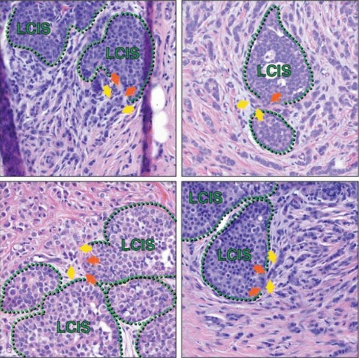
  • Lobular carcinoma in situ (LCIS)

Invasive:

  • Infiltrating ductal (most common)
  • Infiltrating lobular
  • Ductal/lobular
  • Mucinous/ colloid Colloid Colloid solutions include large proteins or cells that do not readily cross capillary membranes. They remain in the ecf and do not distribute into the icf (similar to crystalloids). Intravenous Fluids
  • Tubular
  • Medullary 
  • Micropapillary

Other clinical forms:

  • Paget’s disease of the breast
  • Inflammatory carcinoma

Molecular classification

Based on expression of:

  • Estrogen Estrogen Compounds that interact with estrogen receptors in target tissues to bring about the effects similar to those of estradiol. Estrogens stimulate the female reproductive organs, and the development of secondary female sex characteristics. Estrogenic chemicals include natural, synthetic, steroidal, or non-steroidal compounds. Ovaries: Anatomy receptors Receptors Receptors are proteins located either on the surface of or within a cell that can bind to signaling molecules known as ligands (e.g., hormones) and cause some type of response within the cell. Receptors (ERs)
  • Progesterone Progesterone The major progestational steroid that is secreted primarily by the corpus luteum and the placenta. Progesterone acts on the uterus, the mammary glands and the brain. It is required in embryo implantation; pregnancy maintenance, and the development of mammary tissue for milk production. Progesterone, converted from pregnenolone, also serves as an intermediate in the biosynthesis of gonadal steroid hormones and adrenal corticosteroids. Gonadal Hormones receptors Receptors Receptors are proteins located either on the surface of or within a cell that can bind to signaling molecules known as ligands (e.g., hormones) and cause some type of response within the cell. Receptors ( PRs PRS Pierre robin sequence, also known as pierre robin syndrome or simply robin sequence, is a condition in infants that is characterized by a smaller-than-normal mandible, a tongue that retracts back into the throat, and difficulty breathing. Pierre Robin Sequence)
  • Human epidermal growth factor receptor 2 Human epidermal growth factor receptor 2 A cell surface protein-tyrosine kinase receptor that is overexpressed in a variety of adenocarcinomas. It has extensive homology to and heterodimerizes with the EGF receptor, the ERBB-3 receptor, and the ERBB-4 receptor. Activation of the erbB-2 receptor occurs through heterodimer formation with a ligand-bound erbB receptor family member. Targeted and Other Nontraditional Antineoplastic Therapy ( HER2 HER2 A cell surface protein-tyrosine kinase receptor that is overexpressed in a variety of adenocarcinomas. It has extensive homology to and heterodimerizes with the EGF receptor, the ERBB-3 receptor, and the ERBB-4 receptor. Activation of the erbB-2 receptor occurs through heterodimer formation with a ligand-bound erbB receptor family member. Targeted and Other Nontraditional Antineoplastic Therapy)
  • Cell proliferation regulator/protein (Ki-67)

Molecular types:

  • Luminal A:
    • ER positive, HER2 HER2 A cell surface protein-tyrosine kinase receptor that is overexpressed in a variety of adenocarcinomas. It has extensive homology to and heterodimerizes with the EGF receptor, the ERBB-3 receptor, and the ERBB-4 receptor. Activation of the erbB-2 receptor occurs through heterodimer formation with a ligand-bound erbB receptor family member. Targeted and Other Nontraditional Antineoplastic Therapy negative
    • PR positive, low Ki-67 (low proliferation)
    • Favorable prognosis Prognosis A prediction of the probable outcome of a disease based on a individual’s condition and the usual course of the disease as seen in similar situations. Non-Hodgkin Lymphomas
  • Luminal B:
    • ER positive, HER2 HER2 A cell surface protein-tyrosine kinase receptor that is overexpressed in a variety of adenocarcinomas. It has extensive homology to and heterodimerizes with the EGF receptor, the ERBB-3 receptor, and the ERBB-4 receptor. Activation of the erbB-2 receptor occurs through heterodimer formation with a ligand-bound erbB receptor family member. Targeted and Other Nontraditional Antineoplastic Therapy negative/positive
    • PR positive, high Ki-67 (high proliferation)
    • Worse prognosis Prognosis A prediction of the probable outcome of a disease based on a individual’s condition and the usual course of the disease as seen in similar situations. Non-Hodgkin Lymphomas than luminal A
  • HER2 HER2 A cell surface protein-tyrosine kinase receptor that is overexpressed in a variety of adenocarcinomas. It has extensive homology to and heterodimerizes with the EGF receptor, the ERBB-3 receptor, and the ERBB-4 receptor. Activation of the erbB-2 receptor occurs through heterodimer formation with a ligand-bound erbB receptor family member. Targeted and Other Nontraditional Antineoplastic Therapy amplified:
    • Overexpress HER2 HER2 A cell surface protein-tyrosine kinase receptor that is overexpressed in a variety of adenocarcinomas. It has extensive homology to and heterodimerizes with the EGF receptor, the ERBB-3 receptor, and the ERBB-4 receptor. Activation of the erbB-2 receptor occurs through heterodimer formation with a ligand-bound erbB receptor family member. Targeted and Other Nontraditional Antineoplastic Therapy
    • ER negative, PR negative
    • Grows faster than luminal cancers
  • Basal-like or triple-negative breast cancers (TNBCs):
    • Negative for ER, PR, and HER2 HER2 A cell surface protein-tyrosine kinase receptor that is overexpressed in a variety of adenocarcinomas. It has extensive homology to and heterodimerizes with the EGF receptor, the ERBB-3 receptor, and the ERBB-4 receptor. Activation of the erbB-2 receptor occurs through heterodimer formation with a ligand-bound erbB receptor family member. Targeted and Other Nontraditional Antineoplastic Therapy
    • More common in African American women

Pathophysiology

Familial breast cancer

  • 25%–30% of breast cancers are associated with susceptibility genes Genes A category of nucleic acid sequences that function as units of heredity and which code for the basic instructions for the development, reproduction, and maintenance of organisms. DNA Types and Structure.
  • Tumor Tumor Inflammation suppressor genes Genes A category of nucleic acid sequences that function as units of heredity and which code for the basic instructions for the development, reproduction, and maintenance of organisms. DNA Types and Structure (TSGs):
  • TSG mutations:
    • BRCA1 (on chromosome Chromosome In a prokaryotic cell or in the nucleus of a eukaryotic cell, a structure consisting of or containing DNA which carries the genetic information essential to the cell. Basic Terms of Genetics 17q21) and BRCA2 (on chromosome Chromosome In a prokaryotic cell or in the nucleus of a eukaryotic cell, a structure consisting of or containing DNA which carries the genetic information essential to the cell. Basic Terms of Genetics 13q12.3) mutations (familial breast and ovarian cancer Ovarian cancer Ovarian cancer is a malignant tumor arising from the ovarian tissue and is classified according to the type of tissue from which it originates. The 3 major types of ovarian cancer are epithelial ovarian carcinomas (EOCs), ovarian germ cell tumors (OGCTs), and sex cord-stromal tumors (SCSTs). Ovarian Cancer):
      • 80%–90% of single gene Gene A category of nucleic acid sequences that function as units of heredity and which code for the basic instructions for the development, reproduction, and maintenance of organisms. Basic Terms of Genetics familial breast cancers
      • At-risk population: strong family history Family History Adult Health Maintenance, certain ethnic groups (Ashkenazi Jewish descent)
      • Associated with poorly differentiated cancer
    • TP53 (Li-Fraumeni syndrome)
    • PTEN ( Cowden syndrome Cowden Syndrome Cowden syndrome, also known as multiple hamartoma syndrome, is an autosomal dominant inherited condition that presents with multiple, noncancerous growths on various parts of the body. The syndrome is classified as a phosphatase and tensin homolog (PTEN) hamartoma tumor syndrome that is caused by mutations in the PTEN gene. Cowden Syndrome)
    • STK11 ( Peutz-Jeghers syndrome Peutz-Jeghers Syndrome Peutz-Jeghers syndrome (PJS) is an autosomal-dominant inherited disorder characterized by GI polyps and mucocutaneous-pigmented macules. Peutz-Jeghers syndrome is 1 of the polyposis syndromes, a group of inherited or acquired conditions characterized by the growth of polyps in the GI tract and associated with other extracolonic features. Peutz-Jeghers Syndrome)
    • CDH1 (hereditary diffuse gastric cancer Gastric cancer Gastric cancer is the 3rd-most common cause of cancer-related deaths worldwide. The majority of cases are from adenocarcinoma. The modifiable risk factors include Helicobacter pylori infection, smoking, and nitrate-rich diets. Gastric Cancer syndrome)
    • PALPB2 (hereditary breast cancer)
  • Normally functioning TSGs induce cell cycle Cell cycle The phases of the cell cycle include interphase (G1, S, and G2) and mitosis (prophase, metaphase, anaphase, and telophase). The cell’s progression through these phases is punctuated by checkpoints regulated by cyclins, cyclin-dependent kinases, tumor suppressors, and their antagonists. Cell Cycle arrest and deoxyribonucleic acid Deoxyribonucleic acid A deoxyribonucleotide polymer that is the primary genetic material of all cells. Eukaryotic and prokaryotic organisms normally contain DNA in a double-stranded state, yet several important biological processes transiently involve single-stranded regions. DNA, which consists of a polysugar-phosphate backbone possessing projections of purines (adenine and guanine) and pyrimidines (thymine and cytosine), forms a double helix that is held together by hydrogen bonds between these purines and pyrimidines (adenine to thymine and guanine to cytosine). DNA Types and Structure ( DNA DNA A deoxyribonucleotide polymer that is the primary genetic material of all cells. Eukaryotic and prokaryotic organisms normally contain DNA in a double-stranded state, yet several important biological processes transiently involve single-stranded regions. DNA, which consists of a polysugar-phosphate backbone possessing projections of purines (adenine and guanine) and pyrimidines (thymine and cytosine), forms a double helix that is held together by hydrogen bonds between these purines and pyrimidines (adenine to thymine and guanine to cytosine). DNA Types and Structure) repair in the setting of DNA damage DNA Damage Injuries to DNA that introduce deviations from its normal, intact structure and which may, if left unrepaired, result in a mutation or a block of DNA replication. These deviations may be caused by physical or chemical agents and occur by natural or unnatural, introduced circumstances. They include the introduction of illegitimate bases during replication or by deamination or other modification of bases; the loss of a base from the DNA backbone leaving an abasic site; single-strand breaks; double strand breaks; and intrastrand (pyrimidine dimers) or interstrand crosslinking. Damage can often be repaired (DNA repair). If the damage is extensive, it can induce apoptosis. DNA Repair Mechanisms.
  • Impaired function of TSGs → ↑ DNA damage DNA Damage Injuries to DNA that introduce deviations from its normal, intact structure and which may, if left unrepaired, result in a mutation or a block of DNA replication. These deviations may be caused by physical or chemical agents and occur by natural or unnatural, introduced circumstances. They include the introduction of illegitimate bases during replication or by deamination or other modification of bases; the loss of a base from the DNA backbone leaving an abasic site; single-strand breaks; double strand breaks; and intrastrand (pyrimidine dimers) or interstrand crosslinking. Damage can often be repaired (DNA repair). If the damage is extensive, it can induce apoptosis. DNA Repair Mechanisms → ↑ oncogenic mutations

Sporadic Sporadic Selective IgA Deficiency breast cancer

  • Tumor Tumor Inflammation progression:
    • Breast cancer occurs from malignant transformation Transformation Change brought about to an organism’s genetic composition by unidirectional transfer (transfection; transduction, genetic; conjugation, genetic, etc.) and incorporation of foreign DNA into prokaryotic or eukaryotic cells by recombination of part or all of that DNA into the cell’s genome. Bacteriology of secretory epithelial cells that are part of the normal breast epithelium Epithelium The epithelium is a complex of specialized cellular organizations arranged into sheets and lining cavities and covering the surfaces of the body. The cells exhibit polarity, having an apical and a basal pole. Structures important for the epithelial integrity and function involve the basement membrane, the semipermeable sheet on which the cells rest, and interdigitations, as well as cellular junctions. Surface Epithelium: Histology (2 layers):
      • Basal myo-epithelial layer
      • Luminal epithelial layer
    • Early genetic mutations Genetic Mutations Carcinogenesis cause proliferative epithelial replacement.
    • Additional genetic events + hormone signaling → increased and abnormal overgrowth ( hyperplasia Hyperplasia An increase in the number of cells in a tissue or organ without tumor formation. It differs from hypertrophy, which is an increase in bulk without an increase in the number of cells. Cellular Adaptation, precursor lesion(s)) → carcinoma in situ → eventually invasive ductal carcinoma
    • The morphology and clinical behavior depend on the molecular type.
  • In the luminal type (ER positive/ HER2 HER2 A cell surface protein-tyrosine kinase receptor that is overexpressed in a variety of adenocarcinomas. It has extensive homology to and heterodimerizes with the EGF receptor, the ERBB-3 receptor, and the ERBB-4 receptor. Activation of the erbB-2 receptor occurs through heterodimer formation with a ligand-bound erbB receptor family member. Targeted and Other Nontraditional Antineoplastic Therapy negative):
    • Up to 65% of breast malignancies
    • Estrogen Estrogen Compounds that interact with estrogen receptors in target tissues to bring about the effects similar to those of estradiol. Estrogens stimulate the female reproductive organs, and the development of secondary female sex characteristics. Estrogenic chemicals include natural, synthetic, steroidal, or non-steroidal compounds. Ovaries: Anatomy exposure (major factor):
      • ↑ Growth factors (e.g., transforming growth factor ɑ)
      • Stimulate epithelial cell proliferation ( puberty Puberty Puberty is a complex series of physical, psychosocial, and cognitive transitions usually experienced by adolescents (11-19 years of age). Puberty is marked by a growth in stature and the development of secondary sexual characteristics, achievement of fertility, and changes in most body systems. Puberty, menses Menses The periodic shedding of the endometrium and associated menstrual bleeding in the menstrual cycle of humans and primates. Menstruation is due to the decline in circulating progesterone, and occurs at the late luteal phase when luteolysis of the corpus luteum takes place. Menstrual Cycle, pregnancy Pregnancy The status during which female mammals carry their developing young (embryos or fetuses) in utero before birth, beginning from fertilization to birth. Pregnancy: Diagnosis, Physiology, and Care), increasing the risk of malignant transformation Transformation Change brought about to an organism’s genetic composition by unidirectional transfer (transfection; transduction, genetic; conjugation, genetic, etc.) and incorporation of foreign DNA into prokaryotic or eukaryotic cells by recombination of part or all of that DNA into the cell’s genome. Bacteriology
      • ↑ Age and ↑ estrogen Estrogen Compounds that interact with estrogen receptors in target tissues to bring about the effects similar to those of estradiol. Estrogens stimulate the female reproductive organs, and the development of secondary female sex characteristics. Estrogenic chemicals include natural, synthetic, steroidal, or non-steroidal compounds. Ovaries: Anatomy exposure with more menstrual cycles → ↑ breast cancer risk
    • ER-positive precursor lesions: flat atypia Atypia Fibrocystic Change and atypical ductal and lobular hyperplasia Hyperplasia An increase in the number of cells in a tissue or organ without tumor formation. It differs from hypertrophy, which is an increase in bulk without an increase in the number of cells. Cellular Adaptation (more associated with luminal A)
  • In the HER2-positive/amplified type:
    • Up to 20% of breast cancers
    • Associated with amplification of the HER2 HER2 A cell surface protein-tyrosine kinase receptor that is overexpressed in a variety of adenocarcinomas. It has extensive homology to and heterodimerizes with the EGF receptor, the ERBB-3 receptor, and the ERBB-4 receptor. Activation of the erbB-2 receptor occurs through heterodimer formation with a ligand-bound erbB receptor family member. Targeted and Other Nontraditional Antineoplastic Therapy gene Gene A category of nucleic acid sequences that function as units of heredity and which code for the basic instructions for the development, reproduction, and maintenance of organisms. Basic Terms of Genetics ( chromosome Chromosome In a prokaryotic cell or in the nucleus of a eukaryotic cell, a structure consisting of or containing DNA which carries the genetic information essential to the cell. Basic Terms of Genetics 17q)
    • HER2 HER2 A cell surface protein-tyrosine kinase receptor that is overexpressed in a variety of adenocarcinomas. It has extensive homology to and heterodimerizes with the EGF receptor, the ERBB-3 receptor, and the ERBB-4 receptor. Activation of the erbB-2 receptor occurs through heterodimer formation with a ligand-bound erbB receptor family member. Targeted and Other Nontraditional Antineoplastic Therapy (also called ERBB2 ErbB2 A cell surface protein-tyrosine kinase receptor that is overexpressed in a variety of adenocarcinomas. It has extensive homology to and heterodimerizes with the EGF receptor, the ERBB-3 receptor, and the ERBB-4 receptor. Activation of the erbB-2 receptor occurs through heterodimer formation with a ligand-bound erbB receptor family member. Targeted and Other Nontraditional Antineoplastic Therapy): protein that promotes proliferation, and opposes apoptosis Apoptosis A regulated cell death mechanism characterized by distinctive morphologic changes in the nucleus and cytoplasm, including the endonucleolytic cleavage of genomic DNA, at regularly spaced, internucleosomal sites, I.e., DNA fragmentation. It is genetically-programmed and serves as a balance to mitosis in regulating the size of animal tissues and in mediating pathologic processes associated with tumor growth. Ischemic Cell Damage
    • Used to have a poor outcome but with biologic therapy ( trastuzumab Trastuzumab A humanized monoclonal antibody against the ErbB-2 receptor (HER2). As an antineoplastic agent, it is used to treat breast cancer where HER2 is overexpressed. Targeted and Other Nontraditional Antineoplastic Therapy), prognosis Prognosis A prediction of the probable outcome of a disease based on a individual’s condition and the usual course of the disease as seen in similar situations. Non-Hodgkin Lymphomas has improved
    • No definite precursor lesion
  • In TNBCs:
    • 15% of breast cancers
    • Basal-like: have markers characterizing the basal myoepithelial cells
    • Associated with genomic instability from defects in DNA DNA A deoxyribonucleotide polymer that is the primary genetic material of all cells. Eukaryotic and prokaryotic organisms normally contain DNA in a double-stranded state, yet several important biological processes transiently involve single-stranded regions. DNA, which consists of a polysugar-phosphate backbone possessing projections of purines (adenine and guanine) and pyrimidines (thymine and cytosine), forms a double helix that is held together by hydrogen bonds between these purines and pyrimidines (adenine to thymine and guanine to cytosine). DNA Types and Structure repair
    • Possible precursor lesion: cells with TP53 mutations
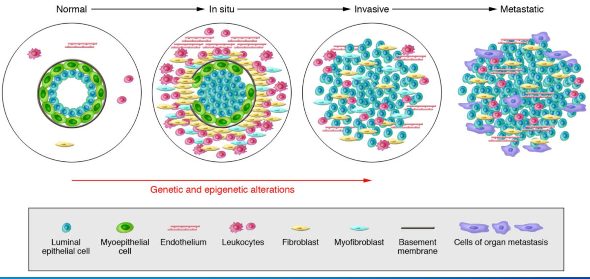
Model of breast tumor progression

Model of breast tumor progression
From left to right: Normal breast ducts are composed of the basement membrane and a layer of luminal epithelial and myoepithelial cells. The stroma includes various leukocytes, fibroblasts, myofibroblasts, and endothelial cells. In in situ carcinomas: The myoepithelial cells are epigenetically and phenotypically altered and their number decreases, potentially due to degradation of the basement membrane. The stromal fibroblasts, myofibroblasts, lymphocytes, and endothelial cells increase. In invasive carcinomas, there is a loss of myoepithelial cells and basement membrane, in which tumor cells can invade surrounding tissues. The tumor cells migrate to distant organs, eventually leading to metastases.

Image: “Hypothetical model of breast tumor progression” by Kornelia Polyak. License: CC BY 4.0.

Clinical Presentation

Clinical findings

In areas with established breast cancer screening Screening Preoperative Care: Most cases of cancer are diagnosed by having an abnormal mammogram Mammogram Fibrocystic Change.

Symptoms:

  • Patient feels a lump/ mass Mass Three-dimensional lesion that occupies a space within the breast Imaging of the Breast.
  • Skin Skin The skin, also referred to as the integumentary system, is the largest organ of the body. The skin is primarily composed of the epidermis (outer layer) and dermis (deep layer). The epidermis is primarily composed of keratinocytes that undergo rapid turnover, while the dermis contains dense layers of connective tissue. Skin: Structure and Functions changes (dimpling, erythema Erythema Redness of the skin produced by congestion of the capillaries. This condition may result from a variety of disease processes. Chalazion, thickening)
  • Nipple Nipple The conic organs which usually give outlet to milk from the mammary glands. Examination of the Breast changes (appearance, discharge)

Signs:

  • Firm or hard mass Mass Three-dimensional lesion that occupies a space within the breast Imaging of the Breast with poorly defined margins, fixed or immovable (on its own not enough to distinguish malignancy Malignancy Hemothorax)
  • Location:
    • Highest frequency: upper outer quadrant (50% of cases)
    • Lowest frequency: lower inner quadrant (5%)
  • Possible to have multifocal Multifocal Retinoblastoma/multiple foci in the same quadrant 
  • Possible to have different quadrants of the breasts Breasts The breasts are found on the anterior thoracic wall and consist of mammary glands surrounded by connective tissue. The mammary glands are modified apocrine sweat glands that produce milk, which serves as nutrition for infants. Breasts are rudimentary and usually nonfunctioning in men. Breasts: Anatomy affected (multicentric)
  • Contralateral breast also affected by the tumor Tumor Inflammation in 5%–10% of cases
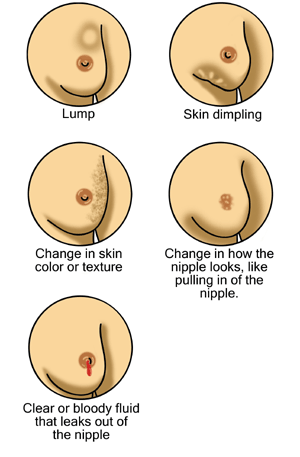
Possible signs of breast cancer

Possible signs of breast cancer
From right to left, top to bottom: a breast lump/mass, skin dimpling, change in skin color/texture, nipple changes including retraction (pulling in of the nipple), and nipple discharge
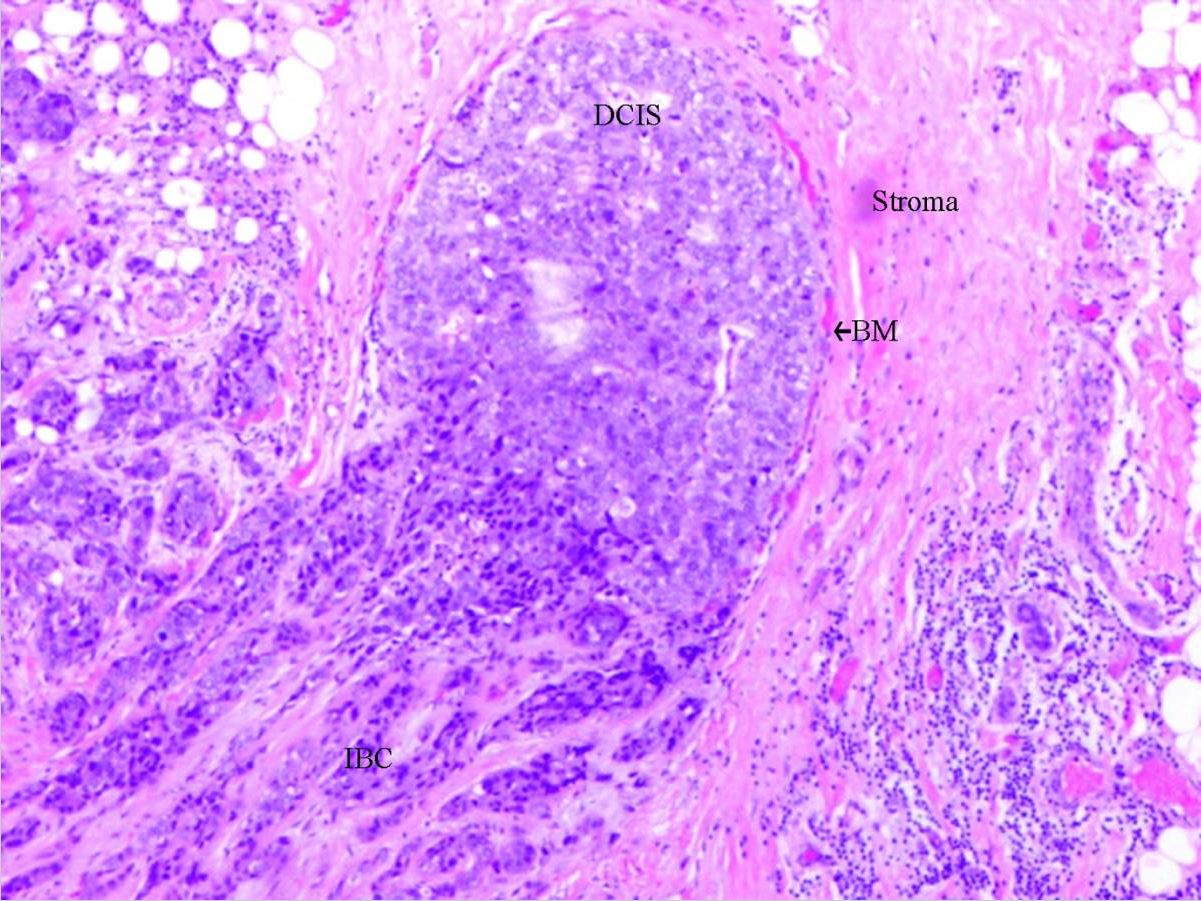
Image: “Early signs of breast cancer” by Morning2k. License: Public domain.

Metastasis Metastasis The transfer of a neoplasm from one organ or part of the body to another remote from the primary site. Grading, Staging, and Metastasis

  • Presentation depends on organ(s) involved
  • Most common sites:
    • Bone Bone Bone is a compact type of hardened connective tissue composed of bone cells, membranes, an extracellular mineralized matrix, and central bone marrow. The 2 primary types of bone are compact and spongy. Bones: Structure and Types (back or leg Leg The lower leg, or just “leg” in anatomical terms, is the part of the lower limb between the knee and the ankle joint. The bony structure is composed of the tibia and fibula bones, and the muscles of the leg are grouped into the anterior, lateral, and posterior compartments by extensions of fascia. Leg: Anatomy pain Pain An unpleasant sensation induced by noxious stimuli which are detected by nerve endings of nociceptive neurons. Pain: Types and Pathways)
    • Liver Liver The liver is the largest gland in the human body. The liver is found in the superior right quadrant of the abdomen and weighs approximately 1.5 kilograms. Its main functions are detoxification, metabolism, nutrient storage (e.g., iron and vitamins), synthesis of coagulation factors, formation of bile, filtration, and storage of blood. Liver: Anatomy ( jaundice Jaundice Jaundice is the abnormal yellowing of the skin and/or sclera caused by the accumulation of bilirubin. Hyperbilirubinemia is caused by either an increase in bilirubin production or a decrease in the hepatic uptake, conjugation, or excretion of bilirubin. Jaundice, abdominal pain Abdominal Pain Acute Abdomen, nausea Nausea An unpleasant sensation in the stomach usually accompanied by the urge to vomit. Common causes are early pregnancy, sea and motion sickness, emotional stress, intense pain, food poisoning, and various enteroviruses. Antiemetics, abnormal liver Liver The liver is the largest gland in the human body. The liver is found in the superior right quadrant of the abdomen and weighs approximately 1.5 kilograms. Its main functions are detoxification, metabolism, nutrient storage (e.g., iron and vitamins), synthesis of coagulation factors, formation of bile, filtration, and storage of blood. Liver: Anatomy tests)
    • Lungs Lungs Lungs are the main organs of the respiratory system. Lungs are paired viscera located in the thoracic cavity and are composed of spongy tissue. The primary function of the lungs is to oxygenate blood and eliminate CO2. Lungs: Anatomy ( shortness of breath Shortness of breath Dyspnea is the subjective sensation of breathing discomfort. Dyspnea is a normal manifestation of heavy physical or psychological exertion, but also may be caused by underlying conditions (both pulmonary and extrapulmonary). Dyspnea, cough, abnormal chest imaging)

Non-invasive Breast Cancer

DCIS

  • Proliferation of cytologically malignant cells within the mammary ductal system, with no invasion of the surrounding stroma
  • ⅓ develop invasive cancer in 5 years
  • Frequently detected by mammography Mammography Radiographic examination of the breast. Breast Cancer Screening

LCIS

  • Proliferation of malignant cells within the lobules Lobules Breasts: Anatomy, growing in an incohesive manner
  • Mucin-positive signet cell rings usually noted
  • Loss of cellular adhesion Adhesion The process whereby platelets adhere to something other than platelets, e.g., collagen; basement membrane; microfibrils; or other ‘foreign’ surfaces. Coagulation Studies also observed (from dysfunction of E-cadherin E-cadherin Calcium-dependent cell adhesion proteins. They are important in the formation of adherens junctions between cells. Cadherins are classified by their distinct immunological and tissue specificities, either by letters (e- for epithelial, n- for neural, and p- for placental cadherins) or by numbers (cadherin-12 or n-cadherin 2 for brain-cadherin). Cadherins promote cell adhesion via a homophilic mechanism as in the construction of tissues and of the whole animal body. Gastric Cancer)
  • Rarely with calcifications ( incidence Incidence The number of new cases of a given disease during a given period in a specified population. It also is used for the rate at which new events occur in a defined population. It is differentiated from prevalence, which refers to all cases in the population at a given time. Measures of Disease Frequency not changed by mammography Mammography Radiographic examination of the breast. Breast Cancer Screening)

Comparison DCIS versus LCIS

Table: Features of DCIS and LCIS
DCIS LCIS
Presentation Unifocal Unifocal Retinoblastoma Multifocal Multifocal Retinoblastoma
Patterns
  • Comedo: central necrosis Necrosis The death of cells in an organ or tissue due to disease, injury or failure of the blood supply. Ischemic Cell Damage, which can be calcified
  • Cribriform: back-to-back glands
  • Micropapillary: small tufts of cells/protrusions, no fibrovascular cores
  • Papillary: with fibrovascular cores
  • Solid: not as well defined
Solid
Calcification Yes/no Usually no
Risk of invasive breast cancer Higher Lower
Location of cancer Ipsilateral breast Ipsilateral or contralateral
DCIS: ductal carcinoma in situ
LCIS: lobular carcinoma in situ

Invasive Breast Cancer

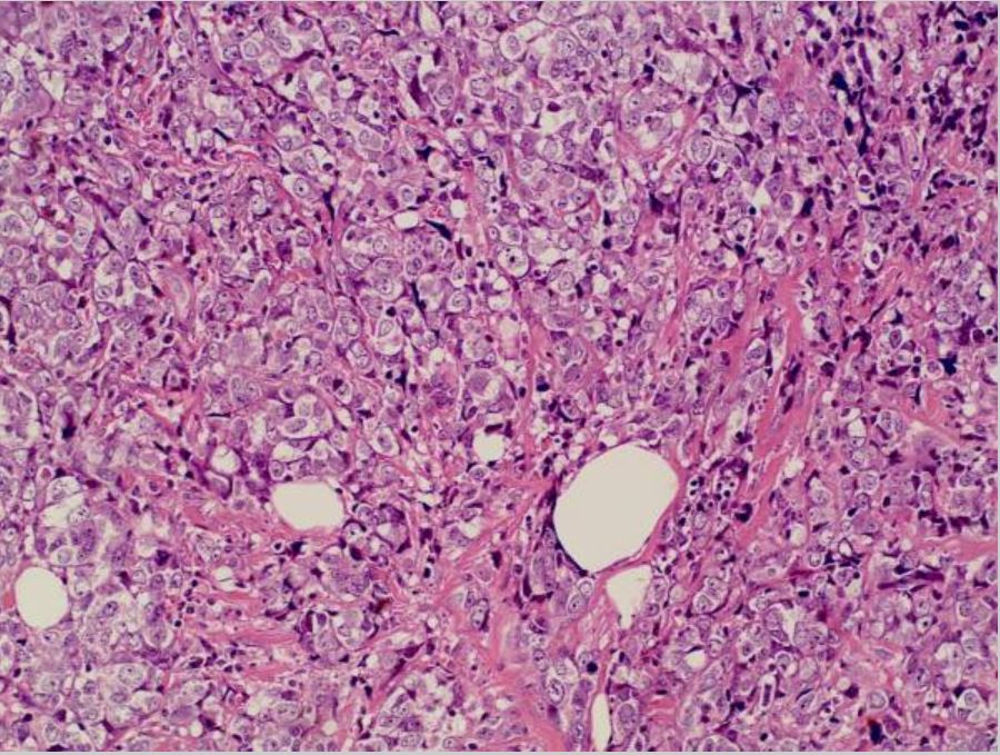
Infiltrating ductal carcinoma

  • Most common invasive breast cancer (76% of all breast carcinomas)
  • Mostly unilateral
  • Gross appearance:
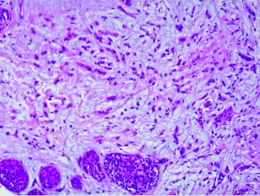
  • Microscopic findings (Nottingham Histologic Score):
    • Well differentiated (grade 1): tubular/cribriform pattern, small uniform nuclei, low proliferative rate
    • Moderately differentiated (grade 2): cells in clusters or single infiltrating cells, nuclear polymorphism Polymorphism The regular and simultaneous occurrence in a single interbreeding population of two or more discontinuous genotypes. The concept includes differences in genotypes ranging in size from a single nucleotide site to large nucleotide sequences visible at a chromosomal level. Basic Terms of Genetics, + mitotic figures
    • Poorly differentiated (grade 3): ragged nest pattern, large irregular nuclei, high proliferative rate

Infiltrating lobular carcinoma

  • 2nd most common invasive breast cancer (5%–10%)
  • Gross appearance:
  • Microscopic findings:
    • Incohesive infiltrating tumor Tumor Inflammation cells (often with signet ring cells)
    • Staining with E-cadherin E-cadherin Calcium-dependent cell adhesion proteins. They are important in the formation of adherens junctions between cells. Cadherins are classified by their distinct immunological and tissue specificities, either by letters (e- for epithelial, n- for neural, and p- for placental cadherins) or by numbers (cadherin-12 or n-cadherin 2 for brain-cadherin). Cadherins promote cell adhesion via a homophilic mechanism as in the construction of tissues and of the whole animal body. Gastric Cancer (negative in infiltrating lobular carcinoma): helps distinguish from infiltrating ductal carcinoma
  • Metastatic pattern: involves the peritoneum Peritoneum The peritoneum is a serous membrane lining the abdominopelvic cavity. This lining is formed by connective tissue and originates from the mesoderm. The membrane lines both the abdominal walls (as parietal peritoneum) and all of the visceral organs (as visceral peritoneum). Peritoneum: Anatomy, retroperitoneum, leptomeninges Leptomeninges Meninges: Anatomy, ovaries Ovaries Ovaries are the paired gonads of the female reproductive system that contain haploid gametes known as oocytes. The ovaries are located intraperitoneally in the pelvis, just posterior to the broad ligament, and are connected to the pelvic sidewall and to the uterus by ligaments. These organs function to secrete hormones (estrogen and progesterone) and to produce the female germ cells (oocytes). Ovaries: Anatomy/ uterus Uterus The uterus, cervix, and fallopian tubes are part of the internal female reproductive system. The uterus has a thick wall made of smooth muscle (the myometrium) and an inner mucosal layer (the endometrium). The most inferior portion of the uterus is the cervix, which connects the uterine cavity to the vagina. Uterus, Cervix, and Fallopian Tubes: Anatomy, and gastrointestinal tract
  • Heterozygous germline mutations in CDH1: increased risk of lobular carcinoma

Tubular carcinoma

  • ≤ 2% of invasive breast cancers
  • Microscopic findings: well-formed tubules; associated with low-grade DCIS
  • Good prognosis Prognosis A prediction of the probable outcome of a disease based on a individual’s condition and the usual course of the disease as seen in similar situations. Non-Hodgkin Lymphomas, metastasis Metastasis The transfer of a neoplasm from one organ or part of the body to another remote from the primary site. Grading, Staging, and Metastasis rare

Mucinous/ colloid Colloid Colloid solutions include large proteins or cells that do not readily cross capillary membranes. They remain in the ecf and do not distribute into the icf (similar to crystalloids). Intravenous Fluids carcinoma

  • 1%–2% of invasive breast cancers
  • More common in older patients Patients Individuals participating in the health care system for the purpose of receiving therapeutic, diagnostic, or preventive procedures. Clinician–Patient Relationship 
  • Favorable prognosis Prognosis A prediction of the probable outcome of a disease based on a individual’s condition and the usual course of the disease as seen in similar situations. Non-Hodgkin Lymphomas
  • Gross appearance: soft, circumscribed, pale gray-blue gelatin
  • Microscopic findings: clusters of tumor Tumor Inflammation cells within lakes of mucin

Medullary carcinoma

  • More frequently found in BRCA1 carcinomas
  • Occurs in younger patients Patients Individuals participating in the health care system for the purpose of receiving therapeutic, diagnostic, or preventive procedures. Clinician–Patient Relationship
  • Better prognosis Prognosis A prediction of the probable outcome of a disease based on a individual’s condition and the usual course of the disease as seen in similar situations. Non-Hodgkin Lymphomas than other poorly differentiated carcinomas
  • Microscopic findings:
    • Well circumscribed, with areas of necrosis Necrosis The death of cells in an organ or tissue due to disease, injury or failure of the blood supply. Ischemic Cell Damage and hemorrhage
    • Sheets of large cells, pleomorphic Pleomorphic Bacteroides nuclei with prominent nucleoli
    • Marked lymphoplasmacytic infiltrate

Micropapillary carcinoma

  • Rare but aggressive cancer
  • High propensity for lymphovascular invasion and lymph Lymph The interstitial fluid that is in the lymphatic system. Secondary Lymphatic Organs node metastasis Metastasis The transfer of a neoplasm from one organ or part of the body to another remote from the primary site. Grading, Staging, and Metastasis
  • Microscopic findings: Clusters of cells float within an empty stromal space.

Paget’s disease of the breast

  • 1%–4% of cases
  • Appearance:
    • Unilateral eczematous, erythematous patches Patches Vitiligo on the nipple Nipple The conic organs which usually give outlet to milk from the mammary glands. Examination of the Breast, and nipple retraction Nipple Retraction Mastitis
    • In > 50%, palpable mass Mass Three-dimensional lesion that occupies a space within the breast Imaging of the Breast present (often invasive carcinoma, ER negative and overexpress HER2 HER2 A cell surface protein-tyrosine kinase receptor that is overexpressed in a variety of adenocarcinomas. It has extensive homology to and heterodimerizes with the EGF receptor, the ERBB-3 receptor, and the ERBB-4 receptor. Activation of the erbB-2 receptor occurs through heterodimer formation with a ligand-bound erbB receptor family member. Targeted and Other Nontraditional Antineoplastic Therapy)
  • Those without a mass Mass Three-dimensional lesion that occupies a space within the breast Imaging of the Breast: Most have DCIS.
  • Paget’s cells: large, round, malignant cells in the epidermis Epidermis The external, nonvascular layer of the skin. It is made up, from within outward, of five layers of epithelium: (1) basal layer (stratum basale epidermidis); (2) spinous layer (stratum spinosum epidermidis); (3) granular layer (stratum granulosum epidermidis); (4) clear layer (stratum lucidum epidermidis); and (5) horny layer (stratum corneum epidermidis). Skin: Structure and Functions 
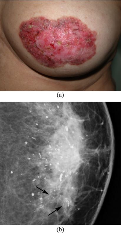
Paget’s disease of the breast

Paget’s disease of the breast: left nipple areolar changes
(a) Photograph of the left breast shows skin thickening, redness, erythema, erosion of the nipple, and scaling around the nipple-areola area.
(b) Mammogram shows scattered rod-like calcifications and groups of pleomorphic, fine, linear microcalcifications in the inner quadrant (arrows). Simple mastectomy revealed DCIS and secretory calcifications in the breast and Paget’s disease of the nipple.
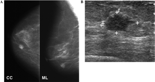
Image: “A 56-year-old woman, presented with left nipple areolar changes” by the Department of Biomedical Imaging, University of Malaya; and the University of Malaya Research Imaging Centre (UMRIC). License: Public domain.

Inflammatory carcinoma

  • Characterized by dermal lymphovascular invasion of tumor Tumor Inflammation cells
  • Appearance:
    • Breast skin Skin The skin, also referred to as the integumentary system, is the largest organ of the body. The skin is primarily composed of the epidermis (outer layer) and dermis (deep layer). The epidermis is primarily composed of keratinocytes that undergo rapid turnover, while the dermis contains dense layers of connective tissue. Skin: Structure and Functions erythema Erythema Redness of the skin produced by congestion of the capillaries. This condition may result from a variety of disease processes. Chalazion and thickening
    • Breast skin Skin The skin, also referred to as the integumentary system, is the largest organ of the body. The skin is primarily composed of the epidermis (outer layer) and dermis (deep layer). The epidermis is primarily composed of keratinocytes that undergo rapid turnover, while the dermis contains dense layers of connective tissue. Skin: Structure and Functions resembles orange peel ( peau d’orange Peau D’Orange Mastitis).
  • Poor prognosis Prognosis A prediction of the probable outcome of a disease based on a individual’s condition and the usual course of the disease as seen in similar situations. Non-Hodgkin Lymphomas 

Diagnosis

Clinical and diagnostic tools

  • Breast exam 
  • Screening Screening Preoperative Care mammography Mammography Radiographic examination of the breast. Breast Cancer Screening:
    • May offer screening Screening Preoperative Care starting at age 40 years.
    • Signs of a malignant finding:
      • Soft-tissue mass Mass Three-dimensional lesion that occupies a space within the breast Imaging of the Breast or density
      • Clustered microcalcifications
      • Spiculated high-density mass Mass Three-dimensional lesion that occupies a space within the breast Imaging of the Breast (most specific for invasive cancer)
    • The presence of a breast lump with a negative mammogram Mammogram Fibrocystic Change still warrants further investigation.
  • Ultrasonography:
    • Complementary test to mammography Mammography Radiographic examination of the breast. Breast Cancer Screening
    • Benefit:
      • No radiation Radiation Emission or propagation of acoustic waves (sound), electromagnetic energy waves (such as light; radio waves; gamma rays; or x-rays), or a stream of subatomic particles (such as electrons; neutrons; protons; or alpha particles). Osteosarcoma exposure
      • Differentiates solid (such as a benign Benign Fibroadenoma fibroadenoma Fibroadenoma Fibroadenomas are the most common benign tumor of the female breast and the most common breast tumor in adolescent and young women. The tumors are well-circumscribed, mobile, and unencapsulated, with a rubbery or firm consistency. Fibroadenoma or cancer) from fluid-filled cystic Cystic Fibrocystic Change (such as a benign Benign Fibroadenoma cyst) lesions
    • Disadvantage: highly operator dependent, not suited for screening Screening Preoperative Care on its own
    • Signs of malignant finding:
  • Magnetic resonance imaging (MRI):
    • Screening Screening Preoperative Care for women at high risk for breast cancer
    • Benefit: high soft-tissue contrast, very high sensitivity
    • Disadvantage: low specificity, no detection of microcalcifications
    • Signs of malignant finding:
      • Spiculated mass Mass Three-dimensional lesion that occupies a space within the breast Imaging of the Breast
      • Enhancing internal septa
      • Heterogenous internal enhancement
  • Biopsy Biopsy Removal and pathologic examination of specimens from the living body. Ewing Sarcoma (confirms diagnosis):
    • Fine-needle aspiration: small sample, with a false-negative rate of 10%
    • Core needle biopsy Core Needle Biopsy Fibrocystic Change (recommended): larger sample, and allows for immunohistochemical testing 

Post-diagnostic tools

  • Breast cancer receptor Receptor Receptors are proteins located either on the surface of or within a cell that can bind to signaling molecules known as ligands (e.g., hormones) and cause some type of response within the cell. Receptors testing:
    • When cancer is confirmed, determine ER/PR receptor Receptor Receptors are proteins located either on the surface of or within a cell that can bind to signaling molecules known as ligands (e.g., hormones) and cause some type of response within the cell. Receptors expression:
      •  If > 1% of the tumor Tumor Inflammation cells stain positive (on immunohistochemistry Immunohistochemistry Histochemical localization of immunoreactive substances using labeled antibodies as reagents. Myeloperoxidase Deficiency or IHC) for ER/PR → hormone receptor Receptor Receptors are proteins located either on the surface of or within a cell that can bind to signaling molecules known as ligands (e.g., hormones) and cause some type of response within the cell. Receptors positive 
      • ER/PR-positive carcinoma is suitable for endocrine therapy → better prognosis Prognosis A prediction of the probable outcome of a disease based on a individual’s condition and the usual course of the disease as seen in similar situations. Non-Hodgkin Lymphomas
    • Overexpression of HER2 HER2 A cell surface protein-tyrosine kinase receptor that is overexpressed in a variety of adenocarcinomas. It has extensive homology to and heterodimerizes with the EGF receptor, the ERBB-3 receptor, and the ERBB-4 receptor. Activation of the erbB-2 receptor occurs through heterodimer formation with a ligand-bound erbB receptor family member. Targeted and Other Nontraditional Antineoplastic Therapy receptor Receptor Receptors are proteins located either on the surface of or within a cell that can bind to signaling molecules known as ligands (e.g., hormones) and cause some type of response within the cell. Receptors:
      • Detected by IHC or fluorescence in situ hybridization Hybridization The genetic process of crossbreeding between genetically dissimilar parents to produce a hybrid. Blotting Techniques ( FISH FISH A type of in situ hybridization in which target sequences are stained with fluorescent dye so their location and size can be determined using fluorescence microscopy. This staining is sufficiently distinct that the hybridization signal can be seen both in metaphase spreads and in interphase nuclei. Chromosome Testing
      • HER2 HER2 A cell surface protein-tyrosine kinase receptor that is overexpressed in a variety of adenocarcinomas. It has extensive homology to and heterodimerizes with the EGF receptor, the ERBB-3 receptor, and the ERBB-4 receptor. Activation of the erbB-2 receptor occurs through heterodimer formation with a ligand-bound erbB receptor family member. Targeted and Other Nontraditional Antineoplastic Therapy positive → treatment with trastuzumab Trastuzumab A humanized monoclonal antibody against the ErbB-2 receptor (HER2). As an antineoplastic agent, it is used to treat breast cancer where HER2 is overexpressed. Targeted and Other Nontraditional Antineoplastic Therapy ( HER2 HER2 A cell surface protein-tyrosine kinase receptor that is overexpressed in a variety of adenocarcinomas. It has extensive homology to and heterodimerizes with the EGF receptor, the ERBB-3 receptor, and the ERBB-4 receptor. Activation of the erbB-2 receptor occurs through heterodimer formation with a ligand-bound erbB receptor family member. Targeted and Other Nontraditional Antineoplastic Therapy antibody)
  • Additional imaging (for metastatic breast cancer):
    • Bone scan Bone Scan Osteosarcoma: for patients Patients Individuals participating in the health care system for the purpose of receiving therapeutic, diagnostic, or preventive procedures. Clinician–Patient Relationship with bone Bone Bone is a compact type of hardened connective tissue composed of bone cells, membranes, an extracellular mineralized matrix, and central bone marrow. The 2 primary types of bone are compact and spongy. Bones: Structure and Types pain Pain An unpleasant sensation induced by noxious stimuli which are detected by nerve endings of nociceptive neurons. Pain: Types and Pathways or elevated alkaline phosphatase Alkaline Phosphatase An enzyme that catalyzes the conversion of an orthophosphoric monoester and water to an alcohol and orthophosphate. Osteosarcoma
    • Computed tomography (CT) of the chest: for patients Patients Individuals participating in the health care system for the purpose of receiving therapeutic, diagnostic, or preventive procedures. Clinician–Patient Relationship with pulmonary symptoms
    • CT of the abdomen and pelvis Pelvis The pelvis consists of the bony pelvic girdle, the muscular and ligamentous pelvic floor, and the pelvic cavity, which contains viscera, vessels, and multiple nerves and muscles. The pelvic girdle, composed of 2 “hip” bones and the sacrum, is a ring-like bony structure of the axial skeleton that links the vertebral column with the lower extremities. Pelvis: Anatomy or MRI: for patients Patients Individuals participating in the health care system for the purpose of receiving therapeutic, diagnostic, or preventive procedures. Clinician–Patient Relationship with abnormal pain Pain An unpleasant sensation induced by noxious stimuli which are detected by nerve endings of nociceptive neurons. Pain: Types and Pathways and/or examination, elevated liver Liver The liver is the largest gland in the human body. The liver is found in the superior right quadrant of the abdomen and weighs approximately 1.5 kilograms. Its main functions are detoxification, metabolism, nutrient storage (e.g., iron and vitamins), synthesis of coagulation factors, formation of bile, filtration, and storage of blood. Liver: Anatomy enzymes Enzymes Enzymes are complex protein biocatalysts that accelerate chemical reactions without being consumed by them. Due to the body’s constant metabolic needs, the absence of enzymes would make life unsustainable, as reactions would occur too slowly without these molecules. Basics of Enzymes/ alkaline phosphatase Alkaline Phosphatase An enzyme that catalyzes the conversion of an orthophosphoric monoester and water to an alcohol and orthophosphate. Osteosarcoma
    • Positron emission tomography-CT (PET-CT): for whole-body screening Screening Preoperative Care for metastasis Metastasis The transfer of a neoplasm from one organ or part of the body to another remote from the primary site. Grading, Staging, and Metastasis (stage III or higher)
  • Tumor Tumor Inflammation markers:
    • CA 15-3 CA 15-3 Serum Tumor Markers, CA CA Condylomata acuminata are a clinical manifestation of genital HPV infection. Condylomata acuminata are described as raised, pearly, flesh-colored, papular, cauliflower-like lesions seen in the anogenital region that may cause itching, pain, or bleeding. Condylomata Acuminata (Genital Warts) 27.29, and carcinoembryonic antigen Carcinoembryonic antigen A glycoprotein that is secreted into the luminal surface of the epithelia in the gastrointestinal tract. It is found in the feces and pancreaticobiliary secretions and is used to monitor the response to colon cancer treatment. Serum Tumor Markers ( CEA CEA A glycoprotein that is secreted into the luminal surface of the epithelia in the gastrointestinal tract. It is found in the feces and pancreaticobiliary secretions and is used to monitor the response to colon cancer treatment. Serum Tumor Markers)
    • Biochemical markers are not specific for breast cancer relapse Relapse Relapsing Fever.
    • Used to monitor treatment response of patients Patients Individuals participating in the health care system for the purpose of receiving therapeutic, diagnostic, or preventive procedures. Clinician–Patient Relationship with metastatic disease
  • Genetic testing Genetic Testing Detection of a mutation; genotype; karyotype; or specific alleles associated with genetic traits, heritable diseases, or predisposition to a disease, or that may lead to the disease in descendants. It includes prenatal genetic testing. Myotonic Dystrophies:
    • United States Preventive Services Task Force recommends the use of approved familial risk assessment Risk assessment The qualitative or quantitative estimation of the likelihood of adverse effects that may result from exposure to specified health hazards or from the absence of beneficial influences. Preoperative Care tools for the following:
      • A personal or family history Family History Adult Health Maintenance of breast, ovarian, tubal, or peritoneal cancer
      • Ancestry associated with BRCA1 or 2 mutations
      • Positive assessment indicates the need for genetic counseling Genetic Counseling An educational process that provides information and advice to individuals or families about a genetic condition that may affect them. The purpose is to help individuals make informed decisions about marriage, reproduction, and other health management issues based on information about the genetic disease, the available diagnostic tests, and management programs. Psychosocial support is usually offered. Myotonic Dystrophies and testing.
    • Other expert groups have varying recommendations.
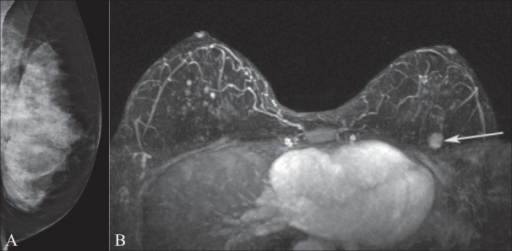
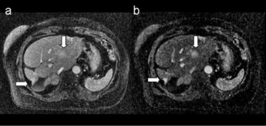
Breast metastasis mri

MRI of breast metastasis: images show metastatic breast lesions in the liver
A. T1-weighted 3D postcontrast image
B. corresponding 3D subtraction image
Arrows indicate the metastatic lesions.

Image: “A method for dynamic subtraction MR imaging of the liver” by Mainardi LT, Passera KM, Lucesoli A, Potepan P, Setti E, Musumeci R. License: CC BY 2.0

Staging

Overview of breast cancer staging Staging Methods which attempt to express in replicable terms the extent of the neoplasm in the patient. Grading, Staging, and Metastasis

  • Based on the American Joint Committee on Cancer (AJCC) and Union for International Cancer Control (UICC) 8th edition
  • Includes the tumor Tumor Inflammation, node, metastasis Metastasis The transfer of a neoplasm from one organ or part of the body to another remote from the primary site. Grading, Staging, and Metastasis (TNM) staging Staging Methods which attempt to express in replicable terms the extent of the neoplasm in the patient. Grading, Staging, and Metastasis system and incorporates biologic factors for prognostic staging Staging Methods which attempt to express in replicable terms the extent of the neoplasm in the patient. Grading, Staging, and Metastasis
  • Information included in staging Staging Methods which attempt to express in replicable terms the extent of the neoplasm in the patient. Grading, Staging, and Metastasis:
    • Tumor Tumor Inflammation
    • Node
    • Metastasis Metastasis The transfer of a neoplasm from one organ or part of the body to another remote from the primary site. Grading, Staging, and Metastasis
    • ER status
    • PR status
    • HER2 HER2 A cell surface protein-tyrosine kinase receptor that is overexpressed in a variety of adenocarcinomas. It has extensive homology to and heterodimerizes with the EGF receptor, the ERBB-3 receptor, and the ERBB-4 receptor. Activation of the erbB-2 receptor occurs through heterodimer formation with a ligand-bound erbB receptor family member. Targeted and Other Nontraditional Antineoplastic Therapy status
    • Histologic grade
    • Recurrence score (Oncotype Dx) considered
  • Anatomic stage grouping relies on TNM.
  • Prognostic stage grouping:
    • Relies on TNM, ER/PR/ HER2 HER2 A cell surface protein-tyrosine kinase receptor that is overexpressed in a variety of adenocarcinomas. It has extensive homology to and heterodimerizes with the EGF receptor, the ERBB-3 receptor, and the ERBB-4 receptor. Activation of the erbB-2 receptor occurs through heterodimer formation with a ligand-bound erbB receptor family member. Targeted and Other Nontraditional Antineoplastic Therapy, grade
    • Primary prognostic staging Staging Methods which attempt to express in replicable terms the extent of the neoplasm in the patient. Grading, Staging, and Metastasis system for those who receive neoadjuvant treatment or for those who do not undergo surgery

TNM staging Staging Methods which attempt to express in replicable terms the extent of the neoplasm in the patient. Grading, Staging, and Metastasis

Table: Tumor Tumor Inflammation (T) staging Staging Methods which attempt to express in replicable terms the extent of the neoplasm in the patient. Grading, Staging, and Metastasis
Tumor Tumor Inflammation stage Description
Tx Primary tumor Tumor Inflammation unable to be assessed
T0 No evidence of primary tumor Tumor Inflammation
Tis
  • Tis (DCIS): ductal carcinoma in situ
  • Tis (Paget): Paget’s disease of the nipple Nipple The conic organs which usually give outlet to milk from the mammary glands. Examination of the Breast, neither associated invasive carcinoma nor DCIS in the underlying breast parenchyma
T1 ≤ 20-mm tumor Tumor Inflammation in greatest dimension
T2 > 20-mm but ≤ 50-mm tumor Tumor Inflammation in greatest dimension
T3 T3 A T3 thyroid hormone normally synthesized and secreted by the thyroid gland in much smaller quantities than thyroxine (T4). Most T3 is derived from peripheral monodeiodination of T4 at the 5′ position of the outer ring of the iodothyronine nucleus. The hormone finally delivered and used by the tissues is mainly t3. Thyroid Hormones > 50-mm tumor Tumor Inflammation in greatest dimension
T4 T4 The major hormone derived from the thyroid gland. Thyroxine is synthesized via the iodination of tyrosines (monoiodotyrosine) and the coupling of iodotyrosines (diiodotyrosine) in the thyroglobulin. Thyroxine is released from thyroglobulin by proteolysis and secreted into the blood. Thyroxine is peripherally deiodinated to form triiodothyronine which exerts a broad spectrum of stimulatory effects on cell metabolism. Thyroid Hormones Tumor Tumor Inflammation of any size, with direct extension Extension Examination of the Upper Limbs to the chest wall Chest wall The chest wall consists of skin, fat, muscles, bones, and cartilage. The bony structure of the chest wall is composed of the ribs, sternum, and thoracic vertebrae. The chest wall serves as armor for the vital intrathoracic organs and provides the stability necessary for the movement of the shoulders and arms. Chest Wall: Anatomy and/or skin Skin The skin, also referred to as the integumentary system, is the largest organ of the body. The skin is primarily composed of the epidermis (outer layer) and dermis (deep layer). The epidermis is primarily composed of keratinocytes that undergo rapid turnover, while the dermis contains dense layers of connective tissue. Skin: Structure and Functions ( ulceration Ulceration Corneal Abrasions, Erosion, and Ulcers or macroscopic nodules)
Note: LCIS is considered a benign entity; not Tis.
Table: Regional lymph Lymph The interstitial fluid that is in the lymphatic system. Secondary Lymphatic Organs node (N) staging Staging Methods which attempt to express in replicable terms the extent of the neoplasm in the patient. Grading, Staging, and Metastasis (axillary, ipsilateral intramammary, internal mammary, and supraclavicular LN)
Node stage Description
cNX Regional LNs cannot be assessed.
cN0 No regional LN metastases
cN1 Metastasis Metastasis The transfer of a neoplasm from one organ or part of the body to another remote from the primary site. Grading, Staging, and Metastasis to movable ipsilateral level I, II axillary LNs
cN2
  • Ipsilateral level I, II axillary LNs (fixed or matted)
  • Ipsilateral internal mammary nodes, in the absence of axillary LNs
cN3
  • Ipsilateral infraclavicular (level III axillary) LNs with or without level I, II axillary LNs
  • Ipsilateral internal mammary nodes with level I, II axillary LNs
  • Ipsilateral supraclavicular LNs with/without axillary or internal mammary LN
LN: lymph nodes
Cn: clinical node
Note: Spread to other LN (cervical or contralateral axillary) is considered metastatic (M1).
Table: Metastasis Metastasis The transfer of a neoplasm from one organ or part of the body to another remote from the primary site. Grading, Staging, and Metastasis (M) staging Staging Methods which attempt to express in replicable terms the extent of the neoplasm in the patient. Grading, Staging, and Metastasis
Metastasis Metastasis The transfer of a neoplasm from one organ or part of the body to another remote from the primary site. Grading, Staging, and Metastasis Description
M0 No evidence of distant metastasis Metastasis The transfer of a neoplasm from one organ or part of the body to another remote from the primary site. Grading, Staging, and Metastasis (clinical or radiographic)
M1 Detectable metastasis Metastasis The transfer of a neoplasm from one organ or part of the body to another remote from the primary site. Grading, Staging, and Metastasis
Table: AJCC UICC 8th edition anatomic stage
Stage Substages Tumor Tumor Inflammation Node Metastasis Metastasis The transfer of a neoplasm from one organ or part of the body to another remote from the primary site. Grading, Staging, and Metastasis
0 Tis N0 M0
I
  • IA: T1N0M0
  • IB: T0-1, N1M0
T0-T1 N1 M0
II
  • IIA: T0-1, N1M0
  • IIA: T2N0M0
  • IIB: T2N1M0
  • IIB: T3N0M0
T0-T3 N0-N1 M0
III
  • IIIA: T0-2, N2M0
  • IIIA: T3 T3 A T3 thyroid hormone normally synthesized and secreted by the thyroid gland in much smaller quantities than thyroxine (T4). Most T3 is derived from peripheral monodeiodination of T4 at the 5′ position of the outer ring of the iodothyronine nucleus. The hormone finally delivered and used by the tissues is mainly t3. Thyroid Hormones, N1-2, M0
  • IIIB: T4 T4 The major hormone derived from the thyroid gland. Thyroxine is synthesized via the iodination of tyrosines (monoiodotyrosine) and the coupling of iodotyrosines (diiodotyrosine) in the thyroglobulin. Thyroxine is released from thyroglobulin by proteolysis and secreted into the blood. Thyroxine is peripherally deiodinated to form triiodothyronine which exerts a broad spectrum of stimulatory effects on cell metabolism. Thyroid Hormones, N0-2, M0
  • IIIC: any T, N3M0
T0-T3 N1-N2 M0
IV Any T Any N M1
AJCC: American Joint Committee on Cancer
UICC: Union for International Cancer Control
T: tumor
N: node
M: metastasis

Categories

  • Early-stage non-metastatic breast cancer: stage I, IIA, a subset of stage IIB (T2N1)
  • Locally advanced non-metastatic breast cancer: subset of stage IIB (T3N0), stage IIIA–IIIC
  • Metastatic breast cancer: stage IV

Management and Prognosis

DCIS

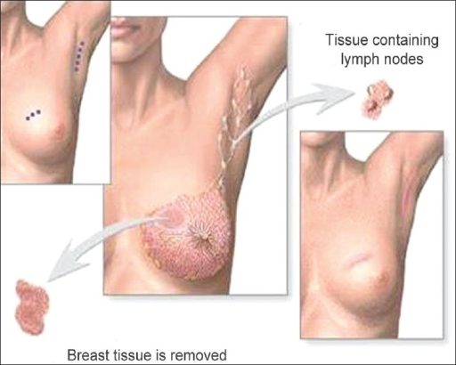
  • Surgery: breast-conserving surgery (BCS) (e.g., lumpectomy Lumpectomy Fat Necrosis of the Breast) or mastectomy Mastectomy Surgical procedure to remove one or both breasts. Fat Necrosis of the Breast
  • Radiation Radiation Emission or propagation of acoustic waves (sound), electromagnetic energy waves (such as light; radio waves; gamma rays; or x-rays), or a stream of subatomic particles (such as electrons; neutrons; protons; or alpha particles). Osteosarcoma therapy (RT) considered in those with high risk of recurrence
  • Endocrine therapy for ER-positive DCIS (for 5 years):
    • Tamoxifen Tamoxifen One of the selective estrogen receptor modulators with tissue-specific activities. Tamoxifen acts as an anti-estrogen (inhibiting agent) in the mammary tissue, but as an estrogen (stimulating agent) in cholesterol metabolism, bone density, and cell proliferation in the endometrium. Antiestrogens:
      • ER antagonist
      • Option for all women
    • Aromatase inhibitors Aromatase inhibitors Compounds that inhibit aromatase in order to reduce production of estrogenic steroid hormones. Antiestrogens (e.g., anastrozole Anastrozole A nitrile and triazole derivative that acts as a selective nonsteroidal aromatase inhibitor. It is used in the treatment of estrogen nuclear receptor-positive breast cancer in postmenopausal women. Antiestrogens):
      • Inactivates aromatase Aromatase An enzyme that catalyzes the desaturation (aromatization) of the ring a of C19 androgens and converts them to C18 estrogens. In this process, the 19-methyl is removed. This enzyme is membrane-bound, located in the endoplasmic reticulum of estrogen-producing cells of ovaries, placenta, testes, adipose, and brain tissues. Aromatase is encoded by the cyp19 gene, and functions in complex with NADPH-ferrihemoprotein reductase in the cytochrome p450 system. Adipose Tissue: Histology, reducing peripheral conversion of androgens Androgens Androgens are naturally occurring steroid hormones responsible for development and maintenance of the male sex characteristics, including penile, scrotal, and clitoral growth, development of sexual hair, deepening of the voice, and musculoskeletal growth. Androgens and Antiandrogens to estrogens
      • Alternative for post-menopausal women
Breast-conserving surgery (lumpectomy)

Breast-conserving surgery (lumpectomy): excision of the tumor to the negative margins and axillary lymph node evaluation

Image: “Phantom breast syndrome” by the Indian Journal of Palliative Care. License: CC BY 2.0.

Early-stage cancer

  • Surgery:
  • RT in most cases (if BCS is done and for those at risk for local recurrence)
  • Adjuvant Adjuvant Substances that augment, stimulate, activate, potentiate, or modulate the immune response at either the cellular or humoral level. The classical agents (freund’s adjuvant, bcg, corynebacterium parvum, et al.) contain bacterial antigens. Some are endogenous (e.g., histamine, interferon, transfer factor, tuftsin, interleukin-1). Their mode of action is either non-specific, resulting in increased immune responsiveness to a wide variety of antigens, or antigen-specific, i.e., affecting a restricted type of immune response to a narrow group of antigens. The therapeutic efficacy of many biological response modifiers is related to their antigen-specific immunoadjuvanticity. Vaccination or systemic therapy given in addition to surgery:
    • Endocrine therapy:
      • For hormone receptor Receptor Receptors are proteins located either on the surface of or within a cell that can bind to signaling molecules known as ligands (e.g., hormones) and cause some type of response within the cell. Receptors (ER/PR)–positive patients Patients Individuals participating in the health care system for the purpose of receiving therapeutic, diagnostic, or preventive procedures. Clinician–Patient Relationship
      • Aromatase inhibitors Aromatase inhibitors Compounds that inhibit aromatase in order to reduce production of estrogenic steroid hormones. Antiestrogens or tamoxifen Tamoxifen One of the selective estrogen receptor modulators with tissue-specific activities. Tamoxifen acts as an anti-estrogen (inhibiting agent) in the mammary tissue, but as an estrogen (stimulating agent) in cholesterol metabolism, bone density, and cell proliferation in the endometrium. Antiestrogens
    • HER2-targeted therapy:
    • Chemotherapy Chemotherapy Osteosarcoma (doxorubicin + cyclophosphamide Cyclophosphamide Precursor of an alkylating nitrogen mustard antineoplastic and immunosuppressive agent that must be activated in the liver to form the active aldophosphamide. It has been used in the treatment of lymphoma and leukemia. Its side effect, alopecia, has been used for defleecing sheep. Cyclophosphamide may also cause sterility, birth defects, mutations, and cancer. Immunosuppressants, then paclitaxel Paclitaxel A cyclodecane isolated from the bark of the pacific yew tree, taxus brevifolia. It stabilizes microtubules in their polymerized form leading to cell death. Microtubule and Topoisomerase Inhibitors):
      • For TNBCs (≥ 0.5 cm)
      • For HER2-positive cancers (> 1 cm) 
      • For hormone receptor Receptor Receptors are proteins located either on the surface of or within a cell that can bind to signaling molecules known as ligands (e.g., hormones) and cause some type of response within the cell. Receptors–positive cancers (with high-risk features: ≥ 2-cm size, + LNs, high-grade lesions) 

Locally advanced breast cancer

  • Neoadjuvant systemic therapy:
    • HER2-directed therapy + chemotherapy Chemotherapy Osteosarcoma: for HER2-positive cancer
    • Chemotherapy Chemotherapy Osteosarcoma given 1st for hormone receptor Receptor Receptors are proteins located either on the surface of or within a cell that can bind to signaling molecules known as ligands (e.g., hormones) and cause some type of response within the cell. Receptors–positive cancer 
    • Chemotherapy Chemotherapy Osteosarcoma given for TNBCs
  • Surgery (BCS or mastectomy Mastectomy Surgical procedure to remove one or both breasts. Fat Necrosis of the Breast + LN assessment) follows neoadjuvant therapy.
  • Adjuvant Adjuvant Substances that augment, stimulate, activate, potentiate, or modulate the immune response at either the cellular or humoral level. The classical agents (freund’s adjuvant, bcg, corynebacterium parvum, et al.) contain bacterial antigens. Some are endogenous (e.g., histamine, interferon, transfer factor, tuftsin, interleukin-1). Their mode of action is either non-specific, resulting in increased immune responsiveness to a wide variety of antigens, or antigen-specific, i.e., affecting a restricted type of immune response to a narrow group of antigens. The therapeutic efficacy of many biological response modifiers is related to their antigen-specific immunoadjuvanticity. Vaccination therapy:
    • Given if there was no neoadjuvant therapy (similar principles as early-stage cancer)
    • For those who had neoadjuvant therapy:
  • RT

Metastatic breast cancer

Prognosis Prognosis A prediction of the probable outcome of a disease based on a individual’s condition and the usual course of the disease as seen in similar situations. Non-Hodgkin Lymphomas

  • 10-year survival:
    • Stage 0 (DCIS): 97%
    • Stage I: 87%
    • Stage II: 65%
    • Stage III: 40%
    • Stage IV: 5%
  • With distant metastasis Metastasis The transfer of a neoplasm from one organ or part of the body to another remote from the primary site. Grading, Staging, and Metastasis, there is no cure.

Differential Diagnosis

  • Fibroadenoma Fibroadenoma Fibroadenomas are the most common benign tumor of the female breast and the most common breast tumor in adolescent and young women. The tumors are well-circumscribed, mobile, and unencapsulated, with a rubbery or firm consistency. Fibroadenoma: benign Benign Fibroadenoma breast tumors that arise in young women. Diagnosis is based on the physical finding of a mobile, non-tender breast mass Mass Three-dimensional lesion that occupies a space within the breast Imaging of the Breast. Some fibroadenoma Fibroadenoma Fibroadenomas are the most common benign tumor of the female breast and the most common breast tumor in adolescent and young women. The tumors are well-circumscribed, mobile, and unencapsulated, with a rubbery or firm consistency. Fibroadenoma are asymptomatic, small in size, and should be treated conservatively. Other fibroadenoma Fibroadenoma Fibroadenomas are the most common benign tumor of the female breast and the most common breast tumor in adolescent and young women. The tumors are well-circumscribed, mobile, and unencapsulated, with a rubbery or firm consistency. Fibroadenoma grow rapidly in size, are symptomatic, and should be surgically excised.
  • Fat necrosis Necrosis The death of cells in an organ or tissue due to disease, injury or failure of the blood supply. Ischemic Cell Damage of the breast: a benign Benign Fibroadenoma condition caused by trauma to the breast, which may go unnoticed, leading to adipocyte Adipocyte Cells in the body that store fats, usually in the form of triglycerides. White adipocytes are the predominant type and found mostly in the abdominal cavity and subcutaneous tissue. Brown adipocytes are thermogenic cells that can be found in newborns of some species and hibernating mammals. Lipoma necrosis Necrosis The death of cells in an organ or tissue due to disease, injury or failure of the blood supply. Ischemic Cell Damage and calcification. Patients Patients Individuals participating in the health care system for the purpose of receiving therapeutic, diagnostic, or preventive procedures. Clinician–Patient Relationship may present with a solid, painless mass Mass Three-dimensional lesion that occupies a space within the breast Imaging of the Breast with or without skin Skin The skin, also referred to as the integumentary system, is the largest organ of the body. The skin is primarily composed of the epidermis (outer layer) and dermis (deep layer). The epidermis is primarily composed of keratinocytes that undergo rapid turnover, while the dermis contains dense layers of connective tissue. Skin: Structure and Functions changes. Fat necrosis Necrosis The death of cells in an organ or tissue due to disease, injury or failure of the blood supply. Ischemic Cell Damage of the breast can mimic malignancy Malignancy Hemothorax and definitive diagnosis is by imaging and biopsy Biopsy Removal and pathologic examination of specimens from the living body. Ewing Sarcoma
  • Mastitis Mastitis Mastitis is inflammation of the breast tissue with or without infection. The most common form of mastitis is associated with lactation in the first few weeks after birth. Non-lactational mastitis includes periductal mastitis and idiopathic granulomatous mastitis (IGM). Mastitis: inflammation Inflammation Inflammation is a complex set of responses to infection and injury involving leukocytes as the principal cellular mediators in the body’s defense against pathogenic organisms. Inflammation is also seen as a response to tissue injury in the process of wound healing. The 5 cardinal signs of inflammation are pain, heat, redness, swelling, and loss of function. Inflammation of the mammary gland Mammary gland Glandular tissue in the breast of human that is under the influence of hormones such as estrogens; progestins; and prolactin. In women, after parturition, the mammary glands secrete milk for the nourishment of the young. Breasts: Anatomy tissue that can be lactational or non-lactational. Mastitis Mastitis Mastitis is inflammation of the breast tissue with or without infection. The most common form of mastitis is associated with lactation in the first few weeks after birth. Non-lactational mastitis includes periductal mastitis and idiopathic granulomatous mastitis (IGM). Mastitis is most common in women of childbearing age. Patients Patients Individuals participating in the health care system for the purpose of receiving therapeutic, diagnostic, or preventive procedures. Clinician–Patient Relationship usually present with high fever Fever Fever is defined as a measured body temperature of at least 38°C (100.4°F). Fever is caused by circulating endogenous and/or exogenous pyrogens that increase levels of prostaglandin E2 in the hypothalamus. Fever is commonly associated with chills, rigors, sweating, and flushing of the skin. Fever, chills Chills The sudden sensation of being cold. It may be accompanied by shivering. Fever, fatigue Fatigue The state of weariness following a period of exertion, mental or physical, characterized by a decreased capacity for work and reduced efficiency to respond to stimuli. Fibromyalgia, malaise Malaise Tick-borne Encephalitis Virus, and myalgia Myalgia Painful sensation in the muscles. Ion Channel Myopathy. Analgesics and antibiotics are given as part of the management.
  • Breast abscess Breast Abscess Benign Breast Conditions: a breast mass Mass Three-dimensional lesion that occupies a space within the breast Imaging of the Breast associated with mastitis Mastitis Mastitis is inflammation of the breast tissue with or without infection. The most common form of mastitis is associated with lactation in the first few weeks after birth. Non-lactational mastitis includes periductal mastitis and idiopathic granulomatous mastitis (IGM). Mastitis. Breast abscess Breast Abscess Benign Breast Conditions presents as a unilateral and fluctuant Fluctuant Dermatologic Examination mass Mass Three-dimensional lesion that occupies a space within the breast Imaging of the Breast, accompanied by fever Fever Fever is defined as a measured body temperature of at least 38°C (100.4°F). Fever is caused by circulating endogenous and/or exogenous pyrogens that increase levels of prostaglandin E2 in the hypothalamus. Fever is commonly associated with chills, rigors, sweating, and flushing of the skin. Fever, and a painful, erythematous, and edematous breast. History, physical findings, and needle aspiration Needle aspiration Using fine needles (finer than 22-gauge) to remove tissue or fluid specimens from the living body for examination in the pathology laboratory and for disease diagnosis. Peritonsillar Abscess (revealing purulent contents) help differentiate a breast abscess Breast Abscess Benign Breast Conditions. Treatment includes incision and drainage Incision And Drainage Chalazion, with antibiotics.
  • Intraductal papillomas: abnormal growths with a papillary configuration of the breast stroma and epithelium Epithelium The epithelium is a complex of specialized cellular organizations arranged into sheets and lining cavities and covering the surfaces of the body. The cells exhibit polarity, having an apical and a basal pole. Structures important for the epithelial integrity and function involve the basement membrane, the semipermeable sheet on which the cells rest, and interdigitations, as well as cellular junctions. Surface Epithelium: Histology within a breast duct. The most common presenting feature is bloody or serous nipple Nipple The conic organs which usually give outlet to milk from the mammary glands. Examination of the Breast discharge. Core needle biopsy Core Needle Biopsy Fibrocystic Change confirms the diagnosis.
  • Simple breast cyst Simple Breast Cyst Benign Breast Conditions: round or oval fluid-filled masses from the terminal duct lobular unit. A cyst is felt as a palpable mass Mass Three-dimensional lesion that occupies a space within the breast Imaging of the Breast that can cause pain Pain An unpleasant sensation induced by noxious stimuli which are detected by nerve endings of nociceptive neurons. Pain: Types and Pathways. Ultrasonography shows a cyst as a well-circumscribed, anechoic Anechoic A structure that produces no echo at all (looks completely black) Ultrasound (Sonography) mass Mass Three-dimensional lesion that occupies a space within the breast Imaging of the Breast with no solid components.

References

  1. Bleiweiss, I., Chagpar, A., & Vora, S. (2020). Pathology of breast cancer. UpToDate. Retrieved Jan 15, 2021, from https://www.uptodate.com/contents/pathology-of-breast-cancer
  2. Hao, S., Zhao, YY., Peng, JJ., et al. (2019). Invasive micropapillary carcinoma of the breast had no difference in prognosis compared with invasive ductal carcinoma: a propensity-matched analysis. Sci Rep 9, 286. https://doi.org/10.1038/s41598-018-36362-8
  3. Hayes D.F., & Lippman M.E. (2018). Breast cancer. In Jameson JL, et al. (Ed.), Harrison’s Principles of Internal Medicine, 20th ed. McGraw-Hill.
  4. Lester, S. (2020). The breast. In Kumar, V., Abbas, A. K., Aster, J.C., (Eds.), Robbins & Cotran Pathologic Basis of Disease. (10th ed. pp. 1037–1064).
  5. Moasser M.M., & Ai W.Z. (2019). Neoplasia. In Hammer G.D., & McPhee S.J. (Eds.), Pathophysiology of Disease: An Introduction to Clinical Medicine, 8th ed. McGraw-Hill.
  6. Nascimento, R., & Otoni, K. (2020). Histological and molecular classification of breast cancer: What do we know? Mastology. https://www.mastology.org/wp-content/uploads/2020/09/MAS_2020024_AOP.pdf
  7. Polyak, K. (2007). Breast cancer: Origins and evolution. J Clin Invest. 117(11), 3155–63. https://doi.org/10.1172/JCI33295
  8. Taghian, A., Meravjer, S., Hayes, D., & Vora, S. (2020). Overview of the treatment of newly diagnosed, invasive, non-metastatic breast cancer. UpToDate. Retrieved Jan 16, 2021, from https://www.uptodate.com/contents/overview-of-the-treatment-of-newly-diagnosed-invasive-non-metastatic-breast-cancer

Create your free account or log in to continue reading!

Sign up now and get free access to Lecturio with concept pages, medical videos, and questions for your medical education.

User Reviews

Details