Body Fluid Compartments

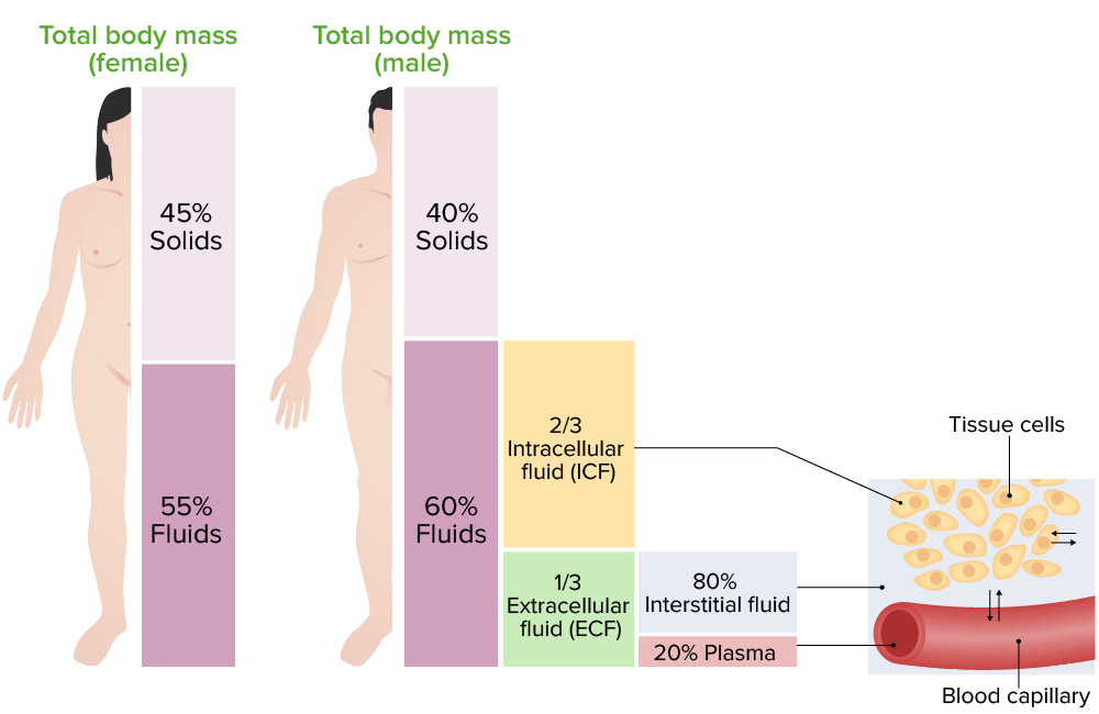
The adult human body is made up of 60% water and is divided into extracellular and intracellular fluid compartments. Extracellular fluid is present outside the cells and makes up ⅓ of the total body water. Intracellular fluid is present inside the cells and makes up ⅔ of the total body water. Intracellular and extracellular fluids are separated into compartments by semipermeable membranes, and the transport of fluid and ions is maintained by channels Channels The Cell: Cell Membrane in the cell membrane Cell Membrane A cell membrane (also known as the plasma membrane or plasmalemma) is a biological membrane that separates the cell contents from the outside environment. A cell membrane is composed of a phospholipid bilayer and proteins that function to protect cellular DNA and mediate the exchange of ions and molecules. The Cell: Cell Membrane. Each compartment contains different concentrations of ions and osmolar molecules. The relative charge and osmolarity Osmolarity The concentration of osmotically active particles in solution expressed in terms of osmoles of solute per liter of solution. Osmolality is expressed in terms of osmoles of solute per kilogram of solvent. Hypernatremia are maintained rigorously by the transport of water and substances between compartments. Hypernatremia Hypernatremia Hypernatremia is an elevated serum sodium concentration > 145 mmol/L. Serum sodium is the greatest contributor to plasma osmolality, which is very tightly controlled by the hypothalamus via the thirst mechanism and antidiuretic hormone (ADH) release. Hypernatremia occurs either from a lack of access to water or an excessive intake of sodium. Hypernatremia, hyponatremia Hyponatremia Hyponatremia is defined as a decreased serum sodium (sNa+) concentration less than 135 mmol/L. Serum sodium is the greatest contributor to plasma osmolality, which is very tightly controlled via antidiuretic hormone (ADH) release from the hypothalamus and by the thirst mechanism. Hyponatremia, and edema Edema Edema is a condition in which excess serous fluid accumulates in the body cavity or interstitial space of connective tissues. Edema is a symptom observed in several medical conditions. It can be categorized into 2 types, namely, peripheral (in the extremities) and internal (in an organ or body cavity). Edema are the clinical conditions arising from disturbances in the maintenance of osmolarity Osmolarity The concentration of osmotically active particles in solution expressed in terms of osmoles of solute per liter of solution. Osmolality is expressed in terms of osmoles of solute per kilogram of solvent. Hypernatremia of the body fluid compartments.

Last updated: May 17, 2024

Editorial responsibility: Stanley Oiseth, Lindsay Jones, Evelin Maza

Overview

Water in the adult human body makes up approximately 60% of the total body weight. The fluid is distributed in various organs, organ systems, and tissues. The sum of the water in these tissues is known as total body water.

Body Fluid Compartments

Overview

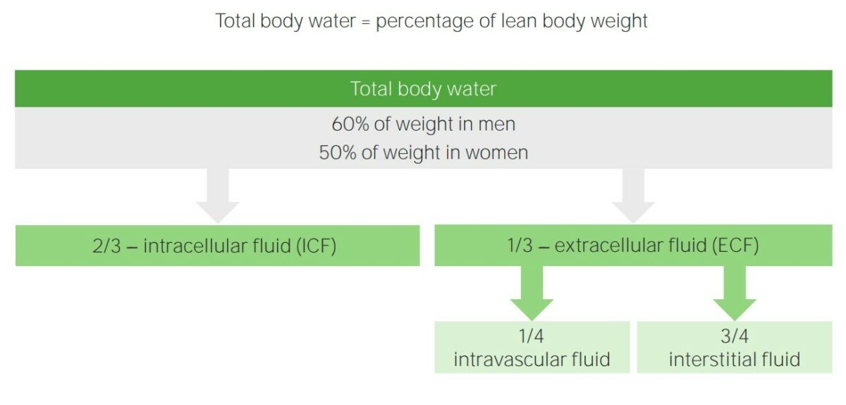
The total body water is distributed primarily between 2 compartments, namely, extracellular fluid (ECF) and intracellular fluid (ICF) compartments.
  • ECF is present outside cells:
    • Makes up about ⅓ of total body water
    • ECF includes:
      • Intravascular fluid (¼ of ECF): primary component of plasma Plasma The residual portion of blood that is left after removal of blood cells by centrifugation without prior blood coagulation. Transfusion Products
      • Interstitial fluid (¾ of ECF): lies outside blood vessels
  • ICF is present inside cells:
    • Makes up about ⅔ of total body water
    • Principal component of the cytoplasm of cells

Movement of fluid between compartments

  • Fluid is separated into compartments by semipermeable membranes. The membranes are highly permeable to water but require energy to transport ions.
  • Distribution of fluid between intracellular and extracellular compartments is determined by the concentration of Na+, chloride Chloride Inorganic compounds derived from hydrochloric acid that contain the Cl- ion. Electrolytes (Cl), and other electrolytes Electrolytes Electrolytes are mineral salts that dissolve in water and dissociate into charged particles called ions, which can be either be positively (cations) or negatively (anions) charged. Electrolytes are distributed in the extracellular and intracellular compartments in different concentrations. Electrolytes are essential for various basic life-sustaining functions. Electrolytes.
  • Water moves between compartments following osmotic gradients.
  • A change in the relative concentration of solute or water will cause water to shift between compartments.
  • 2 processes are responsible for the passive movement of fluid across membranes:
    • Diffusion Diffusion The tendency of a gas or solute to pass from a point of higher pressure or concentration to a point of lower pressure or concentration and to distribute itself throughout the available space. Diffusion, especially facilitated diffusion, is a major mechanism of biological transport. Peritoneal Dialysis and Hemodialysis: A substance passes from an area of higher concentration to an area of lower concentration.
    • Osmosis: Water is drawn across a membrane toward a region where there is a higher solute concentration.

Measurement of body fluid compartments

Compartment volumes can be measured by determining the volume of distribution of an indicator substance. A known amount of an indicator is added to a compartment, and the indicator concentration in that compartment is measured after allowing sufficient time for uniform distribution throughout the compartment. The compartment volume is calculated as follows:

Volume = amount of the indicator / concentration of the indicator

Table: Body fluid compartments and their associated indicators
Volume Indicators
Total body water 3H2O, 2H2O, antipyrine
Extracellular fluid 22Na, 125I-iothalamate, thiosulfate, inulin Inulin A starch found in the tubers and roots of many plants. Since it is hydrolyzable to fructose, it is classified as a fructosan. It has been used in physiologic investigation for determination of the rate of glomerular function. Glomerular Filtration
Intracellular fluid Calculated as: (total body water — extracellular fluid volume)
Plasma Plasma The residual portion of blood that is left after removal of blood cells by centrifugation without prior blood coagulation. Transfusion Products volume 125I-albumin, Evans blue dye (T-1824)
Blood volume 51Cr-labeled RBCs RBCs Erythrocytes, or red blood cells (RBCs), are the most abundant cells in the blood. While erythrocytes in the fetus are initially produced in the yolk sac then the liver, the bone marrow eventually becomes the main site of production. Erythrocytes: Histology, or calculated as: (blood volume = plasma Plasma The residual portion of blood that is left after removal of blood cells by centrifugation without prior blood coagulation. Transfusion Products volume / (1 − hematocrit Hematocrit The volume of packed red blood cells in a blood specimen. The volume is measured by centrifugation in a tube with graduated markings, or with automated blood cell counters. It is an indicator of erythrocyte status in disease. For example, anemia shows a low value; polycythemia, a high value. Neonatal Polycythemia))
Interstitial fluid Calculated as: (extracellular fluid volume − plasma Plasma The residual portion of blood that is left after removal of blood cells by centrifugation without prior blood coagulation. Transfusion Products volume)
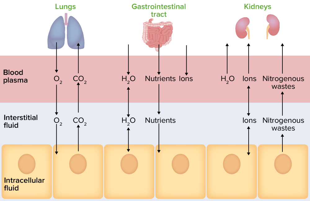
Movement of body fluids between compartments

Illustration representing the movement of body fluids between compartments

Image by Lecturio.
Changes in the Fluid Compartment

Note: This animation does not have sound.

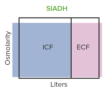
Hypo-osmotic volume expansion

  • Results in an increase in total body water and reduction in osmolality Osmolality Plasma osmolality refers to the combined concentration of all solutes in the blood. Renal Sodium and Water Regulation
  • Can be seen in cases of:
    • Excessive water intake
    • SIADH SIADH Syndrome of inappropriate antidiuretic hormone secretion (SIADH) is a disorder of impaired water excretion due to the inability to suppress the secretion of antidiuretic hormone (ADH). SIADH is characterized by impaired water excretion leading to dilutional hyponatremia, which is mainly asymptomatic but may cause neurologic symptoms. S Syndrome of Inappropriate Antidiuretic Hormone Secretion (SIADH)
  • Changes in ECF constituents:
    • Decrease in Na+ level
    • Decrease in total protein Total protein Liver Function Tests level
    • Hematocrit Hematocrit The volume of packed red blood cells in a blood specimen. The volume is measured by centrifugation in a tube with graduated markings, or with automated blood cell counters. It is an indicator of erythrocyte status in disease. For example, anemia shows a low value; polycythemia, a high value. Neonatal Polycythemia remains the same.
  • Endocrine response:
    • Decrease in aldosterone Aldosterone A hormone secreted by the adrenal cortex that regulates electrolyte and water balance by increasing the renal retention of sodium and the excretion of potassium. Hyperkalemia level (suppressed by hypervolemia Hypervolemia Renal Sodium and Water Regulation)
    • Decrease in antidiuretic hormone Antidiuretic hormone Antidiuretic hormones released by the neurohypophysis of all vertebrates (structure varies with species) to regulate water balance and osmolarity. In general, vasopressin is a nonapeptide consisting of a six-amino-acid ring with a cysteine 1 to cysteine 6 disulfide bridge or an octapeptide containing a cystine. All mammals have arginine vasopressin except the pig with a lysine at position 8. Vasopressin, a vasoconstrictor, acts on the kidney collecting ducts to increase water reabsorption, increase blood volume and blood pressure. Hypernatremia level (e.g., diabetes Diabetes Diabetes mellitus (DM) is a metabolic disease characterized by hyperglycemia and dysfunction of the regulation of glucose metabolism by insulin. Type 1 DM is diagnosed mostly in children and young adults as the result of autoimmune destruction of β cells in the pancreas and the resulting lack of insulin. Type 2 DM has a significant association with obesity and is characterized by insulin resistance. Diabetes Mellitus insipidus)
Hypo-osmotic volume expansion caused by siadh

SIADH causes hypo-osmotic volume expansion:
The body retains too much water and produces excess antidiuretic hormone.
ICF: intracellular fluid
ECF: extracellular fluid
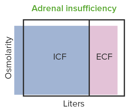
Image by Lecturio.

Hypo-osmotic volume contraction

  • Decrease in total body water along with a decrease in the total osmolality Osmolality Plasma osmolality refers to the combined concentration of all solutes in the blood. Renal Sodium and Water Regulation of solutes
  • Usually seen in the case of adrenal insufficiency Adrenal Insufficiency Conditions in which the production of adrenal corticosteroids falls below the requirement of the body. Adrenal insufficiency can be caused by defects in the adrenal glands, the pituitary gland, or the hypothalamus. Adrenal Insufficiency and Addison Disease
  • Changes in ECF result in:
    • Reduction in blood pressure due to decrease in volume
    • Decrease in Na+ level
    • Increase in total protein Total protein Liver Function Tests level
    • Increase in hematocrit Hematocrit The volume of packed red blood cells in a blood specimen. The volume is measured by centrifugation in a tube with graduated markings, or with automated blood cell counters. It is an indicator of erythrocyte status in disease. For example, anemia shows a low value; polycythemia, a high value. Neonatal Polycythemia
  • Endocrine response:
    • Increase in aldosterone Aldosterone A hormone secreted by the adrenal cortex that regulates electrolyte and water balance by increasing the renal retention of sodium and the excretion of potassium. Hyperkalemia (stimulated by hypovolemia Hypovolemia Sepsis in Children and hypokalemia Hypokalemia Hypokalemia is defined as plasma potassium (K+) concentration < 3.5 mEq/L. Homeostatic mechanisms maintain plasma concentration between 3.5-5.2 mEq/L despite marked variation in dietary intake. Hypokalemia can be due to renal losses, GI losses, transcellular shifts, or poor dietary intake. Hypokalemia)
    • No change in antidiuretic hormone Antidiuretic hormone Antidiuretic hormones released by the neurohypophysis of all vertebrates (structure varies with species) to regulate water balance and osmolarity. In general, vasopressin is a nonapeptide consisting of a six-amino-acid ring with a cysteine 1 to cysteine 6 disulfide bridge or an octapeptide containing a cystine. All mammals have arginine vasopressin except the pig with a lysine at position 8. Vasopressin, a vasoconstrictor, acts on the kidney collecting ducts to increase water reabsorption, increase blood volume and blood pressure. Hypernatremia level
Hypo-osmotic volume contraction seen in adrenal insufficiency

Hypo-osmotic volume contraction seen in adrenal insufficiency:
Low aldosterone leads to a decrease in tubular absorption of sodium, resulting in hypo-osmotic volume contraction.
ICF: intracellular fluid
ECF: extracellular fluid
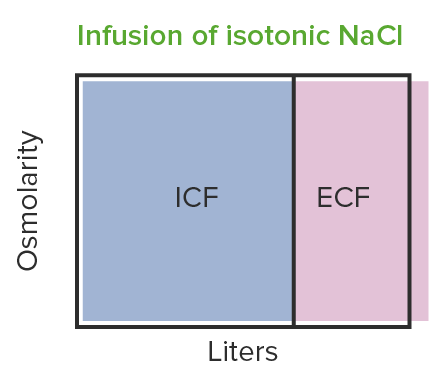
Image by Lecturio.

Iso-osmotic volume expansion

  • Volume expansion where there is no change in osmolality Osmolality Plasma osmolality refers to the combined concentration of all solutes in the blood. Renal Sodium and Water Regulation
  • Seen with IV solution volume expansion
  • Changes in ECF result in:
    • Increase in blood pressure
    • No change in Na+ level
    • Decrease in total protein Total protein Liver Function Tests level
    • Decrease in hematocrit Hematocrit The volume of packed red blood cells in a blood specimen. The volume is measured by centrifugation in a tube with graduated markings, or with automated blood cell counters. It is an indicator of erythrocyte status in disease. For example, anemia shows a low value; polycythemia, a high value. Neonatal Polycythemia
  • Endocrine response to iso-osmolar volume expansion:
    • Decrease in aldosterone Aldosterone A hormone secreted by the adrenal cortex that regulates electrolyte and water balance by increasing the renal retention of sodium and the excretion of potassium. Hyperkalemia (suppressed by volume expansion)
    • No change in antidiuretic hormone Antidiuretic hormone Antidiuretic hormones released by the neurohypophysis of all vertebrates (structure varies with species) to regulate water balance and osmolarity. In general, vasopressin is a nonapeptide consisting of a six-amino-acid ring with a cysteine 1 to cysteine 6 disulfide bridge or an octapeptide containing a cystine. All mammals have arginine vasopressin except the pig with a lysine at position 8. Vasopressin, a vasoconstrictor, acts on the kidney collecting ducts to increase water reabsorption, increase blood volume and blood pressure. Hypernatremia level
Infusion of isotonic nacl results in iso-osmotic volume expansion

Infusion of isotonic NaCl results in iso-osmotic volume expansion:
Changes are observed in ECF (i.e. increase in volume), but the osmolarity remains the same.
ICF: intracellular fluid
ECF: extracellular fluid
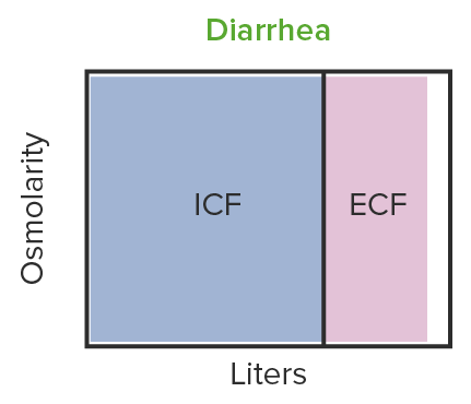
Image by Lecturio.

Iso-osmotic volume contraction

  • Volume contraction occurring without any change in osmolality Osmolality Plasma osmolality refers to the combined concentration of all solutes in the blood. Renal Sodium and Water Regulation
  • Seen in cases of iso-osmotic fluid loss (e.g., diarrhea Diarrhea Diarrhea is defined as ≥ 3 watery or loose stools in a 24-hour period. There are a multitude of etiologies, which can be classified based on the underlying mechanism of disease. The duration of symptoms (acute or chronic) and characteristics of the stools (e.g., watery, bloody, steatorrheic, mucoid) can help guide further diagnostic evaluation. Diarrhea and vomiting Vomiting The forcible expulsion of the contents of the stomach through the mouth. Hypokalemia)
  • Changes in ECF result in:
    • Decrease in blood pressure
    • No change in Na+ level
    • Increase in total protein Total protein Liver Function Tests level
    • Increase in hematocrit Hematocrit The volume of packed red blood cells in a blood specimen. The volume is measured by centrifugation in a tube with graduated markings, or with automated blood cell counters. It is an indicator of erythrocyte status in disease. For example, anemia shows a low value; polycythemia, a high value. Neonatal Polycythemia
  • Endocrine response:
    • Increase in aldosterone Aldosterone A hormone secreted by the adrenal cortex that regulates electrolyte and water balance by increasing the renal retention of sodium and the excretion of potassium. Hyperkalemia levels (stimulated by volume contraction)
    • No change in antidiuretic hormone Antidiuretic hormone Antidiuretic hormones released by the neurohypophysis of all vertebrates (structure varies with species) to regulate water balance and osmolarity. In general, vasopressin is a nonapeptide consisting of a six-amino-acid ring with a cysteine 1 to cysteine 6 disulfide bridge or an octapeptide containing a cystine. All mammals have arginine vasopressin except the pig with a lysine at position 8. Vasopressin, a vasoconstrictor, acts on the kidney collecting ducts to increase water reabsorption, increase blood volume and blood pressure. Hypernatremia level
Iso-osmotic volume contraction caused by diarrhea

Iso-osmotic volume contraction is observed in diarrhea:
Fluid in diarrhea has the same osmolarity as extracellular fluid. The volume of ECF decreases, but not the osmolarity.
ICF: intracellular fluid
ECF: extracellular fluid
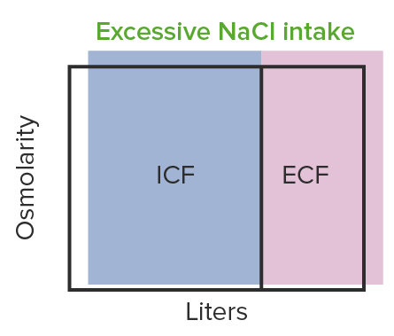
Image by Lecturio.

Hyper-osmotic volume expansion

  • Increase in osmolality Osmolality Plasma osmolality refers to the combined concentration of all solutes in the blood. Renal Sodium and Water Regulation alongside the increase in volume
  • Seen in cases of:
    • Aldosterone Aldosterone A hormone secreted by the adrenal cortex that regulates electrolyte and water balance by increasing the renal retention of sodium and the excretion of potassium. Hyperkalemia hypersecretion
    • Ingestion of salty foods
    • Individuals with a tumor Tumor Inflammation
  • Changes in ECF result in:
    • Increase in blood pressure
    • Increase in Na+ level
    • Reduction in total protein Total protein Liver Function Tests level
    • Reduction in hematocrit Hematocrit The volume of packed red blood cells in a blood specimen. The volume is measured by centrifugation in a tube with graduated markings, or with automated blood cell counters. It is an indicator of erythrocyte status in disease. For example, anemia shows a low value; polycythemia, a high value. Neonatal Polycythemia
  • Endocrine response:
    • Reduction in aldosterone Aldosterone A hormone secreted by the adrenal cortex that regulates electrolyte and water balance by increasing the renal retention of sodium and the excretion of potassium. Hyperkalemia level
    • Increase in antidiuretic hormone Antidiuretic hormone Antidiuretic hormones released by the neurohypophysis of all vertebrates (structure varies with species) to regulate water balance and osmolarity. In general, vasopressin is a nonapeptide consisting of a six-amino-acid ring with a cysteine 1 to cysteine 6 disulfide bridge or an octapeptide containing a cystine. All mammals have arginine vasopressin except the pig with a lysine at position 8. Vasopressin, a vasoconstrictor, acts on the kidney collecting ducts to increase water reabsorption, increase blood volume and blood pressure. Hypernatremia level
Hyper-osmotic volume expansion seen in increased intake of nacl

Hyper-osmotic volume expansion is seen in increased NaCl intake:
An increase in the sodium level causes retention of fluid.
ICF: intracellular fluid
ECF: extracellular fluid
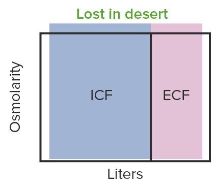
Image by Lecturio.

Hyper-osmotic volume contraction

  • Reduction of total body water but osmolality Osmolality Plasma osmolality refers to the combined concentration of all solutes in the blood. Renal Sodium and Water Regulation increases:
    • Seen in cases of dehydration Dehydration The condition that results from excessive loss of water from a living organism. Volume Depletion and Dehydration
    • Sweat contains more water than solute; thus, excessive sweating results in hyperosmotic volume contraction.
  • Changes in ECF result in:
    • Decrease in blood pressure
    • Increase in Na+ level
    • Increase in total protein Total protein Liver Function Tests level
    • No change in hematocrit Hematocrit The volume of packed red blood cells in a blood specimen. The volume is measured by centrifugation in a tube with graduated markings, or with automated blood cell counters. It is an indicator of erythrocyte status in disease. For example, anemia shows a low value; polycythemia, a high value. Neonatal Polycythemia
  • Endocrine response:
    • Increase in aldosterone Aldosterone A hormone secreted by the adrenal cortex that regulates electrolyte and water balance by increasing the renal retention of sodium and the excretion of potassium. Hyperkalemia level
    • Increase in antidiuretic hormone Antidiuretic hormone Antidiuretic hormones released by the neurohypophysis of all vertebrates (structure varies with species) to regulate water balance and osmolarity. In general, vasopressin is a nonapeptide consisting of a six-amino-acid ring with a cysteine 1 to cysteine 6 disulfide bridge or an octapeptide containing a cystine. All mammals have arginine vasopressin except the pig with a lysine at position 8. Vasopressin, a vasoconstrictor, acts on the kidney collecting ducts to increase water reabsorption, increase blood volume and blood pressure. Hypernatremia level
Hyper-osmotic volume contraction observed in dehydration

Hyper-osmotic volume contraction observed in dehydration causes hypotension:
The hypotension can reach a dangerously low level, depriving the organs of oxygen.
ICF: intracellular fluid
ECF: extracellular fluid

Image by Lecturio.

Body Fluid Composition

  • Body fluid compartments contain water, ions and osmolar molecules at different concentrations.
  • Relative concentration of the components depends on the exchange between cells and tissues in the body which is determined by the activity of transporters on cell membranes.
  • Composition of each fluid compartment is rigorously maintained by the continuous movement of water and other substances between compartments.
Table: Normal levels of ions and osmolar molecules in the plasma Plasma The residual portion of blood that is left after removal of blood cells by centrifugation without prior blood coagulation. Transfusion Products, interstitial fluid, and intracellular fluid
Plasma Plasma The residual portion of blood that is left after removal of blood cells by centrifugation without prior blood coagulation. Transfusion Products Interstitial fluid Intracellular fluid
(mOsm/L) (mOsm/L) (mOsm/L)
Na+ 142 139 14
K+ 4.2 4.0 140
Ca CA Condylomata acuminata are a clinical manifestation of genital HPV infection. Condylomata acuminata are described as raised, pearly, flesh-colored, papular, cauliflower-like lesions seen in the anogenital region that may cause itching, pain, or bleeding. Condylomata Acuminata (Genital Warts)2+ 1.3 1.2 0
Mg2+ 0.8 0.7 20
Chloride Chloride Inorganic compounds derived from hydrochloric acid that contain the Cl- ion. Electrolytes (Cl) 106 108 4
HCO3 24 28.3 10
HPO42–, H2PO4 2 2 11
SO42– 0.5 0.5 1
Phosphocreatine 45
Carnosine 14
Amino acids Amino acids Organic compounds that generally contain an amino (-NH2) and a carboxyl (-COOH) group. Twenty alpha-amino acids are the subunits which are polymerized to form proteins. Basics of Amino Acids 2 2 8
Creatinine 0.2 0.2 9
Lactate 1.2 1.2 1.5
ATP 5
Hexose monophosphate 3.7
Glucose Glucose A primary source of energy for living organisms. It is naturally occurring and is found in fruits and other parts of plants in its free state. It is used therapeutically in fluid and nutrient replacement. Lactose Intolerance 5.6 5.6
Proteins Proteins Linear polypeptides that are synthesized on ribosomes and may be further modified, crosslinked, cleaved, or assembled into complex proteins with several subunits. The specific sequence of amino acids determines the shape the polypeptide will take, during protein folding, and the function of the protein. Energy Homeostasis 1.2 0.2 4
Urea Urea A compound formed in the liver from ammonia produced by the deamination of amino acids. It is the principal end product of protein catabolism and constitutes about one half of the total urinary solids. Urea Cycle 4 1 4
Others 4.8 3.9 10
Total mOsm/L 299.8 300.8 301.2
Corrected osmolar activity 282.0 281.0 281.0

Clinical Relevance

  • Hyponatremia Hyponatremia Hyponatremia is defined as a decreased serum sodium (sNa+) concentration less than 135 mmol/L. Serum sodium is the greatest contributor to plasma osmolality, which is very tightly controlled via antidiuretic hormone (ADH) release from the hypothalamus and by the thirst mechanism. Hyponatremia: a decrease in Na+ levels in the body. Hyponatremia Hyponatremia Hyponatremia is defined as a decreased serum sodium (sNa+) concentration less than 135 mmol/L. Serum sodium is the greatest contributor to plasma osmolality, which is very tightly controlled via antidiuretic hormone (ADH) release from the hypothalamus and by the thirst mechanism. Hyponatremia most often occurs in the setting of severe diarrhea Diarrhea Diarrhea is defined as ≥ 3 watery or loose stools in a 24-hour period. There are a multitude of etiologies, which can be classified based on the underlying mechanism of disease. The duration of symptoms (acute or chronic) and characteristics of the stools (e.g., watery, bloody, steatorrheic, mucoid) can help guide further diagnostic evaluation. Diarrhea and vomiting Vomiting The forcible expulsion of the contents of the stomach through the mouth. Hypokalemia, excessive diuretic use, and Addison disease. Hyponatremia Hyponatremia Hyponatremia is defined as a decreased serum sodium (sNa+) concentration less than 135 mmol/L. Serum sodium is the greatest contributor to plasma osmolality, which is very tightly controlled via antidiuretic hormone (ADH) release from the hypothalamus and by the thirst mechanism. Hyponatremia can also occur less commonly as a result of SIADH SIADH Syndrome of inappropriate antidiuretic hormone secretion (SIADH) is a disorder of impaired water excretion due to the inability to suppress the secretion of antidiuretic hormone (ADH). SIADH is characterized by impaired water excretion leading to dilutional hyponatremia, which is mainly asymptomatic but may cause neurologic symptoms. S Syndrome of Inappropriate Antidiuretic Hormone Secretion (SIADH) and progress to cell edema Edema Edema is a condition in which excess serous fluid accumulates in the body cavity or interstitial space of connective tissues. Edema is a symptom observed in several medical conditions. It can be categorized into 2 types, namely, peripheral (in the extremities) and internal (in an organ or body cavity). Edema that can ultimately lead to brain Brain The part of central nervous system that is contained within the skull (cranium). Arising from the neural tube, the embryonic brain is comprised of three major parts including prosencephalon (the forebrain); mesencephalon (the midbrain); and rhombencephalon (the hindbrain). The developed brain consists of cerebrum; cerebellum; and other structures in the brain stem. Nervous System: Anatomy, Structure, and Classification damage, which presents with neurological symptoms of headache Headache The symptom of pain in the cranial region. It may be an isolated benign occurrence or manifestation of a wide variety of headache disorders. Brain Abscess, nausea Nausea An unpleasant sensation in the stomach usually accompanied by the urge to vomit. Common causes are early pregnancy, sea and motion sickness, emotional stress, intense pain, food poisoning, and various enteroviruses. Antiemetics, and lethargy Lethargy A general state of sluggishness, listless, or uninterested, with being tired, and having difficulty concentrating and doing simple tasks. It may be related to depression or drug addiction. Hyponatremia. Treatment is with gradual Na+ repletion, as a rapid replacement can lead to osmotic demyelination Demyelination Multiple Sclerosis syndrome.
  • Hypernatremia Hypernatremia Hypernatremia is an elevated serum sodium concentration > 145 mmol/L. Serum sodium is the greatest contributor to plasma osmolality, which is very tightly controlled by the hypothalamus via the thirst mechanism and antidiuretic hormone (ADH) release. Hypernatremia occurs either from a lack of access to water or an excessive intake of sodium. Hypernatremia: an increase in Na+ levels in the body. Hypernatremia Hypernatremia Hypernatremia is an elevated serum sodium concentration > 145 mmol/L. Serum sodium is the greatest contributor to plasma osmolality, which is very tightly controlled by the hypothalamus via the thirst mechanism and antidiuretic hormone (ADH) release. Hypernatremia occurs either from a lack of access to water or an excessive intake of sodium. Hypernatremia most often occurs as a result of severe dehydration Severe Dehydration Fluid Replacement Therapy in Children. Affected individuals are often asymptomatic, as symptoms typically occur when the Na+ level is approximately 160 mmol/L. Hypernatremia Hypernatremia Hypernatremia is an elevated serum sodium concentration > 145 mmol/L. Serum sodium is the greatest contributor to plasma osmolality, which is very tightly controlled by the hypothalamus via the thirst mechanism and antidiuretic hormone (ADH) release. Hypernatremia occurs either from a lack of access to water or an excessive intake of sodium. Hypernatremia can be corrected by administering hypo-osmotic NaCl or dextrose Dextrose Intravenous Fluids solution.
  • Edema Edema Edema is a condition in which excess serous fluid accumulates in the body cavity or interstitial space of connective tissues. Edema is a symptom observed in several medical conditions. It can be categorized into 2 types, namely, peripheral (in the extremities) and internal (in an organ or body cavity). Edema: the presence of excess fluid in body tissues. Both ECF and ICF compartments may be involved. Edema Edema Edema is a condition in which excess serous fluid accumulates in the body cavity or interstitial space of connective tissues. Edema is a symptom observed in several medical conditions. It can be categorized into 2 types, namely, peripheral (in the extremities) and internal (in an organ or body cavity). Edema is often clinically apparent in the lower extremities and occurs when the osmotic gradient between the intravascular space and interstitial space promotes the flow Flow Blood flows through the heart, arteries, capillaries, and veins in a closed, continuous circuit. Flow is the movement of volume per unit of time. Flow is affected by the pressure gradient and the resistance fluid encounters between 2 points. Vascular resistance is the opposition to flow, which is caused primarily by blood friction against vessel walls. Vascular Resistance, Flow, and Mean Arterial Pressure of water from the vasculature into the tissues. Edema Edema Edema is a condition in which excess serous fluid accumulates in the body cavity or interstitial space of connective tissues. Edema is a symptom observed in several medical conditions. It can be categorized into 2 types, namely, peripheral (in the extremities) and internal (in an organ or body cavity). Edema can be treated through diuresis or the addition of osmolar molecules (e.g., albumin Albumin Serum albumin from humans. It is an essential carrier of both endogenous substances, such as fatty acids and bilirubin, and of xenobiotics in the blood. Liver Function Tests) to the vascular space.

References

  1. Brinkman, J.E., Dorius, B., Sharma, S. (2021). Physiology, Body Fluids. Retrieved November 22, 2021, from https://www.ncbi.nlm.nih.gov/books/NBK482447
  2. Lobo, D.N. (2004). Fluid and electrolytes in the clinical setting. Nestle Nutr Workshop Ser Clin Perform Programme. 9, 187–203. https://pubmed.ncbi.nlm.nih.gov/15361687/
  3. Bedogni, G., Borghi, A., Battistini, N. (2003). Body water distribution and disease. Acta Diabetol. 40 (Suppl 1), S200-S202. https://pubmed.ncbi.nlm.nih.gov/14618472/
  4. Mathew, J., Sankar, P., Varacallo, M. (2020). StatPearls. StatPearls Publishing. Physiology, Blood Plasma. Retrieved November 22, 2021, from https://pubmed.ncbi.nlm.nih.gov/30285399/
  5. Davids, M.R., et al. (2002). Facilitating an understanding of integrative physiology: emphasis on the composition of body fluid compartments. Can J Physiol Pharmacol. 80, 835–850. https://pubmed.ncbi.nlm.nih.gov/12430978/
  6. Wiig, H., Swartz, M.A. (2012). Interstitial fluid and lymph formation and transport: physiological regulation and roles in inflammation and cancer. Physiol Rev. 92, 1005–1060. https://pubmed.ncbi.nlm.nih.gov/22811424/
  7. Benjamin, R.J., McLaughlin, L.S. (2021). Plasma components: properties, differences, and uses. Transfusion. 52 (Suppl 1), 9S-19S. https://pubmed.ncbi.nlm.nih.gov/22578375/
  8. Woodcock, T.E., Woodcock, T.M. (2012). Revised Starling equation and the glycocalyx model of transvascular fluid exchange: an improved paradigm for prescribing intravenous fluid therapy. Br J Anaesth. 108, 384–394. https://pubmed.ncbi.nlm.nih.gov/22290457/
  9. Levick, J.R., Michel, C.C. (2010). Microvascular fluid exchange and the revised Starling principle. Cardiovasc Res. 87, 198–210. https://pubmed.ncbi.nlm.nih.gov/20200043/
  10. Zierler, K. (2000). Indicator dilution methods for measuring blood flow, volume, and other properties of biological systems: a brief history and memoir. Ann Biomed Eng. 28, 836–848. https://pubmed.ncbi.nlm.nih.gov/11144667/
  11. Henriksen, J.H., Jensen, G.B., Larsson H.B. (2014). A century of indicator dilution techniques. Clin Physiol Funct Imaging. 34, 1–9. https://pubmed.ncbi.nlm.nih.gov/23869947/
  12. Kaptein, E.M., et al. (2016). A systematic literature search and review of sodium concentrations of body fluids. Clin Nephrol. 86, 203–228. https://pubmed.ncbi.nlm.nih.gov/27616761/
  13. Lu, H.A. (2017). Diabetes Insipidus. Adv Exp Med Biol. 969, 213–225. https://pubmed.ncbi.nlm.nih.gov/28258576/
  14. Kortenoeven, M.L., Fenton, R.A. (2014). Renal aquaporins and water balance disorders. Biochim Biophys Acta. 1840, 1533–1549.
  15. Schwartz, M.J., Kokko, J.P. (1980). Urinary concentrating defect of adrenal insufficiency. Permissive role of adrenal steroids on the hydroosmotic response across the rabbit cortical collecting tubule. J Clin Invest. 66,234–242. https://pubmed.ncbi.nlm.nih.gov/6156951/
  16. Lopez-Almaraz, E., Correa-Rotter, R. (2008). Dialysis disequilibrium syndrome and other treatment complications of extreme uremia: a rare occurrence yet not vanished. Hemodial Int. 12, 301–306. https://pubmed.ncbi.nlm.nih.gov/18638082/
  17. Abraham, S.F., et al. (1976). Aldosterone secretion during high sodium cerebrospinal fluid perfusion of the brain ventricles. Acta Endocrinol. 81, 120–132. https://pubmed.ncbi.nlm.nih.gov/946137/
  18. Aguilera, G., Catt, K.J. (1979). Loci of action of regulators of aldosterone biosynthesis in isolated glomerulosa cells. Endocrinology 104, 1046–1052. https://pubmed.ncbi.nlm.nih.gov/220023/
  19. Aguilera, G., Catt, K.J. (1983). Regulation of aldosterone secretion during altered sodium intake. J Steroid Biochem. 19, 525–530. https://pubmed.ncbi.nlm.nih.gov/6136631/
  20. Balla, T., Nagy, K., Tarjan, E., Renczes, G., Spät, A. (1981). Effects of reduced extracellular sodium concentration on the function of adrenal zona glomerulosa: studies in conscious rats. J Endocrinol. 89, 411–416. https://pubmed.ncbi.nlm.nih.gov/7019371/
  21. Birkhäuser, M., et al. (1973). Effect of volume expansion by hyperosmolar and hyperoncotic solutions under constant infusion of angiotensin II on plasma aldosterone in man and its counterbalance by potassium administration. Eur J Clin Invest. 3, 307–331. https://pubmed.ncbi.nlm.nih.gov/4357275/

Create your free account or log in to continue reading!

Sign up now and get free access to Lecturio with concept pages, medical videos, and questions for your medical education.

User Reviews

Details