Blount’s Disease

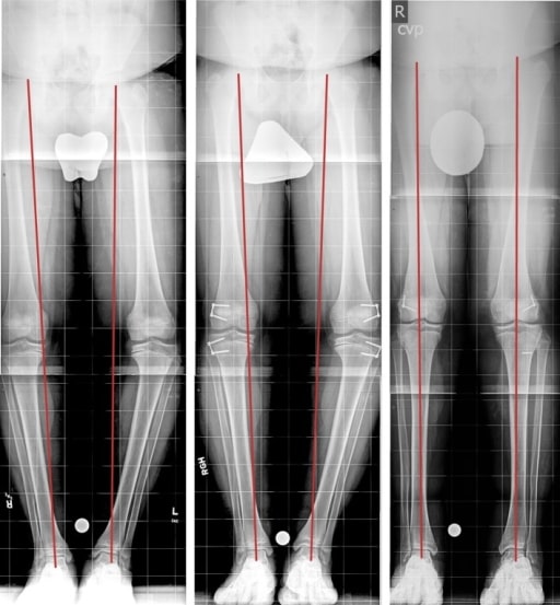
Blount’s disease (BD) is an orthopedic childhood disorder characterized by outward bowing of the leg Leg The lower leg, or just "leg" in anatomical terms, is the part of the lower limb between the knee and the ankle joint. The bony structure is composed of the tibia and fibula bones, and the muscles of the leg are grouped into the anterior, lateral, and posterior compartments by extensions of fascia. Leg: Anatomy due to abnormal ossification Ossification The process of bone formation. Histogenesis of bone including ossification. Bones: Development and Ossification of the medial aspect of the tibial epiphysis Epiphysis The head of a long bone that is separated from the shaft by the epiphyseal plate until bone growth stops. At that time, the plate disappears and the head and shaft are united. Bones: Structure and Types. Blount’s disease mostly affects children of African descent and tends to debut at approximately 1–3 years of age. Diagnosis is made by clinical findings and is confirmed by imaging. The goal of treatment is to correct the anatomy through bracing or surgical repair, according to the severity and age of the patient at diagnosis. The prognosis Prognosis A prediction of the probable outcome of a disease based on a individual's condition and the usual course of the disease as seen in similar situations. Non-Hodgkin Lymphomas is excellent if treatment is promptly started.

Last updated: Oct 6, 2022

Editorial responsibility: Stanley Oiseth, Lindsay Jones, Evelin Maza

Definition and Epidemiology

Definition

Blount’s disease (BD) is the progressive bowing of the legs produced by abnormal ossification Ossification The process of bone formation. Histogenesis of bone including ossification. Bones: Development and Ossification at the medial aspect of the tibial epiphysis Epiphysis The head of a long bone that is separated from the shaft by the epiphyseal plate until bone growth stops. At that time, the plate disappears and the head and shaft are united. Bones: Structure and Types.

Classification

  • Early or infantile: debuts at 1–3 years of age 
  • Late; subdivided into:

Epidemiology

  • Increased incidence Incidence The number of new cases of a given disease during a given period in a specified population. It also is used for the rate at which new events occur in a defined population. It is differentiated from prevalence, which refers to all cases in the population at a given time. Measures of Disease Frequency in people of African descent
  • Early BD: 
    • Boys > girls 
    • Bilateral in 50% of cases 
  • Late BD:
    • Less common and less severe than the early form
    • More likely to be unilateral
  • Prevalent in areas with higher rates of obesity Obesity Obesity is a condition associated with excess body weight, specifically with the deposition of excessive adipose tissue. Obesity is considered a global epidemic. Major influences come from the western diet and sedentary lifestyles, but the exact mechanisms likely include a mixture of genetic and environmental factors. Obesity and vitamin D deficiency Vitamin D Deficiency A nutritional condition produced by a deficiency of vitamin D in the diet, insufficient production of vitamin D in the skin, inadequate absorption of vitamin D from the diet, or abnormal conversion of vitamin D to its bioactive metabolites. It is manifested clinically as rickets in children and osteomalacia in adults. Fat-soluble Vitamins and their Deficiencies

Etiology and Pathophysiology

Etiology

The exact cause of BD is not known and is believed to be multifactorial or due to a combination of hereditary and developmental factors:
  • Genetic susceptibility 
  • Mechanical overload of the joint (associated with obesity Obesity Obesity is a condition associated with excess body weight, specifically with the deposition of excessive adipose tissue. Obesity is considered a global epidemic. Major influences come from the western diet and sedentary lifestyles, but the exact mechanisms likely include a mixture of genetic and environmental factors. Obesity)

Pathophysiology

  • Basis of the development of BD:
    • Excessive force
    • Altered endochondral ossification Ossification The process of bone formation. Histogenesis of bone including ossification. Bones: Development and Ossification of the leg Leg The lower leg, or just “leg” in anatomical terms, is the part of the lower limb between the knee and the ankle joint. The bony structure is composed of the tibia and fibula bones, and the muscles of the leg are grouped into the anterior, lateral, and posterior compartments by extensions of fascia. Leg: Anatomy
  • Mechanical forces ( obesity Obesity Obesity is a condition associated with excess body weight, specifically with the deposition of excessive adipose tissue. Obesity is considered a global epidemic. Major influences come from the western diet and sedentary lifestyles, but the exact mechanisms likely include a mixture of genetic and environmental factors. Obesity and early ambulation) on the medial physis of the tibia Tibia The second longest bone of the skeleton. It is located on the medial side of the lower leg, articulating with the fibula laterally, the talus distally, and the femur proximally. Knee Joint: Anatomy growth retardation Growth Retardation Failure of a fetus to attain expected growth. Fetal Alcohol Spectrum Disorder
  • Histological changes can be seen on the tibial growth plate:
    • Cellular hypertrophy Hypertrophy General increase in bulk of a part or organ due to cell enlargement and accumulation of fluids and secretions, not due to tumor formation, nor to an increase in the number of cells (hyperplasia). Cellular Adaptation and fibrocartilage Fibrocartilage A type of cartilage whose matrix contains large bundles of collagen type I. Fibrocartilage is typically found in the intervertebral disk; pubic symphysis; tibial menisci; and articular discs in synovial joints. Cartilage: Histology islands
    • Signifies damaged cartilage Cartilage Cartilage is a type of connective tissue derived from embryonic mesenchyme that is responsible for structural support, resilience, and the smoothness of physical actions. Perichondrium (connective tissue membrane surrounding cartilage) compensates for the absence of vasculature in cartilage by providing nutrition and support. Cartilage: Histology
  • Damaged cartilage Cartilage Cartilage is a type of connective tissue derived from embryonic mesenchyme that is responsible for structural support, resilience, and the smoothness of physical actions. Perichondrium (connective tissue membrane surrounding cartilage) compensates for the absence of vasculature in cartilage by providing nutrition and support. Cartilage: Histology delays bone Bone Bone is a compact type of hardened connective tissue composed of bone cells, membranes, an extracellular mineralized matrix, and central bone marrow. The 2 primary types of bone are compact and spongy. Bones: Structure and Types growth on the medial side → varus deformity Deformity Examination of the Upper Limbs
  • The procurvatum (backward bending of the tibia Tibia The second longest bone of the skeleton. It is located on the medial side of the lower leg, articulating with the fibula laterally, the talus distally, and the femur proximally. Knee Joint: Anatomy) is mainly due to the greatest inhibitory effect being on the posteromedial aspect of the tibial metaphysis Metaphysis Bones: Structure and Types.
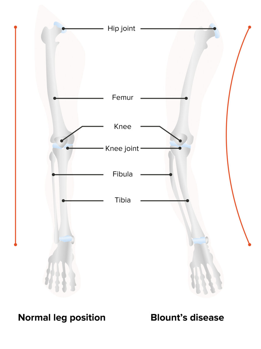
Blount’s disease

Blount’s disease: varus deformity and backward bending of the leg

Image by Lecturio.
Clinical Presentation

  • Patients Patients Individuals participating in the health care system for the purpose of receiving therapeutic, diagnostic, or preventive procedures. Clinician–Patient Relationship are usually overweight or obese.
  • 3-dimensional deformity Deformity Examination of the Upper Limbs:
    1. Varus (bowing)
    2. Procurvatum (backward bending of the tibia Tibia The second longest bone of the skeleton. It is located on the medial side of the lower leg, articulating with the fibula laterally, the talus distally, and the femur proximally. Knee Joint: Anatomy)
    3. Internal tibial rotation Rotation Motion of an object in which either one or more points on a line are fixed. It is also the motion of a particle about a fixed point. X-rays (resulting in in-toeing)
  • Length discrepancy in lower limbs
  • Non-tender bony protuberance at the medial aspect of the proximal tibia Tibia The second longest bone of the skeleton. It is located on the medial side of the lower leg, articulating with the fibula laterally, the talus distally, and the femur proximally. Knee Joint: Anatomy
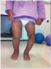
Blount's disease in a child

Left leg with varus deformity

Image: “Blount’s disease” by Universidade de São Paulo, Faculdade de Medicina, Hospital das Clínicas, Instituto de Ortopedia e Traumatologia, São Paulo, SP, Brazil. License: CC BY 4.0, edited by Lecturio.
Table: Clinical features of early BD versus late BD
Early BD Late BD
No pain Pain An unpleasant sensation induced by noxious stimuli which are detected by nerve endings of nociceptive neurons. Pain: Types and Pathways Pain Pain An unpleasant sensation induced by noxious stimuli which are detected by nerve endings of nociceptive neurons. Pain: Types and Pathways in the tibial area
Bilateral Unilateral
Significant deformity Deformity Examination of the Upper Limbs Mild deformity Deformity Examination of the Upper Limbs
Associated with early ambulation Associated with obesity Obesity Obesity is a condition associated with excess body weight, specifically with the deposition of excessive adipose tissue. Obesity is considered a global epidemic. Major influences come from the western diet and sedentary lifestyles, but the exact mechanisms likely include a mixture of genetic and environmental factors. Obesity

Diagnosis

The diagnosis of BD is achieved through medical history, physical examination, and confirmation via imaging studies.

Imaging

  • Long leg Leg The lower leg, or just “leg” in anatomical terms, is the part of the lower limb between the knee and the ankle joint. The bony structure is composed of the tibia and fibula bones, and the muscles of the leg are grouped into the anterior, lateral, and posterior compartments by extensions of fascia. Leg: Anatomy radiograph:
    • Varus angulation Angulation Buckle or Torus Fracture and sloping at the tibial epiphysis Epiphysis The head of a long bone that is separated from the shaft by the epiphyseal plate until bone growth stops. At that time, the plate disappears and the head and shaft are united. Bones: Structure and Types with asymmetrical bowing
    • Medial beaking: fragmentation Fragmentation Chronic Apophyseal Injury of the medial epiphysis Epiphysis The head of a long bone that is separated from the shaft by the epiphyseal plate until bone growth stops. At that time, the plate disappears and the head and shaft are united. Bones: Structure and Types of the tibia Tibia The second longest bone of the skeleton. It is located on the medial side of the lower leg, articulating with the fibula laterally, the talus distally, and the femur proximally. Knee Joint: Anatomy
    • Irregular ossification Ossification The process of bone formation. Histogenesis of bone including ossification. Bones: Development and Ossification and widening at the medial epiphysis Epiphysis The head of a long bone that is separated from the shaft by the epiphyseal plate until bone growth stops. At that time, the plate disappears and the head and shaft are united. Bones: Structure and Types
  • Magnetic resonance imaging (MRI): mostly used in cases of infantile Blount’s disease to assess extratibial tissues

Severity

Severity is based on the metaphyseal-diaphyseal angle (MDA):
  • Measures the angle formed by a line going through the metaphyseal beaks and a perpendicular line going through the longitudinal axis of the tibia Tibia The second longest bone of the skeleton. It is located on the medial side of the lower leg, articulating with the fibula laterally, the talus distally, and the femur proximally. Knee Joint: Anatomy
  • MDA > 16 degrees is diagnostic of BD.
  • MDA between 11 and 15 degrees is a probable diagnosis and warrants follow-up.
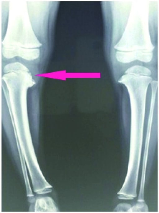
Depression of medial tibial plateau

Knee X-ray shows depression of the medial tibial plateau with beaking of the posteromedial tibial physis (pink arrow).

Image: “X-ray” by US National Library of Medicine. License: CC BY 4.0

Management and Prognosis

Management

The age of the patient and the severity of the deformity Deformity Examination of the Upper Limbs determine the type of management.
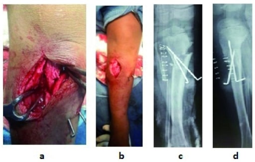
  • Bracing:
    • Indicated if younger than 3 years of age
    • Only used in non-obese patients Patients Individuals participating in the health care system for the purpose of receiving therapeutic, diagnostic, or preventive procedures. Clinician–Patient Relationship
    • Mainly worn at night
  • Surgical management:
    • Indicated in severe cases when the disease progresses despite bracing
    • Osteotomy is performed prior to the age of 4.
    • Hemiepiphysiodesis (gradual angular correction) is performed after the age of 4.
    • Fixation methods

Prognosis Prognosis A prediction of the probable outcome of a disease based on a individual’s condition and the usual course of the disease as seen in similar situations. Non-Hodgkin Lymphomas

  • Early infantile BD:
    • Must be treated in order to avoid permanent deformity Deformity Examination of the Upper Limbs
    • With timely treatment → excellent prognosis Prognosis A prediction of the probable outcome of a disease based on a individual’s condition and the usual course of the disease as seen in similar situations. Non-Hodgkin Lymphomas with rare recurrences
    • Mild form may regress spontaneously.
  • Late adolescent BD:

Differential Diagnosis

  • Physiological genu varum Genu varum Genu varum is a deformation of the knee joint(s) that creates angulation of the lower limb(s) away from the midline in the coronal plane. Children ages 1-5 years are commonly affected. Genu Varum: Children under the age of 2 have physiological, symmetrical Symmetrical Dermatologic Examination, painless bowing with in-toeing that spontaneously regresses with natural growth. Follow-up is usually performed to ensure that the condition does not progress further or has an underlying pathology.
  • Rickets Rickets Disorders caused by interruption of bone mineralization manifesting as osteomalacia in adults and characteristic deformities in infancy and childhood due to disturbances in normal bone formation. The mineralization process may be interrupted by disruption of vitamin d; phosphorus; or calcium homeostasis, resulting from dietary deficiencies, or acquired, or inherited metabolic, or hormonal disturbances. Osteomalacia and Rickets: distortion Distortion Defense Mechanisms and softening of bones due to vitamin D deficiency Vitamin D Deficiency A nutritional condition produced by a deficiency of vitamin D in the diet, insufficient production of vitamin D in the skin, inadequate absorption of vitamin D from the diet, or abnormal conversion of vitamin D to its bioactive metabolites. It is manifested clinically as rickets in children and osteomalacia in adults. Fat-soluble Vitamins and their Deficiencies or resistance Resistance Physiologically, the opposition to flow of air caused by the forces of friction. As a part of pulmonary function testing, it is the ratio of driving pressure to the rate of air flow. Ventilation: Mechanics of Breathing. Patients Patients Individuals participating in the health care system for the purpose of receiving therapeutic, diagnostic, or preventive procedures. Clinician–Patient Relationship usually present with bowing of the legs. Can be distinguished from BD via blood work and a lack of beaking on X-ray X-ray Penetrating electromagnetic radiation emitted when the inner orbital electrons of an atom are excited and release radiant energy. X-ray wavelengths range from 1 pm to 10 nm. Hard x-rays are the higher energy, shorter wavelength x-rays. Soft x-rays or grenz rays are less energetic and longer in wavelength. The short wavelength end of the x-ray spectrum overlaps the gamma rays wavelength range. The distinction between gamma rays and x-rays is based on their radiation source. Pulmonary Function Tests.

References

  1. Canale, S. T. (2017). Osteochondrosis or epiphysitis and other miscellaneous affections. In F. M. Azar MD, J. H. Beaty MD & S. T. Canale MD (Eds.), Campbell’s operative orthopaedics (pp. 1175-1248.e11). https://www.clinicalkey.es/#!/content/3-s2.0-B978032337462000032X
  2. Deeney, V. F., & Arnold, J. (2018). Orthopedics. In B. J. Zitelli MD, S. C. McIntire MD & Nowalk, Andrew J., MD, Ph.D. (Eds.), Zitelli and Davis’ atlas of pediatric physical diagnosis (pp. 759-844). https://www.clinicalkey.es/#!/content/3-s2.0-B9780323393034000220
  3. Janoyer, M. (2019). Blount disease. Orthopaedics & Traumatology: Surgery & Research, 105(1), S111-S121. doi:http://dx.doi.org/10.1016/j.otsr.2018.01.009
  4. Montgomery CO, Young KL, Austen M, Jo CH, Blasier RD, Ilyas M. Increased risk of Blount disease in obese children and adolescents with vitamin D deficiency. J Pediatr Orthop. 2010;30(8):879–882. DOI:10.1097/BPO.0b013e3181f5a0b3
  5. S DMTS, De Leucio A.(2020) Blount Disease.Treasure Island (FL): StatPearls Publishing; https://www.ncbi.nlm.nih.gov/books/NBK560923/
  6. Lauren LaMont, (2019).Blount Disease (Tibia vara). Medscape. Retrieved nov 24, 2020, from https://emedicine.medscape.com/article/1250420-overview#a1

Create your free account or log in to continue reading!

Sign up now and get free access to Lecturio with concept pages, medical videos, and questions for your medical education.

User Reviews

Details