Advertisement

Advertisement

Advertisement

Advertisement

Benign Liver Tumors

The most common benign Benign Fibroadenoma liver Liver The liver is the largest gland in the human body. The liver is found in the superior right quadrant of the abdomen and weighs approximately 1.5 kilograms. Its main functions are detoxification, metabolism, nutrient storage (e.g., iron and vitamins), synthesis of coagulation factors, formation of bile, filtration, and storage of blood. Liver: Anatomy tumors include hepatic hemangiomas, focal nodular hyperplasia Hyperplasia An increase in the number of cells in a tissue or organ without tumor formation. It differs from hypertrophy, which is an increase in bulk without an increase in the number of cells. Cellular Adaptation, and hepatic adenomas. These tumors are mostly asymptomatic and/or found incidentally on abdominal imaging. While these tumors are benign Benign Fibroadenoma, large lesions can cause symptoms such as upper abdominal pain Abdominal Pain Acute Abdomen, or produce complications such as bleeding. Malignant potential is a concern for hepatic adenoma, depending on risk factors. The diagnosis is based on imaging studies, with characteristic findings defining the tumor Tumor Inflammation. Biopsy Biopsy Removal and pathologic examination of specimens from the living body. Ewing Sarcoma generally is reserved for equivocal cases. Management is observation for most small, asymptomatic, and non-growing tumors. However, high-risk factors, symptoms, increasing tumor Tumor Inflammation size, and complications dictate the need for surgical intervention.

Last updated: Jan 2, 2024

Editorial responsibility: Stanley Oiseth, Lindsay Jones, Evelin Maza

Advertisement

Advertisement

Advertisement

Advertisement

Advertisement

Advertisement

Overview

Introduction

  • Most common benign Benign Fibroadenoma liver Liver The liver is the largest gland in the human body. The liver is found in the superior right quadrant of the abdomen and weighs approximately 1.5 kilograms. Its main functions are detoxification, metabolism, nutrient storage (e.g., iron and vitamins), synthesis of coagulation factors, formation of bile, filtration, and storage of blood. Liver: Anatomy tumors:
    • Hepatic hemangioma Hemangioma A vascular anomaly due to proliferation of blood vessels that forms a tumor-like mass. The common types involve capillaries and veins. It can occur anywhere in the body but is most frequently noticed in the skin and subcutaneous tissue. Imaging of the Liver and Biliary Tract
    • Focal nodular hyperplasia Hyperplasia An increase in the number of cells in a tissue or organ without tumor formation. It differs from hypertrophy, which is an increase in bulk without an increase in the number of cells. Cellular Adaptation (FNH)
    • Hepatocellular adenoma (HCA) or hepatic adenoma
  • Clinical significance:
    • These lesions can cause symptoms and complications.
    • Some lesions have a risk of developing hepatocellular carcinoma Hepatocellular carcinoma Hepatocellular carcinoma (HCC) typically arises in a chronically diseased or cirrhotic liver and is the most common primary liver cancer. Diagnosis may include ultrasound, CT, MRI, biopsy (if inconclusive imaging), and/or biomarkers. Hepatocellular Carcinoma (HCC) and Liver Metastases ( HCC HCC Hepatocellular carcinoma (HCC) typically arises in a chronically diseased or cirrhotic liver and is the most common primary liver cancer. Diagnosis may include ultrasound, CT, MRI, biopsy (if inconclusive imaging), and/or biomarkers. Hepatocellular Carcinoma (HCC) and Liver Metastases).

Epidemiology

  • Advances in imaging have led to many incidental findings.
  • Liver Liver The liver is the largest gland in the human body. The liver is found in the superior right quadrant of the abdomen and weighs approximately 1.5 kilograms. Its main functions are detoxification, metabolism, nutrient storage (e.g., iron and vitamins), synthesis of coagulation factors, formation of bile, filtration, and storage of blood. Liver: Anatomy lesions are noted in the following imaging studies:
    • Ultrasound (US)
    • Computed tomography (CT) scan 
    • Magnetic resonance imaging (MRI) 
  • Incidental hepatobiliary detection in screening Screening Preoperative Care chest CT: prevalence Prevalence The total number of cases of a given disease in a specified population at a designated time. It is differentiated from incidence, which refers to the number of new cases in the population at a given time. Measures of Disease Frequency of 6%
  • Epidemiologic findings for benign Benign Fibroadenoma tumors:
    • Hepatic or cavernous hemangioma Hemangioma A vascular anomaly due to proliferation of blood vessels that forms a tumor-like mass. The common types involve capillaries and veins. It can occur anywhere in the body but is most frequently noticed in the skin and subcutaneous tissue. Imaging of the Liver and Biliary Tract:
      • Most common benign Benign Fibroadenoma liver Liver The liver is the largest gland in the human body. The liver is found in the superior right quadrant of the abdomen and weighs approximately 1.5 kilograms. Its main functions are detoxification, metabolism, nutrient storage (e.g., iron and vitamins), synthesis of coagulation factors, formation of bile, filtration, and storage of blood. Liver: Anatomy tumor Tumor Inflammation
      • Prevalence Prevalence The total number of cases of a given disease in a specified population at a designated time. It is differentiated from incidence, which refers to the number of new cases in the population at a given time. Measures of Disease Frequency: 0.4%–20% of the population
      • Women > men
      • Often found at 30–50 years of age
    • FNH:
      • 2nd-most common benign Benign Fibroadenoma liver Liver The liver is the largest gland in the human body. The liver is found in the superior right quadrant of the abdomen and weighs approximately 1.5 kilograms. Its main functions are detoxification, metabolism, nutrient storage (e.g., iron and vitamins), synthesis of coagulation factors, formation of bile, filtration, and storage of blood. Liver: Anatomy tumor Tumor Inflammation
      • Prevalence Prevalence The total number of cases of a given disease in a specified population at a designated time. It is differentiated from incidence, which refers to the number of new cases in the population at a given time. Measures of Disease Frequency: 2%–3% of the population
      • Women > men
      • Often found at 35–50 years of age
    • HCA:
      • Rare
      • Women > men
      • Diagnosed frequently at 35–40 years of age

General comparison of hepatic tumors

  • It is important to distinguish benign Benign Fibroadenoma tumors from malignancy Malignancy Hemothorax, as management and prognosis Prognosis A prediction of the probable outcome of a disease based on a individual’s condition and the usual course of the disease as seen in similar situations. Non-Hodgkin Lymphomas are affected.
  • An outline of differentiating characteristics is summarized below.
Table: Comparison of liver Liver The liver is the largest gland in the human body. The liver is found in the superior right quadrant of the abdomen and weighs approximately 1.5 kilograms. Its main functions are detoxification, metabolism, nutrient storage (e.g., iron and vitamins), synthesis of coagulation factors, formation of bile, filtration, and storage of blood. Liver: Anatomy tumors
Hepatic hemangioma Hemangioma A vascular anomaly due to proliferation of blood vessels that forms a tumor-like mass. The common types involve capillaries and veins. It can occur anywhere in the body but is most frequently noticed in the skin and subcutaneous tissue. Imaging of the Liver and Biliary Tract Focal nodular hyperplasia Hyperplasia An increase in the number of cells in a tissue or organ without tumor formation. It differs from hypertrophy, which is an increase in bulk without an increase in the number of cells. Cellular Adaptation Hepatocellular adenoma Hepatocellular carcinoma Hepatocellular carcinoma Hepatocellular carcinoma (HCC) typically arises in a chronically diseased or cirrhotic liver and is the most common primary liver cancer. Diagnosis may include ultrasound, CT, MRI, biopsy (if inconclusive imaging), and/or biomarkers. Hepatocellular Carcinoma (HCC) and Liver Metastases
Characteristics and pathologic features Cavernous vascular spaces Central stellate scar Scar Dermatologic Examination + portal tracts, bile ductules Bile ductules Gallbladder and Biliary Tract: Anatomy, Kupffer cells Sheets of enlarged hepatocytes Hepatocytes The main structural component of the liver. They are specialized epithelial cells that are organized into interconnected plates called lobules. Liver: Anatomy; no portal tracts or bile ductules Bile ductules Gallbladder and Biliary Tract: Anatomy Well-differentiated (similar to normal hepatocytes Hepatocytes The main structural component of the liver. They are specialized epithelial cells that are organized into interconnected plates called lobules. Liver: Anatomy); poorly differentiated (marked cytologic atypia Atypia Fibrocystic Change)
Predominant gender Gender Gender Dysphoria affected Women Women Women Men
Clinical history Oral contraceptive pills (OCPs) may affect growth. OCP effect not proven; little or no effect on development or growth OCPs, anabolic steroids a factor History of cirrhosis Cirrhosis Cirrhosis is a late stage of hepatic parenchymal necrosis and scarring (fibrosis) most commonly due to hepatitis C infection and alcoholic liver disease. Patients may present with jaundice, ascites, and hepatosplenomegaly. Cirrhosis can also cause complications such as hepatic encephalopathy, portal hypertension, portal vein thrombosis, and hepatorenal syndrome. Cirrhosis and risk factors (e.g., hepatitis B Hepatitis B Hepatitis B virus (HBV) is a partially double-stranded DNA virus, which belongs to the Orthohepadnavirus genus and the Hepadnaviridae family. Most individuals with acute HBV infection are asymptomatic or have mild, self-limiting symptoms. Chronic infection can be asymptomatic or create hepatic inflammation, leading to liver cirrhosis and hepatocellular carcinoma (HCC). Hepatitis B Virus)
Malignancy Malignancy Hemothorax potential None None Yes N/A
MRI (contrast): arterial phase Peripheral nodular enhancement Early homogeneous Homogeneous Imaging of the Spleen/diffuse enhancement Well-demarcated enhancement; heterogeneous (due to hemorrhage, necrosis Necrosis The death of cells in an organ or tissue due to disease, injury or failure of the blood supply. Ischemic Cell Damage, steatosis Steatosis Nonalcoholic Fatty Liver Disease) Hyperenhancement
MRI (contrast): venous phase Progressive centripetal fill-in Isointense with central scar Scar Dermatologic Examination enhancement Variable Variable Variables represent information about something that can change. The design of the measurement scales, or of the methods for obtaining information, will determine the data gathered and the characteristics of that data. As a result, a variable can be qualitative or quantitative, and may be further classified into subgroups. Types of Variables late phase Late Phase Sepsis in Children
  • Inflammatory: Enhancement persists.
  • HNF1-ɑ mutation Mutation Genetic mutations are errors in DNA that can cause protein misfolding and dysfunction. There are various types of mutations, including chromosomal, point, frameshift, and expansion mutations. Types of Mutations: Arterial enhancement does not persist.
Portal venous washout
Additional key points
  • Can use technetium-99m pertechnetate-labeled red blood cell scan
  • Biopsy Biopsy Removal and pathologic examination of specimens from the living body. Ewing Sarcoma not recommended (high risk of hemorrhage)
60%–70% positive uptake in sulfur colloid Colloid Colloid solutions include large proteins or cells that do not readily cross capillary membranes. They remain in the ecf and do not distribute into the icf (similar to crystalloids). Intravenous Fluids scan No MRI-specific pattern was identified for β-catenin–mutated HCA. Rim enhancement on delayed post-contrast images causing a capsule-appearance: relatively specific for HCC HCC Hepatocellular carcinoma (HCC) typically arises in a chronically diseased or cirrhotic liver and is the most common primary liver cancer. Diagnosis may include ultrasound, CT, MRI, biopsy (if inconclusive imaging), and/or biomarkers. Hepatocellular Carcinoma (HCC) and Liver Metastases

Hemangioma

Features

  • Gross examination:
    • Often solitary, but may present as multiple lesions
    • Red-brown spongy mass Mass Three-dimensional lesion that occupies a space within the breast Imaging of the Breast, frequently with a capsule Capsule An envelope of loose gel surrounding a bacterial cell which is associated with the virulence of pathogenic bacteria. Some capsules have a well-defined border, whereas others form a slime layer that trails off into the medium. Most capsules consist of relatively simple polysaccharides but there are some bacteria whose capsules are made of polypeptides. Bacteroides
    • Size:
      • Most are < 5 cm
      • If the lesion is ≥ 10 cm: giant hemangioma Hemangioma A vascular anomaly due to proliferation of blood vessels that forms a tumor-like mass. The common types involve capillaries and veins. It can occur anywhere in the body but is most frequently noticed in the skin and subcutaneous tissue. Imaging of the Liver and Biliary Tract
    • Location: frequently in the right lobe
  • Microscopic examination:
    • Consists of cavernous vascular spaces, with a layer of endothelium Endothelium A layer of epithelium that lines the heart, blood vessels (vascular endothelium), lymph vessels (lymphatic endothelium), and the serous cavities of the body. Arteries: Histology
    • May contain thrombi
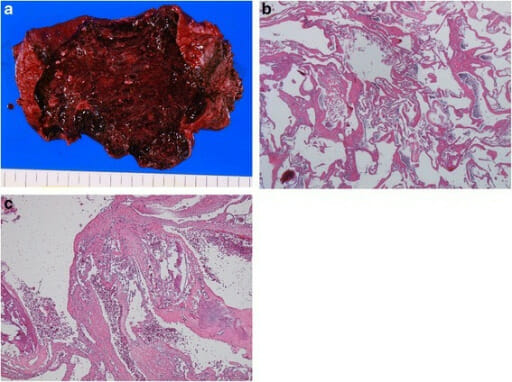
Gross and histological findings of hepatic hemangioma

Gross and histological findings of hepatic hemangioma:
a: wine-colored, elastic tumor
b: tumor composed of blood-filled spaces lined by a single layer of endothelial cells without smooth muscle (H&E stain, 40x objective)
c: fibrotic, hyalinized, calcified lesions (H&E stain, 40x objective)

Image: “Fig2” by Yano T, Kobayashi T et al. License: CC BY 4.0

Pathogenesis

  • Unclear pathogenetic process
  • Believed to result from a congenital Congenital Chorioretinitis vascular malformation, with dilation as the growth pattern 
  • Risk factors: Estrogen Estrogen Compounds that interact with estrogen receptors in target tissues to bring about the effects similar to those of estradiol. Estrogens stimulate the female reproductive organs, and the development of secondary female sex characteristics. Estrogenic chemicals include natural, synthetic, steroidal, or non-steroidal compounds. Ovaries: Anatomy such as in OCPs or pregnancy Pregnancy The status during which female mammals carry their developing young (embryos or fetuses) in utero before birth, beginning from fertilization to birth. Pregnancy: Diagnosis, Physiology, and Care may promote its growth.

Clinical presentation

  • Typically asymptomatic
  • Frequently discovered incidentally on imaging studies
  • May cause right upper quadrant Right upper quadrant Anterior Abdominal Wall: Anatomy (RUQ) pain Pain An unpleasant sensation induced by noxious stimuli which are detected by nerve endings of nociceptive neurons. Pain: Types and Pathways/fullness
  • If large enough (> 10 cm), may compress other organs and manifest as nausea Nausea An unpleasant sensation in the stomach usually accompanied by the urge to vomit. Common causes are early pregnancy, sea and motion sickness, emotional stress, intense pain, food poisoning, and various enteroviruses. Antiemetics, early satiety Early Satiety Bariatric Surgery, and bloating Bloating Constipation
  • Physical examination often normal, but may show a palpable liver Liver The liver is the largest gland in the human body. The liver is found in the superior right quadrant of the abdomen and weighs approximately 1.5 kilograms. Its main functions are detoxification, metabolism, nutrient storage (e.g., iron and vitamins), synthesis of coagulation factors, formation of bile, filtration, and storage of blood. Liver: Anatomy mass Mass Three-dimensional lesion that occupies a space within the breast Imaging of the Breast
  • Kasabach-Merritt syndrome:
    • A giant hemangioma Hemangioma A vascular anomaly due to proliferation of blood vessels that forms a tumor-like mass. The common types involve capillaries and veins. It can occur anywhere in the body but is most frequently noticed in the skin and subcutaneous tissue. Imaging of the Liver and Biliary Tract may cause this rare coagulation disorder.
    • Associated with thrombocytopenia Thrombocytopenia Thrombocytopenia occurs when the platelet count is < 150,000 per microliter. The normal range for platelets is usually 150,000-450,000/µL of whole blood. Thrombocytopenia can be a result of decreased production, increased destruction, or splenic sequestration of platelets. Patients are often asymptomatic until platelet counts are < 50,000/µL. Thrombocytopenia, consumption coagulopathy, and bleeding
    • Mortality Mortality All deaths reported in a given population. Measures of Health Status rate of up to 37%

Diagnosis

Management

  • Observation if asymptomatic:
    • ≤ 5 cm: no further imaging
    • > 5 cm:
      • Repeat imaging in 6–12 months. 
      • If lesion is stable (growth rate ≤ 3 mm/year), no further testing
  • Consider surgery ( liver Liver The liver is the largest gland in the human body. The liver is found in the superior right quadrant of the abdomen and weighs approximately 1.5 kilograms. Its main functions are detoxification, metabolism, nutrient storage (e.g., iron and vitamins), synthesis of coagulation factors, formation of bile, filtration, and storage of blood. Liver: Anatomy resection or enucleation):
    • Persistent symptoms and/or rapid enlargement (> 3 mm/year)
    • Symptomatic patients Patients Individuals participating in the health care system for the purpose of receiving therapeutic, diagnostic, or preventive procedures. Clinician–Patient Relationship
  • Transcatheter arterial embolization Embolization A method of hemostasis utilizing various agents such as gelfoam, silastic, metal, glass, or plastic pellets, autologous clot, fat, and muscle as emboli. It has been used in the treatment of spinal cord and intracranial arteriovenous malformations, renal arteriovenous fistulas, gastrointestinal bleeding, epistaxis, hypersplenism, certain highly vascular tumors, traumatic rupture of blood vessels, and control of operative hemorrhage. Gastrointestinal Bleeding can be used to decrease lesion size prior to surgery.

Focal Nodular Hyperplasia

Features

  • Gross examination:
    • Firm, solitary lesion, without a capsule Capsule An envelope of loose gel surrounding a bacterial cell which is associated with the virulence of pathogenic bacteria. Some capsules have a well-defined border, whereas others form a slime layer that trails off into the medium. Most capsules consist of relatively simple polysaccharides but there are some bacteria whose capsules are made of polypeptides. Bacteroides
    • Characteristic central stellate scar Scar Dermatologic Examination (multiple branches from an artery radiating to the periphery)
    • Size: often < 5 cm
    • Location: usually subcapsular 
    • FNH has atypical variants.
  • Microscopic examination:
    • Grouped hepatocytes Hepatocytes The main structural component of the liver. They are specialized epithelial cells that are organized into interconnected plates called lobules. Liver: Anatomy, divided by fibrous Fibrous Fibrocystic Change septa, radiating from the central scar Scar Dermatologic Examination
    • Septa contain arteries Arteries Arteries are tubular collections of cells that transport oxygenated blood and nutrients from the heart to the tissues of the body. The blood passes through the arteries in order of decreasing luminal diameter, starting in the largest artery (the aorta) and ending in the small arterioles. Arteries are classified into 3 types: large elastic arteries, medium muscular arteries, and small arteries and arterioles. Arteries: Histology, portal veins Veins Veins are tubular collections of cells, which transport deoxygenated blood and waste from the capillary beds back to the heart. Veins are classified into 3 types: small veins/venules, medium veins, and large veins. Each type contains 3 primary layers: tunica intima, tunica media, and tunica adventitia. Veins: Histology, bile ductules Bile ductules Gallbladder and Biliary Tract: Anatomy, and Kupffer cells.
    • Strong cytoplasmic glutamine synthetase Glutamine synthetase Synthesis of Nonessential Amino Acids staining within groups of hepatocytes Hepatocytes The main structural component of the liver. They are specialized epithelial cells that are organized into interconnected plates called lobules. Liver: Anatomy
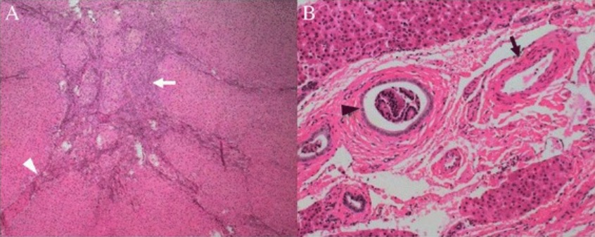
Classic focal nodular hyperplasia

Histologic features of classic FNH: Tumor was subdivided into nodules by fibrous septa (A, white arrowhead) originating from a central scar (A, white arrow). Malformed arteries (B, black arrow) and bile ductular proliferation are demonstrated (B, black arrowhead).

Image: “Histological features of the classic focal nodular hyperplasia” by Department of Radiology, PLA General Hospital, #28 Fuxing Road, Beijing, 100853, China. License: CC BY 4.0
Pathogenesis

  • Regenerative response of hepatocytes Hepatocytes The main structural component of the liver. They are specialized epithelial cells that are organized into interconnected plates called lobules. Liver: Anatomy to altered perfusion from anomalous arteries Arteries Arteries are tubular collections of cells that transport oxygenated blood and nutrients from the heart to the tissues of the body. The blood passes through the arteries in order of decreasing luminal diameter, starting in the largest artery (the aorta) and ending in the small arterioles. Arteries are classified into 3 types: large elastic arteries, medium muscular arteries, and small arteries and arterioles. Arteries: Histology (in the center of the nodule Nodule Chalazion
  • No malignant potential
  • Increased risk of FNH in hereditary hemorrhagic telangiectasia Telangiectasia Permanent dilation of preexisting blood vessels creating small focal red lesions, most commonly in the skin or mucous membranes. It is characterized by the prominence of skin blood vessels, such as vascular spiders. Chronic Venous Insufficiency

Clinical presentation

  • Asymptomatic
  • Often found incidentally on imaging
  • If symptomatic, commonly the symptom is abdominal pain Abdominal Pain Acute Abdomen.
  • Physical examination: usually normal, but may show an abdominal mass Mass Three-dimensional lesion that occupies a space within the breast Imaging of the Breast

Diagnosis

  • US:
    • Non-contrast: isoechoic Isoechoic A structure that produces an echo of a very similar amplitude to its environment and is very difficult to distinguish Ultrasound (Sonography) mass Mass Three-dimensional lesion that occupies a space within the breast Imaging of the Breast
    • With contrast:
      • FNH: enhancement sustained in arterial phase and early venous phase
      • Central arteries Arteries Arteries are tubular collections of cells that transport oxygenated blood and nutrients from the heart to the tissues of the body. The blood passes through the arteries in order of decreasing luminal diameter, starting in the largest artery (the aorta) and ending in the small arterioles. Arteries are classified into 3 types: large elastic arteries, medium muscular arteries, and small arteries and arterioles. Arteries: Histology seen with spoke-wheel pattern on arterial phase (centrifugal)
  • MRI scan: 
  • CT scan:
    • Pre-contrast: isodense
    • With contrast:
      • Arterial phase: homogeneous Homogeneous Imaging of the Spleen hyperdense lesion 
      • Venous phase: isodense, similar to liver Liver The liver is the largest gland in the human body. The liver is found in the superior right quadrant of the abdomen and weighs approximately 1.5 kilograms. Its main functions are detoxification, metabolism, nutrient storage (e.g., iron and vitamins), synthesis of coagulation factors, formation of bile, filtration, and storage of blood. Liver: Anatomy parenchyma; the central scar Scar Dermatologic Examination becomes hyperdense.
  • Nuclear medicine Nuclear medicine A specialty field of radiology concerned with diagnostic, therapeutic, and investigative use of radioactive compounds. Nuclear Imaging (technetium-99m sulfur colloid Colloid Colloid solutions include large proteins or cells that do not readily cross capillary membranes. They remain in the ecf and do not distribute into the icf (similar to crystalloids). Intravenous Fluids scan): 
    • Increased uptake of sulfur colloid Colloid Colloid solutions include large proteins or cells that do not readily cross capillary membranes. They remain in the ecf and do not distribute into the icf (similar to crystalloids). Intravenous Fluids (Kupffer cell activity) is seen in 60%–70% of patients Patients Individuals participating in the health care system for the purpose of receiving therapeutic, diagnostic, or preventive procedures. Clinician–Patient Relationship. 
    • Helps to differentiate FNH from adenoma (which has no Kupffer cell activity)
  • If imaging findings are not typical of FNH, a biopsy Biopsy Removal and pathologic examination of specimens from the living body. Ewing Sarcoma may be required.
Focal nodular hyperplasia

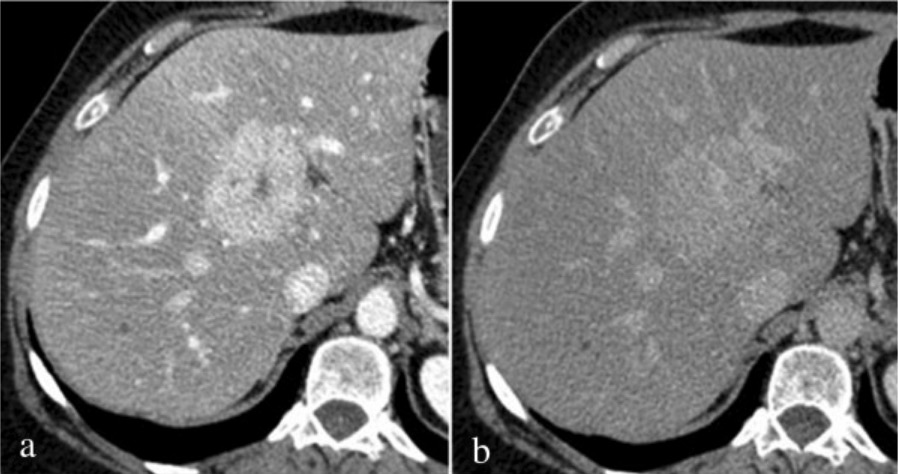
Computed tomography findings of FNH: Mixed phase (hepatic arterial phase/portal venous phase during hepatic enhancement) shows intense homogeneous enhancement with hypodense focal central scar (a); on delayed phase (b), the lesion appears substantially isodense to liver parenchyma with persistent enhancement of central scar.

Image: “Fig3” by Department of Surgical and Biomedical Sciences, Division of Radiology 2, Perugia University, S, Maria della Misericordia Hospital, S, Andrea delle Fratte, 06134 Perugia, Italy. License: CC BY 4.0
Management

  • For asymptomatic patients Patients Individuals participating in the health care system for the purpose of receiving therapeutic, diagnostic, or preventive procedures. Clinician–Patient Relationship: no routine surveillance Surveillance Developmental Milestones and Normal Growth recommended as lesion rarely grows
  • For symptomatic patients Patients Individuals participating in the health care system for the purpose of receiving therapeutic, diagnostic, or preventive procedures. Clinician–Patient Relationship: may undergo transarterial embolization Embolization A method of hemostasis utilizing various agents such as gelfoam, silastic, metal, glass, or plastic pellets, autologous clot, fat, and muscle as emboli. It has been used in the treatment of spinal cord and intracranial arteriovenous malformations, renal arteriovenous fistulas, gastrointestinal bleeding, epistaxis, hypersplenism, certain highly vascular tumors, traumatic rupture of blood vessels, and control of operative hemorrhage. Gastrointestinal Bleeding, radiofrequency ablation Radiofrequency ablation Removal of tissue using heat generated from electrodes delivering an alternating electrical current in the frequency of radio waves. Hepatocellular Carcinoma (HCC) and Liver Metastases, or surgical resection

Hepatocellular Adenoma

Features

  • Gross examination:
    • Typically a solitary lesion with well-defined margin, but can occur as multiple lesions
    • Varied sizes, from small to several centimeters
    • Lacks a fibrous capsule Fibrous capsule Hip Joint: Anatomy (risk for rupture and bleeding)
    • Location: often in the right lobe of the liver Liver The liver is the largest gland in the human body. The liver is found in the superior right quadrant of the abdomen and weighs approximately 1.5 kilograms. Its main functions are detoxification, metabolism, nutrient storage (e.g., iron and vitamins), synthesis of coagulation factors, formation of bile, filtration, and storage of blood. Liver: Anatomy
  • Microscopic examination:
    • Sheets of enlarged hepatocytes Hepatocytes The main structural component of the liver. They are specialized epithelial cells that are organized into interconnected plates called lobules. Liver: Anatomy with small nuclei, glycogen, and lipid
    • Large arteries Arteries Arteries are tubular collections of cells that transport oxygenated blood and nutrients from the heart to the tissues of the body. The blood passes through the arteries in order of decreasing luminal diameter, starting in the largest artery (the aorta) and ending in the small arterioles. Arteries are classified into 3 types: large elastic arteries, medium muscular arteries, and small arteries and arterioles. Arteries: Histology present
    • Generally, no portal tracts or bile ductules Bile ductules Gallbladder and Biliary Tract: Anatomy (differentiates HCA from FNH); inflammatory subtype is an exception
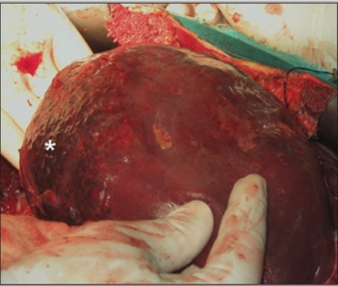
Hepatic adenoma

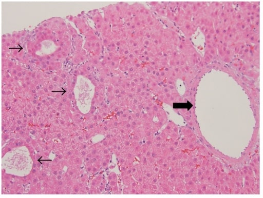
Hepatic adenoma: well-differentiated neoplasm composed of normal-appearing hepatocytes arranged in sheets and thin cords with patchy pseudoacinar growth pattern (thin arrows), scattered inflammatory foci, and bands of fibrosis with unpaired large arteries (thick arrow)

Image: “Hepatic adenoma” by M. I. Montenovo. License: CC BY 4.0
Pathogenesis

  • Benign Benign Fibroadenoma glandular epithelial tumor Tumor Inflammation, with the following risk factors:
    • Use of OCPs, anabolic steroids 
    • Genetic syndromes such as glycogen storage diseases and familial adenomatous polyposis Familial Adenomatous Polyposis Familial adenomatous polyposis (FAP) is an autosomal dominant inherited genetic disorder that presents with numerous adenomatous polyps in the colon. Familial adenomatous polyposis is the most common of the polyposis syndromes, which is a group of inherited or acquired conditions characterized by the growth of polyps in the GI tract, associated with other extracolonic features. Familial Adenomatous Polyposis
    • Obesity Obesity Obesity is a condition associated with excess body weight, specifically with the deposition of excessive adipose tissue. Obesity is considered a global epidemic. Major influences come from the western diet and sedentary lifestyles, but the exact mechanisms likely include a mixture of genetic and environmental factors. Obesity, metabolic syndrome Metabolic syndrome Metabolic syndrome is a cluster of conditions that significantly increases the risk for several secondary diseases, notably cardiovascular disease, type 2 diabetes, and nonalcoholic fatty liver. In general, it is agreed that hypertension, insulin resistance/hyperglycemia, and hyperlipidemia, along with central obesity, are components of the metabolic syndrome. Metabolic Syndrome
  • Subtypes based on the molecular behavior of HCA:
    • Adenomas with hepatocyte nuclear factor 1 alpha (HNF1-ɑ) mutation Mutation Genetic mutations are errors in DNA that can cause protein misfolding and dysfunction. There are various types of mutations, including chromosomal, point, frameshift, and expansion mutations. Types of Mutations:
      • 35%–40%
      • Have loss-of-function mutations in the HNF1-ɑ gene Gene A category of nucleic acid sequences that function as units of heredity and which code for the basic instructions for the development, reproduction, and maintenance of organisms. Basic Terms of Genetics
      • Findings: steatosis Steatosis Nonalcoholic Fatty Liver Disease or prominent fat in hepatocytes Hepatocytes The main structural component of the liver. They are specialized epithelial cells that are organized into interconnected plates called lobules. Liver: Anatomy
      • Predominantly affects women 
      • Low risk of malignant transformation Transformation Change brought about to an organism’s genetic composition by unidirectional transfer (transfection; transduction, genetic; conjugation, genetic, etc.) and incorporation of foreign DNA into prokaryotic or eukaryotic cells by recombination of part or all of that DNA into the cell’s genome. Bacteriology
      • Associated with mature-onset diabetes Diabetes Diabetes mellitus (DM) is a metabolic disease characterized by hyperglycemia and dysfunction of the regulation of glucose metabolism by insulin. Type 1 DM is diagnosed mostly in children and young adults as the result of autoimmune destruction of β cells in the pancreas and the resulting lack of insulin. Type 2 DM has a significant association with obesity and is characterized by insulin resistance. Diabetes Mellitus of the young ( MODY MODY Diabetes Mellitus 3)
    • β-catenin–activated HCA:
      • 10%–20% 
      • Associated with mutations of the CTNNB1 (β-catenin) gene Gene A category of nucleic acid sequences that function as units of heredity and which code for the basic instructions for the development, reproduction, and maintenance of organisms. Basic Terms of Genetics or other Wnt pathway components
      • Frequently in men
      • Linked to anabolic steroid use
      • Findings: atypical cytologic features
      • High risk for HCC HCC Hepatocellular carcinoma (HCC) typically arises in a chronically diseased or cirrhotic liver and is the most common primary liver cancer. Diagnosis may include ultrasound, CT, MRI, biopsy (if inconclusive imaging), and/or biomarkers. Hepatocellular Carcinoma (HCC) and Liver Metastases development
    • Inflammatory adenomas:
      • 40%–50%
      • Findings: inflammatory infiltrates, sinusoidal dilation
      • More prevalent in women with obesity Obesity Obesity is a condition associated with excess body weight, specifically with the deposition of excessive adipose tissue. Obesity is considered a global epidemic. Major influences come from the western diet and sedentary lifestyles, but the exact mechanisms likely include a mixture of genetic and environmental factors. Obesity or diabetes Diabetes Diabetes mellitus (DM) is a metabolic disease characterized by hyperglycemia and dysfunction of the regulation of glucose metabolism by insulin. Type 1 DM is diagnosed mostly in children and young adults as the result of autoimmune destruction of β cells in the pancreas and the resulting lack of insulin. Type 2 DM has a significant association with obesity and is characterized by insulin resistance. Diabetes Mellitus
    • Unclassified: up to 10%

Clinical presentation

  • May be asymptomatic, but 25% of patients Patients Individuals participating in the health care system for the purpose of receiving therapeutic, diagnostic, or preventive procedures. Clinician–Patient Relationship present with RUQ pain Pain An unpleasant sensation induced by noxious stimuli which are detected by nerve endings of nociceptive neurons. Pain: Types and Pathways or mass Mass Three-dimensional lesion that occupies a space within the breast Imaging of the Breast
  • Hemorrhage: higher risk when size is > 5 cm
    • Bleeding into the lesion can manifest as abdominal pain Abdominal Pain Acute Abdomen.
    • In severe cases, rupture into the peritoneum Peritoneum The peritoneum is a serous membrane lining the abdominopelvic cavity. This lining is formed by connective tissue and originates from the mesoderm. The membrane lines both the abdominal walls (as parietal peritoneum) and all of the visceral organs (as visceral peritoneum). Peritoneum: Anatomy presents with severe pain Pain An unpleasant sensation induced by noxious stimuli which are detected by nerve endings of nociceptive neurons. Pain: Types and Pathways and hypotension Hypotension Hypotension is defined as low blood pressure, specifically < 90/60 mm Hg, and is most commonly a physiologic response. Hypotension may be mild, serious, or life threatening, depending on the cause. Hypotension.

Diagnosis

  • US:
    • Without contrast: nonspecific heterogeneous mass Mass Three-dimensional lesion that occupies a space within the breast Imaging of the Breast
    • With contrast: hyperenhancement from periphery to center (centripetal)
  • CT scan:
    • Without contrast: isodense lesion
    • With contrast:
      • Arterial phase: may have peripheral enhancement 
      • Portal venous phase: with centripetal flow Flow Blood flows through the heart, arteries, capillaries, and veins in a closed, continuous circuit. Flow is the movement of volume per unit of time. Flow is affected by the pressure gradient and the resistance fluid encounters between 2 points. Vascular resistance is the opposition to flow, which is caused primarily by blood friction against vessel walls. Vascular Resistance, Flow, and Mean Arterial Pressure
    • If with areas of hemorrhage, necrosis Necrosis The death of cells in an organ or tissue due to disease, injury or failure of the blood supply. Ischemic Cell Damage, and calcification, adenoma appears heterogeneous.
  • MRI with contrast:
    • Superior to other modalities for HCA diagnosis
    • Arterial phase: well-demarcated, enhanced lesion (heterogeneous due to hemorrhage, necrosis Necrosis The death of cells in an organ or tissue due to disease, injury or failure of the blood supply. Ischemic Cell Damage, steatosis Steatosis Nonalcoholic Fatty Liver Disease)
    • Pattern in later phases correlates with molecular subtypes in majority of cases (inflammatory and HNF1-ɑ type):
      • Inflammatory adenoma: Arterial enhancement persists into the portal venous and delayed phases.
      • HCA with HNF1-ɑ mutation Mutation Genetic mutations are errors in DNA that can cause protein misfolding and dysfunction. There are various types of mutations, including chromosomal, point, frameshift, and expansion mutations. Types of Mutations: Arterial enhancement does not persist.
      • β-catenin–activated HCA: no specific MRI characteristics
  • Core needle biopsy Core Needle Biopsy Fibrocystic Change: for clarification if imaging is equivocal
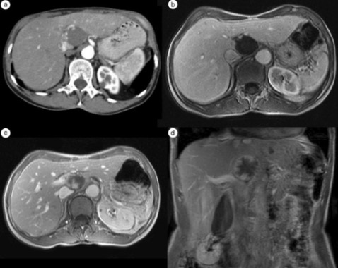
Benign liver disease mri with adenoma with bleeding

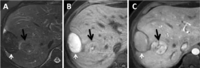
Magnetic resonance imaging in a patient with adenoma with bleeding: white arrow, hematoma; black arrow, hepatic adenoma.
A: T2 sequence with fat saturation shows a hepatic subcapsular nodule with hyposignal.
B: Pre-contrast T1 sequence shows a lesion with hypersignal, signifying products of hemoglobin degradation.
C: Post-contrast T1 sequence in the arterial phase emphasizes the hepatic lesion.

Image: “MRI” by Enio Campos AMICO, José Roberto ALVES et al. License: CC BY 4.0

Management

  • Discontinue anabolic steroids or OCPs.
  • Weight loss Weight loss Decrease in existing body weight. Bariatric Surgery
  • Observation and surveillance Surveillance Developmental Milestones and Normal Growth:
  • Surgical resection:
    • Tumor Tumor Inflammation is > 5 cm: surgical resection due to increased risk of rupture, bleeding, or malignant transformation Transformation Change brought about to an organism’s genetic composition by unidirectional transfer (transfection; transduction, genetic; conjugation, genetic, etc.) and incorporation of foreign DNA into prokaryotic or eukaryotic cells by recombination of part or all of that DNA into the cell’s genome. Bacteriology
    • Men with HCA (regardless of size)
    • Proven β-catenin mutation Mutation Genetic mutations are errors in DNA that can cause protein misfolding and dysfunction. There are various types of mutations, including chromosomal, point, frameshift, and expansion mutations. Types of Mutations
  • Transarterial embolization Embolization A method of hemostasis utilizing various agents such as gelfoam, silastic, metal, glass, or plastic pellets, autologous clot, fat, and muscle as emboli. It has been used in the treatment of spinal cord and intracranial arteriovenous malformations, renal arteriovenous fistulas, gastrointestinal bleeding, epistaxis, hypersplenism, certain highly vascular tumors, traumatic rupture of blood vessels, and control of operative hemorrhage. Gastrointestinal Bleeding:
    • Tumor Tumor Inflammation complicated by bleeding and hemodynamic instability
    • If after the procedure there is residual lesion noted on follow-up imaging, perform surgical resection.

Differential Diagnosis

  • HCC HCC Hepatocellular carcinoma (HCC) typically arises in a chronically diseased or cirrhotic liver and is the most common primary liver cancer. Diagnosis may include ultrasound, CT, MRI, biopsy (if inconclusive imaging), and/or biomarkers. Hepatocellular Carcinoma (HCC) and Liver Metastases: a type of cancer that develops in patients Patients Individuals participating in the health care system for the purpose of receiving therapeutic, diagnostic, or preventive procedures. Clinician–Patient Relationship with liver Liver The liver is the largest gland in the human body. The liver is found in the superior right quadrant of the abdomen and weighs approximately 1.5 kilograms. Its main functions are detoxification, metabolism, nutrient storage (e.g., iron and vitamins), synthesis of coagulation factors, formation of bile, filtration, and storage of blood. Liver: Anatomy cirrhosis Cirrhosis Cirrhosis is a late stage of hepatic parenchymal necrosis and scarring (fibrosis) most commonly due to hepatitis C infection and alcoholic liver disease. Patients may present with jaundice, ascites, and hepatosplenomegaly. Cirrhosis can also cause complications such as hepatic encephalopathy, portal hypertension, portal vein thrombosis, and hepatorenal syndrome. Cirrhosis. Typically presents with abdominal pain Abdominal Pain Acute Abdomen and is found on imaging of the liver Liver The liver is the largest gland in the human body. The liver is found in the superior right quadrant of the abdomen and weighs approximately 1.5 kilograms. Its main functions are detoxification, metabolism, nutrient storage (e.g., iron and vitamins), synthesis of coagulation factors, formation of bile, filtration, and storage of blood. Liver: Anatomy. Treatment is with surgery, radiation Radiation Emission or propagation of acoustic waves (sound), electromagnetic energy waves (such as light; radio waves; gamma rays; or x-rays), or a stream of subatomic particles (such as electrons; neutrons; protons; or alpha particles). Osteosarcoma, and chemotherapy Chemotherapy Osteosarcoma. There are specific imaging features of hepatic hemangioma Hemangioma A vascular anomaly due to proliferation of blood vessels that forms a tumor-like mass. The common types involve capillaries and veins. It can occur anywhere in the body but is most frequently noticed in the skin and subcutaneous tissue. Imaging of the Liver and Biliary Tract (peripheral enhancement) and FNH (central stellate scar Scar Dermatologic Examination) that can help to differentiate these tumors from HCC HCC Hepatocellular carcinoma (HCC) typically arises in a chronically diseased or cirrhotic liver and is the most common primary liver cancer. Diagnosis may include ultrasound, CT, MRI, biopsy (if inconclusive imaging), and/or biomarkers. Hepatocellular Carcinoma (HCC) and Liver Metastases. Further differentiation may require a biopsy Biopsy Removal and pathologic examination of specimens from the living body. Ewing Sarcoma of the lesion.
  • Liver Liver The liver is the largest gland in the human body. The liver is found in the superior right quadrant of the abdomen and weighs approximately 1.5 kilograms. Its main functions are detoxification, metabolism, nutrient storage (e.g., iron and vitamins), synthesis of coagulation factors, formation of bile, filtration, and storage of blood. Liver: Anatomy metastases: present as masses in the liver Liver The liver is the largest gland in the human body. The liver is found in the superior right quadrant of the abdomen and weighs approximately 1.5 kilograms. Its main functions are detoxification, metabolism, nutrient storage (e.g., iron and vitamins), synthesis of coagulation factors, formation of bile, filtration, and storage of blood. Liver: Anatomy parenchyma. These lesions are seen as multiple ring-enhancing lesions of the liver Liver The liver is the largest gland in the human body. The liver is found in the superior right quadrant of the abdomen and weighs approximately 1.5 kilograms. Its main functions are detoxification, metabolism, nutrient storage (e.g., iron and vitamins), synthesis of coagulation factors, formation of bile, filtration, and storage of blood. Liver: Anatomy on contrast CT scan, which help distinguish them from benign Benign Fibroadenoma lesions. Patients Patients Individuals participating in the health care system for the purpose of receiving therapeutic, diagnostic, or preventive procedures. Clinician–Patient Relationship often have a history of extrahepatic malignancy Malignancy Hemothorax that makes metastasis Metastasis The transfer of a neoplasm from one organ or part of the body to another remote from the primary site. Grading, Staging, and Metastasis a more likely cause of liver Liver The liver is the largest gland in the human body. The liver is found in the superior right quadrant of the abdomen and weighs approximately 1.5 kilograms. Its main functions are detoxification, metabolism, nutrient storage (e.g., iron and vitamins), synthesis of coagulation factors, formation of bile, filtration, and storage of blood. Liver: Anatomy lesions. 
  • Liver Liver The liver is the largest gland in the human body. The liver is found in the superior right quadrant of the abdomen and weighs approximately 1.5 kilograms. Its main functions are detoxification, metabolism, nutrient storage (e.g., iron and vitamins), synthesis of coagulation factors, formation of bile, filtration, and storage of blood. Liver: Anatomy abscess Abscess Accumulation of purulent material in tissues, organs, or circumscribed spaces, usually associated with signs of infection. Chronic Granulomatous Disease: presents as a mass Mass Three-dimensional lesion that occupies a space within the breast Imaging of the Breast in the liver Liver The liver is the largest gland in the human body. The liver is found in the superior right quadrant of the abdomen and weighs approximately 1.5 kilograms. Its main functions are detoxification, metabolism, nutrient storage (e.g., iron and vitamins), synthesis of coagulation factors, formation of bile, filtration, and storage of blood. Liver: Anatomy parenchymal due to infection in the liver Liver The liver is the largest gland in the human body. The liver is found in the superior right quadrant of the abdomen and weighs approximately 1.5 kilograms. Its main functions are detoxification, metabolism, nutrient storage (e.g., iron and vitamins), synthesis of coagulation factors, formation of bile, filtration, and storage of blood. Liver: Anatomy. Patients Patients Individuals participating in the health care system for the purpose of receiving therapeutic, diagnostic, or preventive procedures. Clinician–Patient Relationship often present with diarrhea Diarrhea Diarrhea is defined as ≥ 3 watery or loose stools in a 24-hour period. There are a multitude of etiologies, which can be classified based on the underlying mechanism of disease. The duration of symptoms (acute or chronic) and characteristics of the stools (e.g., watery, bloody, steatorrheic, mucoid) can help guide further diagnostic evaluation. Diarrhea (in cases of amebic liver abscess Amebic liver abscess Single or multiple areas of pus due to infection by any ameboid protozoa (amebiasis). A common form is caused by the ingestion of entamoeba histolytica. Pyogenic Liver Abscess), abdominal pain Abdominal Pain Acute Abdomen, and/or fever Fever Fever is defined as a measured body temperature of at least 38°C (100.4°F). Fever is caused by circulating endogenous and/or exogenous pyrogens that increase levels of prostaglandin E2 in the hypothalamus. Fever is commonly associated with chills, rigors, sweating, and flushing of the skin. Fever. Systemic symptoms help differentiate abscesses from other tumors. 
  • Hepatic cysts Cysts Any fluid-filled closed cavity or sac that is lined by an epithelium. Cysts can be of normal, abnormal, non-neoplastic, or neoplastic tissues. Fibrocystic Change: benign Benign Fibroadenoma liquid-filled lesions on the liver Liver The liver is the largest gland in the human body. The liver is found in the superior right quadrant of the abdomen and weighs approximately 1.5 kilograms. Its main functions are detoxification, metabolism, nutrient storage (e.g., iron and vitamins), synthesis of coagulation factors, formation of bile, filtration, and storage of blood. Liver: Anatomy, often presenting as incidental liver Liver The liver is the largest gland in the human body. The liver is found in the superior right quadrant of the abdomen and weighs approximately 1.5 kilograms. Its main functions are detoxification, metabolism, nutrient storage (e.g., iron and vitamins), synthesis of coagulation factors, formation of bile, filtration, and storage of blood. Liver: Anatomy masses. On ultrasound, the cyst is an anechoic Anechoic A structure that produces no echo at all (looks completely black) Ultrasound (Sonography), round lesion with dorsal acoustic enhancement. On CT scan, the cyst is seen as a well-delimited lesion with no contrast enhancement, which helps to differentiate hepatic cysts Cysts Any fluid-filled closed cavity or sac that is lined by an epithelium. Cysts can be of normal, abnormal, non-neoplastic, or neoplastic tissues. Fibrocystic Change from benign Benign Fibroadenoma liver Liver The liver is the largest gland in the human body. The liver is found in the superior right quadrant of the abdomen and weighs approximately 1.5 kilograms. Its main functions are detoxification, metabolism, nutrient storage (e.g., iron and vitamins), synthesis of coagulation factors, formation of bile, filtration, and storage of blood. Liver: Anatomy tumors.

References

  1. Chopra, S. (2019). Focal nodular hyperplasia. Runyon, B., Robson, K. (Eds). UpToDate. Retrieved 22 Nov 2020 from https://www.uptodate.com/contents/focal-nodular-hyperplasia
  2. Colombo, M. (2020). EASL Clinical Practice Guidelines on the Management of Benign Liver Tumors. Retrieved 22 Nov 2020 from https://aasldpubs.onlinelibrary.wiley.com/doi/full/10.1002/cld.933
  3. Curry, M., Chopra, S. (2019). Hepatic hemangioma. Lindor, K., Robson, K. (Eds). UpToDate. Retrieved 22 Nov 2020 from https://www.uptodate.com/contents/hepatic-hemangioma
  4. Friedman L.S. (2021). Benign liver neoplasms. Papadakis M.A., & McPhee S.J., & Rabow M.W.(Eds.), Current Medical Diagnosis & Treatment 2021. McGraw-Hill.
  5. Gill, R., Kakar, S. (2020). In Kumar, V., Abbas, A. K., Aster, J.C., (Eds.), Robbins & Cotran Pathologic Basis of Disease. (10th ed., pp. 865-867). Elsevier, Inc
  6. Leon, M., Chavez, L., Surani, S. (2020). Hepatic hemangioma: what internists need to know. World J Gastroenterol. 26(1): 11–20. doi: 10.3748/wjg.v26.i1.11
  7. Llovet J.M. (2018). Tumors of the liver and biliary tree. Jameson J, & Fauci A.S., & Kasper D.L., & Hauser S.L., & Longo D.L., & Loscalzo J (Eds.), Harrison’s Principles of Internal Medicine, 20e. McGraw-Hill.
  8. Raveendran, S., Lu, Z. (2018). A short review on early HCC: MRI findings and pathological diagnosis. Radiology of Infectious Diseases, Volume 5, Issue 2, pp. 91-97. https://doi.org/10.1016/j.jrid.2017.08.008
  9. Shreenath, A., Kahloon, A. (2020). Hepatic Adenoma. StatPearls. https://www.ncbi.nlm.nih.gov/books/NBK513264/

Create your free account or log in to continue reading!

Sign up now and get free access to Lecturio with concept pages, medical videos, and questions for your medical education.

User Reviews

Details