Bartonella

Bartonella is a genus of gram-negative bacteria gram-negative bacteria Bacteria which lose crystal violet stain but are stained pink when treated by gram's method. Bacteriology in the family Bartonellaceae. As a facultative intracellular Facultative intracellular Yersinia spp./Yersiniosis parasite, Bartonella can infect healthy people as well as act as an opportunistic pathogen Opportunistic pathogen Moraxella. Bartonella species are transmitted by vectors such as ticks Ticks Blood-sucking acarid parasites of the order ixodida comprising two families: the softbacked ticks (argasidae) and hardbacked ticks (ixodidae). Ticks are larger than their relatives, the mites. They penetrate the skin of their host by means of highly specialized, hooked mouth parts and feed on its blood. Ticks attack all groups of terrestrial vertebrates. In humans they are responsible for many tick-borne diseases, including the transmission of rocky mountain spotted fever; tularemia; babesiosis; african swine fever; and relapsing fever. Coxiella/Q Fever, fleas, sandflies, and mosquitoes. B. henselae is the most common of the 3 species known to cause human disease; it is a zoonosis that causes cat-scratch disease and bacillary angiomatosis Bacillary Angiomatosis A reactive vascular proliferation that is characterized by the multiple tumor-like lesions in skin, bone, brain, and other organs. Bacillary angiomatosis is caused by infection with gram-negative Bartonella bacilli (such as Bartonella henselae), and is often seen in AIDS patients and other immunocompromised hosts. AIDS-defining Conditions (BA). The other 2 species are human-specific: B. bacilliformis causes trench fever Fever Fever is defined as a measured body temperature of at least 38°C (100.4°F). Fever is caused by circulating endogenous and/or exogenous pyrogens that increase levels of prostaglandin E2 in the hypothalamus. Fever is commonly associated with chills, rigors, sweating, and flushing of the skin. Fever and BA, and B. quintana causes Oroya fever Fever Fever is defined as a measured body temperature of at least 38°C (100.4°F). Fever is caused by circulating endogenous and/or exogenous pyrogens that increase levels of prostaglandin E2 in the hypothalamus. Fever is commonly associated with chills, rigors, sweating, and flushing of the skin. Fever, verruga peruana, and Carrion’s disease.

Last updated: Dec 5, 2024

Editorial responsibility: Stanley Oiseth, Lindsay Jones, Evelin Maza

General Characteristics

Basic features

  • Genus: Bartonella, closely related to the genera Brucella Brucella Brucellosis (also known as undulant fever, Mediterranean fever, or Malta fever) is a zoonotic infection that spreads predominantly through ingestion of unpasteurized dairy products or direct contact with infected animal products. Clinical manifestations include fever, arthralgias, malaise, lymphadenopathy, and hepatosplenomegaly. Brucella/Brucellosis and Agrobacterium
  • Family: Bartonellaceae
  • Facultative intracellular Facultative intracellular Yersinia spp./Yersiniosis parasitic bacteria Bacteria Bacteria are prokaryotic single-celled microorganisms that are metabolically active and divide by binary fission. Some of these organisms play a significant role in the pathogenesis of diseases. Bacteriology
  • Stain very poorly using Gram stain Gram stain Klebsiella:
    • Sometimes classified as gram-negative
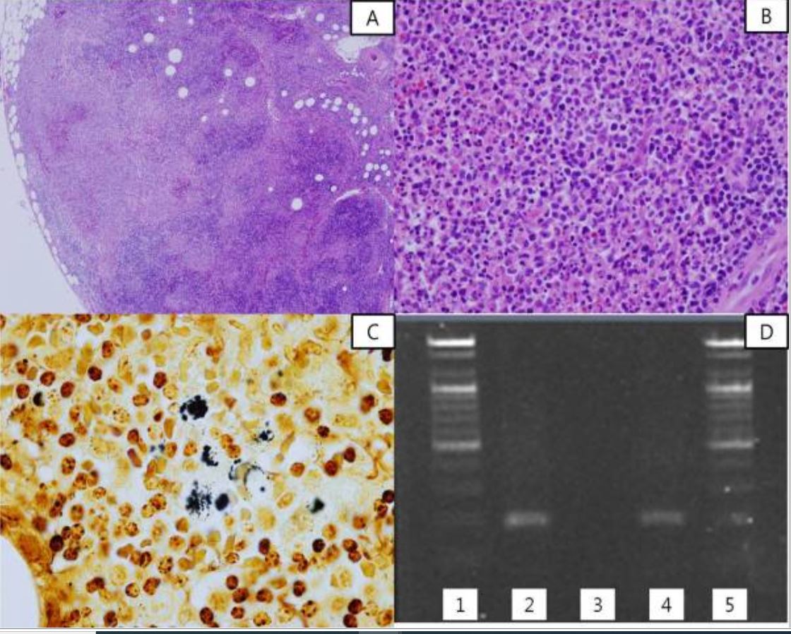
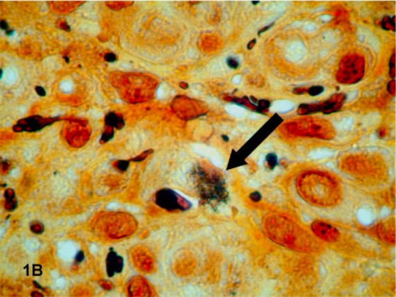
    • Stain black with silver-impregnated stains (e.g., Warthin-Starry stain)
  • At least 22 named species; the 3 most relevant medically are:
    • B. henselae
    • B. quintana (formerly known as Rickettsiae quintana)
    • B. bacilliformis
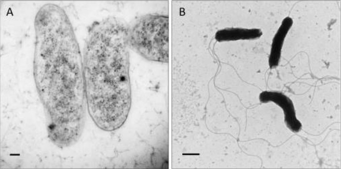
Transmission electron micrographs showing morphology of b. Bacilliformis

Transmission electron micrographs showing morphology of Bartonella bacilliformis:
Scale bars represent 100 nm in panel A and 500 nm in panel B.
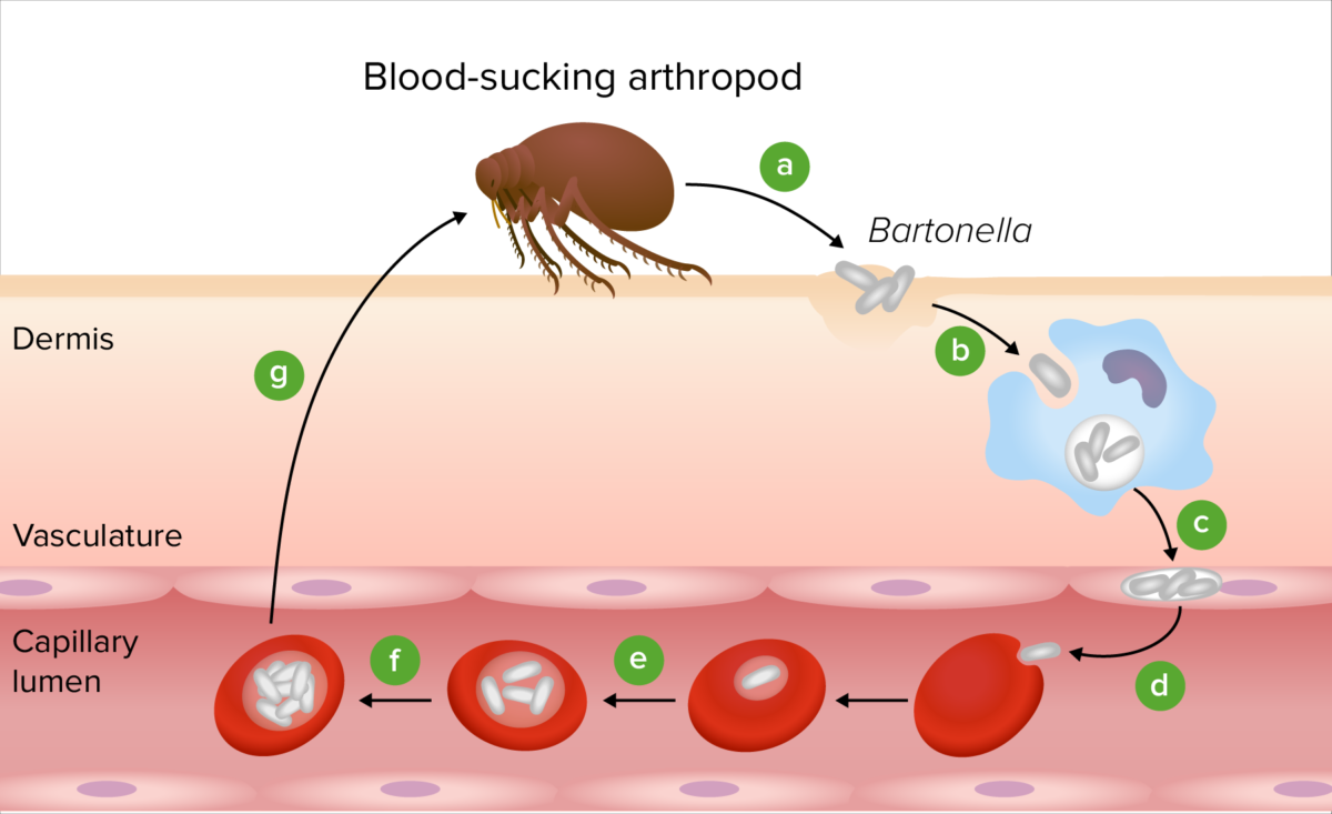
Image: “Transmission electron micrographs showing morphology of B. bacilliformis” by Michael F. Minnick, Burt E. Anderson, Amorce Lima, James M. Battisti, Phillip G. Lawyer, and Richard J. Birtles. License: Public Domain

Epidemiology and Pathogenesis

Epidemiology

  • Bartonella spp. have a worldwide distribution.
  • No types of bartonellosis have a sex Sex The totality of characteristics of reproductive structure, functions, phenotype, and genotype, differentiating the male from the female organism. Gender Dysphoria predilection
  • Cat-scratch disease (B. henselae):
    • Worldwide distribution
    • Approximately 12,000 cases are diagnosed annually in the outpatient setting, with 500 persons hospitalized each year
    • More common in children and adolescents than in adults (except for immunocompromised immunocompromised A human or animal whose immunologic mechanism is deficient because of an immunodeficiency disorder or other disease or as the result of the administration of immunosuppressive drugs or radiation. Gastroenteritis adults)
  • Carrion’s disease (B. bacilliformis)
    • Very rare infectious disease
    • Found only in Peru (endemic), Ecuador, and Colombia or associated with a history of recent travel to these regions
  • Trench fever Fever Fever is defined as a measured body temperature of at least 38°C (100.4°F). Fever is caused by circulating endogenous and/or exogenous pyrogens that increase levels of prostaglandin E2 in the hypothalamus. Fever is commonly associated with chills, rigors, sweating, and flushing of the skin. Fever (B. quintana) is most common in populations prone to infestations with lice, such as the homeless

Transmission

  • Mainly transmitted by vectors like fleas, lice, sandflies, or ticks Ticks Blood-sucking acarid parasites of the order ixodida comprising two families: the softbacked ticks (argasidae) and hardbacked ticks (ixodidae). Ticks are larger than their relatives, the mites. They penetrate the skin of their host by means of highly specialized, hooked mouth parts and feed on its blood. Ticks attack all groups of terrestrial vertebrates. In humans they are responsible for many tick-borne diseases, including the transmission of rocky mountain spotted fever; tularemia; babesiosis; african swine fever; and relapsing fever. Coxiella/Q Fever
  • Zoonotic disease (transmitted from animals Animals Unicellular or multicellular, heterotrophic organisms, that have sensation and the power of voluntary movement. Under the older five kingdom paradigm, animalia was one of the kingdoms. Under the modern three domain model, animalia represents one of the many groups in the domain eukaryota. Cell Types: Eukaryotic versus Prokaryotic to people)
  • Reservoirs:
    • Humans
    • Domestic animals Animals Unicellular or multicellular, heterotrophic organisms, that have sensation and the power of voluntary movement. Under the older five kingdom paradigm, animalia was one of the kingdoms. Under the modern three domain model, animalia represents one of the many groups in the domain eukaryota. Cell Types: Eukaryotic versus Prokaryotic, especially cats (asymptomatic carriers Carriers The Cell: Cell Membrane)
    • Wild animals Animals Unicellular or multicellular, heterotrophic organisms, that have sensation and the power of voluntary movement. Under the older five kingdom paradigm, animalia was one of the kingdoms. Under the modern three domain model, animalia represents one of the many groups in the domain eukaryota. Cell Types: Eukaryotic versus Prokaryotic, especially rodents, coyotes, or foxes

Host risk factors

  • Immunocompromised immunocompromised A human or animal whose immunologic mechanism is deficient because of an immunodeficiency disorder or other disease or as the result of the administration of immunosuppressive drugs or radiation. Gastroenteritis patients Patients Individuals participating in the health care system for the purpose of receiving therapeutic, diagnostic, or preventive procedures. Clinician–Patient Relationship:
    • Chemotherapy Chemotherapy Osteosarcoma
    • Organ transplant
    • HIV HIV Anti-HIV Drugs/AIDS → bacillary angiomatosis Bacillary Angiomatosis A reactive vascular proliferation that is characterized by the multiple tumor-like lesions in skin, bone, brain, and other organs. Bacillary angiomatosis is caused by infection with gram-negative Bartonella bacilli (such as Bartonella henselae), and is often seen in AIDS patients and other immunocompromised hosts. AIDS-defining Conditions is an AIDS-defining condition
  • Homelessness
  • Poor hygiene
  • Alcoholism Alcoholism A primary, chronic disease with genetic, psychosocial, and environmental factors influencing its development and manifestations. The disease is often progressive and fatal. It is characterized by impaired control over drinking, preoccupation with the drug alcohol, use of alcohol despite adverse consequences, and distortions in thinking, most notably denial. Each of these symptoms may be continuous or periodic. Wernicke Encephalopathy and Korsakoff Syndrome

Pathogenesis

  • After exposure from a reservoir Reservoir Animate or inanimate sources which normally harbor disease-causing organisms and thus serve as potential sources of disease outbreaks. Reservoirs are distinguished from vectors (disease vectors) and carriers, which are agents of disease transmission rather than continuing sources of potential disease outbreaks. Humans may serve both as disease reservoirs and carriers. Escherichia coli or vector, bacteria Bacteria Bacteria are prokaryotic single-celled microorganisms that are metabolically active and divide by binary fission. Some of these organisms play a significant role in the pathogenesis of diseases. Bacteriology colonize the endothelial cells of the new host.
  • From the endothelial cells, the bacteria Bacteria Bacteria are prokaryotic single-celled microorganisms that are metabolically active and divide by binary fission. Some of these organisms play a significant role in the pathogenesis of diseases. Bacteriology are released into the bloodstream, where they infect erythrocytes Erythrocytes Erythrocytes, or red blood cells (RBCs), are the most abundant cells in the blood. While erythrocytes in the fetus are initially produced in the yolk sac then the liver, the bone marrow eventually becomes the main site of production. Erythrocytes: Histology.
  • Bacteria Bacteria Bacteria are prokaryotic single-celled microorganisms that are metabolically active and divide by binary fission. Some of these organisms play a significant role in the pathogenesis of diseases. Bacteriology invade and replicate in erythrocytes Erythrocytes Erythrocytes, or red blood cells (RBCs), are the most abundant cells in the blood. While erythrocytes in the fetus are initially produced in the yolk sac then the liver, the bone marrow eventually becomes the main site of production. Erythrocytes: Histology and in endothelial cells.
  • Erythrocytes Erythrocytes Erythrocytes, or red blood cells (RBCs), are the most abundant cells in the blood. While erythrocytes in the fetus are initially produced in the yolk sac then the liver, the bone marrow eventually becomes the main site of production. Erythrocytes: Histology lack the major histocompatibility complex Major histocompatibility complex The genetic region which contains the loci of genes which determine the structure of the serologically defined (sd) and lymphocyte-defined (ld) transplantation antigens, genes which control the structure of the immune response-associated antigens, human; the immune response genes which control the ability of an animal to respond immunologically to antigenic stimuli, and genes which determine the structure and/or level of the first four components of complement. Innate Immunity: Phagocytes and Antigen Presentation (MHC), which prevents antigen Antigen Substances that are recognized by the immune system and induce an immune reaction. Vaccination presentation and renders MHC-dependent cytotoxicity ineffective.
    • All Bartonella spp. induce host tissues to produce hypoxia-inducible factor-1α, which drives the production of vascular endothelial growth factor Vascular endothelial growth factor A family of angiogenic proteins that are closely-related to vascular endothelial growth factor a. They play an important role in the growth and differentiation of vascular as well as lymphatic endothelial cells. Wound Healing, causing vascular proliferation (angiogenesis).
    • Microscopic pathology of angiogenesis: capillary proliferation with prominent epithelioid endothelial cells showing nuclear atypia Atypia Fibrocystic Change and mitosis Mitosis A type of cell nucleus division by means of which the two daughter nuclei normally receive identical complements of the number of chromosomes of the somatic cells of the species. Cell Cycle, associated with neutrophils Neutrophils Granular leukocytes having a nucleus with three to five lobes connected by slender threads of chromatin, and cytoplasm containing fine inconspicuous granules and stainable by neutral dyes. Innate Immunity: Phagocytes and Antigen Presentation and bacteria Bacteria Bacteria are prokaryotic single-celled microorganisms that are metabolically active and divide by binary fission. Some of these organisms play a significant role in the pathogenesis of diseases. Bacteriology
    • Facultative intracellular Facultative intracellular Yersinia spp./Yersiniosis properties help in the evasion of a host immune response.
  • Immune response:
    • Autoimmune response:
      • B. henselae triggers CD8+ T cell-mediated responses, leading to autoimmune reactions, nerve damage, and inflammation Inflammation Inflammation is a complex set of responses to infection and injury involving leukocytes as the principal cellular mediators in the body’s defense against pathogenic organisms. Inflammation is also seen as a response to tissue injury in the process of wound healing. The 5 cardinal signs of inflammation are pain, heat, redness, swelling, and loss of function. Inflammation.
      • Disruption of the blood-nerve barrier causes focal inflammation Inflammation Inflammation is a complex set of responses to infection and injury involving leukocytes as the principal cellular mediators in the body’s defense against pathogenic organisms. Inflammation is also seen as a response to tissue injury in the process of wound healing. The 5 cardinal signs of inflammation are pain, heat, redness, swelling, and loss of function. Inflammation, ischemia Ischemia A hypoperfusion of the blood through an organ or tissue caused by a pathologic constriction or obstruction of its blood vessels, or an absence of blood circulation. Ischemic Cell Damage, and neurological damage.
      • Chronic Bartonella infection sustains inflammation Inflammation Inflammation is a complex set of responses to infection and injury involving leukocytes as the principal cellular mediators in the body’s defense against pathogenic organisms. Inflammation is also seen as a response to tissue injury in the process of wound healing. The 5 cardinal signs of inflammation are pain, heat, redness, swelling, and loss of function. Inflammation, contributing to neurological dysfunction.
    • Innate immune response Innate Immune Response Immunity to pathogens is divided into innate and adaptive immune responses. The innate immune response is the 1st line of defense against a variety of pathogens, including bacteria, fungi, viruses, and parasites. In essentially the same form, the innate type of immunity is present in all multicellular organisms. Innate Immunity: Barriers, Complement, and Cytokines:
      • Macrophages Macrophages The relatively long-lived phagocytic cell of mammalian tissues that are derived from blood monocytes. Main types are peritoneal macrophages; alveolar macrophages; histiocytes; kupffer cells of the liver; and osteoclasts. They may further differentiate within chronic inflammatory lesions to epithelioid cells or may fuse to form foreign body giant cells or langhans giant cells. Innate Immunity: Phagocytes and Antigen Presentation: rapidly internalize B. henselae (saturation within 4 hours) and produce pro-inflammatory cytokines Cytokines Non-antibody proteins secreted by inflammatory leukocytes and some non-leukocytic cells, that act as intercellular mediators. They differ from classical hormones in that they are produced by a number of tissue or cell types rather than by specialized glands. They generally act locally in a paracrine or autocrine rather than endocrine manner. Adaptive Immune Response: TNF-α, IL-1β, and IL-6.
      • Dendritic cells Dendritic cells Specialized cells of the hematopoietic system that have branch-like extensions. They are found throughout the lymphatic system, and in non-lymphoid tissues such as skin and the epithelia of the intestinal, respiratory, and reproductive tracts. They trap and process antigens, and present them to T-cells, thereby stimulating cell-mediated immunity. They are different from the non-hematopoietic follicular dendritic cells, which have a similar morphology and immune system function, but with respect to humoral immunity (antibody production). Skin: Structure and Functions (DCs): upregulate CXCL8, CXCL1, and CXCL13, recruiting neutrophils Neutrophils Granular leukocytes having a nucleus with three to five lobes connected by slender threads of chromatin, and cytoplasm containing fine inconspicuous granules and stainable by neutral dyes. Innate Immunity: Phagocytes and Antigen Presentation and B cells B cells Lymphoid cells concerned with humoral immunity. They are short-lived cells resembling bursa-derived lymphocytes of birds in their production of immunoglobulin upon appropriate stimulation. B cells: Types and Functions to infection sites while promoting granuloma formation (characteristic of CSD) to limit Limit A value (e.g., pressure or time) that should not be exceeded and which is specified by the operator to protect the lung Invasive Mechanical Ventilation bacterial spread.
    • Humoral immune response:
      • Antibodies Antibodies Immunoglobulins (Igs), also known as antibodies, are glycoprotein molecules produced by plasma cells that act in immune responses by recognizing and binding particular antigens. The various Ig classes are IgG (the most abundant), IgM, IgE, IgD, and IgA, which differ in their biologic features, structure, target specificity, and distribution. Immunoglobulins: Types and Functions: block bacterial attachment to erythrocytes Erythrocytes Erythrocytes, or red blood cells (RBCs), are the most abundant cells in the blood. While erythrocytes in the fetus are initially produced in the yolk sac then the liver, the bone marrow eventually becomes the main site of production. Erythrocytes: Histology, suppress bacteremia Bacteremia The presence of viable bacteria circulating in the blood. Fever, chills, tachycardia, and tachypnea are common acute manifestations of bacteremia. The majority of cases are seen in already hospitalized patients, most of whom have underlying diseases or procedures which render their bloodstreams susceptible to invasion. Glycopeptides, neutralize pathogens, and target Bartonella surface determinants (e.g., CFA) with IgG2a and IgG3.
  • The intraerythrocytic life cycle causes hemolytic anemia Hemolytic Anemia Hemolytic anemia (HA) is the term given to a large group of anemias that are caused by the premature destruction/hemolysis of circulating red blood cells (RBCs). Hemolysis can occur within (intravascular hemolysis) or outside the blood vessels (extravascular hemolysis). Hemolytic Anemia.
  • Endothelial cell proliferation causes microvascular thrombosis Thrombosis Formation and development of a thrombus or blood clot in the blood vessel. Epidemic Typhus and vessel occlusion, leading to tissue ischemia Ischemia A hypoperfusion of the blood through an organ or tissue caused by a pathologic constriction or obstruction of its blood vessels, or an absence of blood circulation. Ischemic Cell Damage 
  • Erythrocytes Erythrocytes Erythrocytes, or red blood cells (RBCs), are the most abundant cells in the blood. While erythrocytes in the fetus are initially produced in the yolk sac then the liver, the bone marrow eventually becomes the main site of production. Erythrocytes: Histology are taken in by another vector (blood-sucking arthropod) to be transmitted to the next host.
  • Types of Bartonella:
    • B. bacilliformis:
    • B. henselae and B. quintana:
      • Invade endothelial cells
      • Make a protein binder that adheres to feline RBC membranes
    • B. quintana:
      • Invades endothelial cells
      • Forms bacterial aggregates that are internally taken in by the invasome, a unique phagosomal structure
      • Invasomes proliferate and make intracellular blebs Blebs Ischemic Cell Damage.
Pathogenesis of bartonella

Pathogenesis of Bartonella:
Following transmission by an arthropod vector (a), the Bartonella bacteria colonize the skin (b). The bacteria are then transported to the vascular endothelium (c) and are seeded into the bloodstream to invade erythrocytes (d). After replication inside the RBC (e), the bacteria persist in the intraerythrocytic niche (f), making them competent for transmission by another bloodsucking arthropod (g).

Image by Lecturio. License: CC BY-NC-SA 4.0

Diseases Caused by Bartonella

Bartonellosis in humans is caused by 3 main species of Bartonella bacteria Bacteria Bacteria are prokaryotic single-celled microorganisms that are metabolically active and divide by binary fission. Some of these organisms play a significant role in the pathogenesis of diseases. Bacteriology and produces a wide range of symptoms and diseases depending on the species and the immune state of the infected individual.

Table: Diseases caused by Bartonella species
Species Disease
B. bacilliformis Carrion’s disease, also known as:
  • Oroya fever Fever Fever is defined as a measured body temperature of at least 38°C (100.4°F). Fever is caused by circulating endogenous and/or exogenous pyrogens that increase levels of prostaglandin E2 in the hypothalamus. Fever is commonly associated with chills, rigors, sweating, and flushing of the skin. Fever: initial bacteremic form
  • Verruga peruana: late-onset, eruptive form
B. quintana
  • Trench fever Fever Fever is defined as a measured body temperature of at least 38°C (100.4°F). Fever is caused by circulating endogenous and/or exogenous pyrogens that increase levels of prostaglandin E2 in the hypothalamus. Fever is commonly associated with chills, rigors, sweating, and flushing of the skin. Fever (“5-day fever Fever Fever is defined as a measured body temperature of at least 38°C (100.4°F). Fever is caused by circulating endogenous and/or exogenous pyrogens that increase levels of prostaglandin E2 in the hypothalamus. Fever is commonly associated with chills, rigors, sweating, and flushing of the skin. Fever”)
  • BA: if severely immunosuppressed; an AIDS-defining condition
  • Endocarditis Endocarditis Endocarditis is an inflammatory disease involving the inner lining (endometrium) of the heart, most commonly affecting the cardiac valves. Both infectious and noninfectious etiologies lead to vegetations on the valve leaflets. Patients may present with nonspecific symptoms such as fever and fatigue. Endocarditis (often culture-negative)
B. henselae
  • CSD
  • BA: if severely immunosuppressed; an AIDS-defining condition
  • Peliosis hepatis: BA in liver Liver The liver is the largest gland in the human body. The liver is found in the superior right quadrant of the abdomen and weighs approximately 1.5 kilograms. Its main functions are detoxification, metabolism, nutrient storage (e.g., iron and vitamins), synthesis of coagulation factors, formation of bile, filtration, and storage of blood. Liver: Anatomy, spleen Spleen The spleen is the largest lymphoid organ in the body, located in the LUQ of the abdomen, superior to the left kidney and posterior to the stomach at the level of the 9th-11th ribs just below the diaphragm. The spleen is highly vascular and acts as an important blood filter, cleansing the blood of pathogens and damaged erythrocytes. Spleen: Anatomy, lymph nodes Lymph Nodes They are oval or bean shaped bodies (1 – 30 mm in diameter) located along the lymphatic system. Lymphatic Drainage System: Anatomy
  • Endocarditis Endocarditis Endocarditis is an inflammatory disease involving the inner lining (endometrium) of the heart, most commonly affecting the cardiac valves. Both infectious and noninfectious etiologies lead to vegetations on the valve leaflets. Patients may present with nonspecific symptoms such as fever and fatigue. Endocarditis: with atypical CSD; often culture-negative
  • Neuroretinitis: with atypical CSD
  • Meningitis Meningitis Meningitis is inflammation of the meninges, the protective membranes of the brain, and spinal cord. The causes of meningitis are varied, with the most common being bacterial or viral infection. The classic presentation of meningitis is a triad of fever, altered mental status, and nuchal rigidity. Meningitis/ encephalitis Encephalitis Encephalitis is inflammation of the brain parenchyma caused by an infection, usually viral. Encephalitis may present with mild symptoms such as headache, fever, fatigue, and muscle and joint pain or with severe symptoms such as seizures, altered consciousness, and paralysis. Encephalitis: with atypical CSD
BA: bacillary angiomatosis
CSD: cat-scatch disease

Carrion’s disease (South American bartonellosis)

Transmission:

  • Carried by night-biting sandflies of the genus Lutzomyia (formerly Phlebotomus)

Clinical presentation:

  • Biphasic clinical course that used to be considered 2 separate diseases
  • Acute phase Acute phase Short Bowel Syndrome:
    • Also known as Oroya fever Fever Fever is defined as a measured body temperature of at least 38°C (100.4°F). Fever is caused by circulating endogenous and/or exogenous pyrogens that increase levels of prostaglandin E2 in the hypothalamus. Fever is commonly associated with chills, rigors, sweating, and flushing of the skin. Fever
    • Develops 3–12 weeks after initial exposure
    • Sudden-onset high fever Fever Fever is defined as a measured body temperature of at least 38°C (100.4°F). Fever is caused by circulating endogenous and/or exogenous pyrogens that increase levels of prostaglandin E2 in the hypothalamus. Fever is commonly associated with chills, rigors, sweating, and flushing of the skin. Fever and chills Chills The sudden sensation of being cold. It may be accompanied by shivering. Fever
    • Profuse sweating
    • Severe headache Headache The symptom of pain in the cranial region. It may be an isolated benign occurrence or manifestation of a wide variety of headache disorders. Brain Abscess
    • Weakness
    • Paleness of the skin Skin The skin, also referred to as the integumentary system, is the largest organ of the body. The skin is primarily composed of the epidermis (outer layer) and dermis (deep layer). The epidermis is primarily composed of keratinocytes that undergo rapid turnover, while the dermis contains dense layers of connective tissue. Skin: Structure and Functions
    • Abdominal, muscular, or joint pain Pain An unpleasant sensation induced by noxious stimuli which are detected by nerve endings of nociceptive neurons. Pain: Types and Pathways
    • Meningoencephalitis Meningoencephalitis Encephalitis and/or seizures Seizures A seizure is abnormal electrical activity of the neurons in the cerebral cortex that can manifest in numerous ways depending on the region of the brain affected. Seizures consist of a sudden imbalance that occurs between the excitatory and inhibitory signals in cortical neurons, creating a net excitation. The 2 major classes of seizures are focal and generalized. Seizures
    • Severe hemolytic anemia Hemolytic Anemia Hemolytic anemia (HA) is the term given to a large group of anemias that are caused by the premature destruction/hemolysis of circulating red blood cells (RBCs). Hemolysis can occur within (intravascular hemolysis) or outside the blood vessels (extravascular hemolysis). Hemolytic Anemia may lead to:
      • Confusion, disorientation Disorientation St. Louis Encephalitis Virus, or coma Coma Coma is defined as a deep state of unarousable unresponsiveness, characterized by a score of 3 points on the GCS. A comatose state can be caused by a multitude of conditions, making the precise epidemiology and prognosis of coma difficult to determine. Coma
      • Chest pain Pain An unpleasant sensation induced by noxious stimuli which are detected by nerve endings of nociceptive neurons. Pain: Types and Pathways
      • Thrombocytopenia Thrombocytopenia Thrombocytopenia occurs when the platelet count is < 150,000 per microliter. The normal range for platelets is usually 150,000-450,000/µL of whole blood. Thrombocytopenia can be a result of decreased production, increased destruction, or splenic sequestration of platelets. Patients are often asymptomatic until platelet counts are < 50,000/µL. Thrombocytopenia
      • Dyspnea Dyspnea Dyspnea is the subjective sensation of breathing discomfort. Dyspnea is a normal manifestation of heavy physical or psychological exertion, but also may be caused by underlying conditions (both pulmonary and extrapulmonary). Dyspnea
      • Organ dysfunction
  • Chronic phase Chronic phase HIV Infection and AIDS:
    • Also known as verruga peruana
    • Usually develops within weeks or months in untreated individuals who may or may not have manifested the acute phase Acute phase Short Bowel Syndrome of the disease
    • Characteristic skin Skin The skin, also referred to as the integumentary system, is the largest organ of the body. The skin is primarily composed of the epidermis (outer layer) and dermis (deep layer). The epidermis is primarily composed of keratinocytes that undergo rapid turnover, while the dermis contains dense layers of connective tissue. Skin: Structure and Functions lesions
      • Reddish-purple in color
      • Develop in outbreaks Outbreaks Sudden increase in the incidence of a disease. The concept includes epidemics and pandemics. Influenza Viruses/Influenza over several regions of the body at once or in a migratory pattern
      • Usually located on exposed skin Skin The skin, also referred to as the integumentary system, is the largest organ of the body. The skin is primarily composed of the epidermis (outer layer) and dermis (deep layer). The epidermis is primarily composed of keratinocytes that undergo rapid turnover, while the dermis contains dense layers of connective tissue. Skin: Structure and Functions, such as the face, arms, and legs, but may also develop in mucous membranes and internal organs
      • Lesions are initially small and become nodular (0.2 to 4 cm in diameter)
      • Can potentially bleed, ulcerate, or become pustules

Trench fever Fever Fever is defined as a measured body temperature of at least 38°C (100.4°F). Fever is caused by circulating endogenous and/or exogenous pyrogens that increase levels of prostaglandin E2 in the hypothalamus. Fever is commonly associated with chills, rigors, sweating, and flushing of the skin. Fever

Transmission:

  • Mainly carried by the human body louse Human Body Louse Epidemic Typhus vector Pediculus humanus var. corporis (along with other species of lice, ticks Ticks Blood-sucking acarid parasites of the order ixodida comprising two families: the softbacked ticks (argasidae) and hardbacked ticks (ixodidae). Ticks are larger than their relatives, the mites. They penetrate the skin of their host by means of highly specialized, hooked mouth parts and feed on its blood. Ticks attack all groups of terrestrial vertebrates. In humans they are responsible for many tick-borne diseases, including the transmission of rocky mountain spotted fever; tularemia; babesiosis; african swine fever; and relapsing fever. Coxiella/Q Fever, and fleas)
  • Transmission via inoculation of infected louse feces into abrasions Abrasions Corneal Abrasions, Erosion, and Ulcers of the skin Skin The skin, also referred to as the integumentary system, is the largest organ of the body. The skin is primarily composed of the epidermis (outer layer) and dermis (deep layer). The epidermis is primarily composed of keratinocytes that undergo rapid turnover, while the dermis contains dense layers of connective tissue. Skin: Structure and Functions or into conjunctivae 

Clinical presentation:

  • Incubation Incubation The amount time between exposure to an infectious agent and becoming symptomatic. Rabies Virus period: 5–20 days (average, 7.7)
  • Classic trench fever Fever Fever is defined as a measured body temperature of at least 38°C (100.4°F). Fever is caused by circulating endogenous and/or exogenous pyrogens that increase levels of prostaglandin E2 in the hypothalamus. Fever is commonly associated with chills, rigors, sweating, and flushing of the skin. Fever can range from mild flu-like illness to a debilitating illness 
  • Acute illness usually presents with malaise Malaise Tick-borne Encephalitis Virus, fever Fever Fever is defined as a measured body temperature of at least 38°C (100.4°F). Fever is caused by circulating endogenous and/or exogenous pyrogens that increase levels of prostaglandin E2 in the hypothalamus. Fever is commonly associated with chills, rigors, sweating, and flushing of the skin. Fever, headache Headache The symptom of pain in the cranial region. It may be an isolated benign occurrence or manifestation of a wide variety of headache disorders. Brain Abscess, dizziness Dizziness An imprecise term which may refer to a sense of spatial disorientation, motion of the environment, or lightheadedness. Lateral Medullary Syndrome (Wallenberg Syndrome), nausea Nausea An unpleasant sensation in the stomach usually accompanied by the urge to vomit. Common causes are early pregnancy, sea and motion sickness, emotional stress, intense pain, food poisoning, and various enteroviruses. Antiemetics and vomiting Vomiting The forcible expulsion of the contents of the stomach through the mouth. Hypokalemia, bone Bone Bone is a compact type of hardened connective tissue composed of bone cells, membranes, an extracellular mineralized matrix, and central bone marrow. The 2 primary types of bone are compact and spongy. Bones: Structure and Types pain Pain An unpleasant sensation induced by noxious stimuli which are detected by nerve endings of nociceptive neurons. Pain: Types and Pathways, and sometimes a macular truncal rash Rash Rocky Mountain Spotted Fever 
  • The fever Fever Fever is defined as a measured body temperature of at least 38°C (100.4°F). Fever is caused by circulating endogenous and/or exogenous pyrogens that increase levels of prostaglandin E2 in the hypothalamus. Fever is commonly associated with chills, rigors, sweating, and flushing of the skin. Fever is usually episodic, presenting every 5 days (“5-day fever Fever Fever is defined as a measured body temperature of at least 38°C (100.4°F). Fever is caused by circulating endogenous and/or exogenous pyrogens that increase levels of prostaglandin E2 in the hypothalamus. Fever is commonly associated with chills, rigors, sweating, and flushing of the skin. Fever” or “quintan fever Fever Fever is defined as a measured body temperature of at least 38°C (100.4°F). Fever is caused by circulating endogenous and/or exogenous pyrogens that increase levels of prostaglandin E2 in the hypothalamus. Fever is commonly associated with chills, rigors, sweating, and flushing of the skin. Fever”).

Cat-scratch disease (CSD)

Transmission:

  • Cats are the most important carriers Carriers The Cell: Cell Membrane, though there are cases of transmission from dogs.
  • Routes of transmission:
    • Scratch or bite from an infected cat
    • Bite from cat fleas
    • Contact with cat saliva Saliva The clear, viscous fluid secreted by the salivary glands and mucous glands of the mouth. It contains mucins, water, organic salts, and ptyalin. Salivary Glands: Anatomy through broken skin Skin The skin, also referred to as the integumentary system, is the largest organ of the body. The skin is primarily composed of the epidermis (outer layer) and dermis (deep layer). The epidermis is primarily composed of keratinocytes that undergo rapid turnover, while the dermis contains dense layers of connective tissue. Skin: Structure and Functions or mucosal surfaces

Clinical presentation:

  • May not manifest until several days or weeks after the initial exposure
  • Characteristic papular lesions
  • Gradual enlargement of regional lymph nodes Lymph Nodes They are oval or bean shaped bodies (1 – 30 mm in diameter) located along the lymphatic system. Lymphatic Drainage System: Anatomy
    • Usually located in the axilla Axilla The axilla is a pyramid-shaped space located between the upper thorax and the arm. The axilla has a base, an apex, and 4 walls (anterior, medial, lateral, posterior). The base of the pyramid is made up of the axillary skin. The apex is the axillary inlet, located between the 1st rib, superior border of the scapula, and clavicle. Axilla and Brachial Plexus: Anatomy, neck Neck The part of a human or animal body connecting the head to the rest of the body. Peritonsillar Abscess, or groin Groin The external junctural region between the lower part of the abdomen and the thigh. Male Genitourinary Examination regions
    • Swollen nodes are tender and can last 2–3 months or longer
    • May appear red and warm to the touch
    • Suppuration may occur
  • General or flu-like symptoms Flu-Like Symptoms Babesia/Babesiosis:
    • Malaise Malaise Tick-borne Encephalitis Virus, back and body aches
    • Fatigue Fatigue The state of weariness following a period of exertion, mental or physical, characterized by a decreased capacity for work and reduced efficiency to respond to stimuli. Fibromyalgia
    • Headache Headache The symptom of pain in the cranial region. It may be an isolated benign occurrence or manifestation of a wide variety of headache disorders. Brain Abscess
    • Low-grade fever Low-Grade Fever Erythema Infectiosum
    • Loss of appetite and weight loss Weight loss Decrease in existing body weight. Bariatric Surgery
    • Sore throat Sore throat Pharyngitis is an inflammation of the back of the throat (pharynx). Pharyngitis is usually caused by an upper respiratory tract infection, which is viral in most cases. It typically results in a sore throat and fever. Other symptoms may include a runny nose, cough, headache, and hoarseness. Pharyngitis
  • Complications (“atypical CSD”), which usually present in immunocompromised immunocompromised A human or animal whose immunologic mechanism is deficient because of an immunodeficiency disorder or other disease or as the result of the administration of immunosuppressive drugs or radiation. Gastroenteritis individuals:
    • Granulomatous hepatitis and/or splenitis
    • Osteomyelitis Osteomyelitis Osteomyelitis is an infection of the bone that results from the spread of microorganisms from the blood (hematogenous), nearby infected tissue, or open wounds (non-hematogenous). Infections are most commonly caused by Staphylococcus aureus. Osteomyelitis
    • Bacillary angiomatosis Bacillary Angiomatosis A reactive vascular proliferation that is characterized by the multiple tumor-like lesions in skin, bone, brain, and other organs. Bacillary angiomatosis is caused by infection with gram-negative Bartonella bacilli (such as Bartonella henselae), and is often seen in AIDS patients and other immunocompromised hosts. AIDS-defining Conditions
    • Meningitis Meningitis Meningitis is inflammation of the meninges, the protective membranes of the brain, and spinal cord. The causes of meningitis are varied, with the most common being bacterial or viral infection. The classic presentation of meningitis is a triad of fever, altered mental status, and nuchal rigidity. Meningitis and/or encephalitis Encephalitis Encephalitis is inflammation of the brain parenchyma caused by an infection, usually viral. Encephalitis may present with mild symptoms such as headache, fever, fatigue, and muscle and joint pain or with severe symptoms such as seizures, altered consciousness, and paralysis. Encephalitis 
    • Neuroretinitis
    • Parinaud’s oculoglandular syndrome (similar to conjunctivitis Conjunctivitis Conjunctivitis is a common inflammation of the bulbar and/or palpebral conjunctiva. It can be classified into infectious (mostly viral) and noninfectious conjunctivitis, which includes allergic causes. Patients commonly present with red eyes, increased tearing, burning, foreign body sensation, and photophobia. Conjunctivitis plus lymphadenopathy Lymphadenopathy Lymphadenopathy is lymph node enlargement (> 1 cm) and is benign and self-limited in most patients. Etiologies include malignancy, infection, and autoimmune disorders, as well as iatrogenic causes such as the use of certain medications. Generalized lymphadenopathy often indicates underlying systemic disease. Lymphadenopathy)
    • Parotitis Parotitis Inflammation of the parotid gland. Mumps Virus/Mumps
    • Endocarditis Endocarditis Endocarditis is an inflammatory disease involving the inner lining (endometrium) of the heart, most commonly affecting the cardiac valves. Both infectious and noninfectious etiologies lead to vegetations on the valve leaflets. Patients may present with nonspecific symptoms such as fever and fatigue. Endocarditis
Clinical presentation of a patient with bacillary angiomatosis

Clinical presentation of a patient with bacillary angiomatosis:
A: Multiple red papules on the chest and abdomen
B: 2 nodules on the back
C: Papules on the face with a subcutaneous mass lesion over the left zygomatic arch
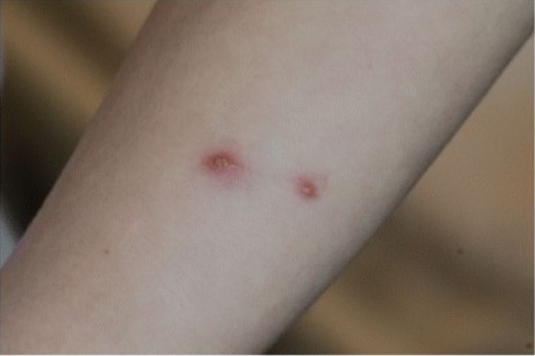
Image: “Bacillary angiomatosis presenting with facial tumor and multiple abscesses” by Markowicz M, Käser S, Müller A, Lang G, Lang S, Mayerhöfer M, Stanek G, Rieger A. License: CC BY 4.0

Bacillary angiomatosis Bacillary Angiomatosis A reactive vascular proliferation that is characterized by the multiple tumor-like lesions in skin, bone, brain, and other organs. Bacillary angiomatosis is caused by infection with gram-negative Bartonella bacilli (such as Bartonella henselae), and is often seen in AIDS patients and other immunocompromised hosts. AIDS-defining Conditions

  • Occurs primarily in immunocompromised immunocompromised A human or animal whose immunologic mechanism is deficient because of an immunodeficiency disorder or other disease or as the result of the administration of immunosuppressive drugs or radiation. Gastroenteritis individuals
    • Especially in patients Patients Individuals participating in the health care system for the purpose of receiving therapeutic, diagnostic, or preventive procedures. Clinician–Patient Relationship with AIDS and a CD4 count < 100 cells/μL
    • Constitutes an AIDS-defining condition
  • Vascular proliferative disease that mainly affects the skin Skin The skin, also referred to as the integumentary system, is the largest organ of the body. The skin is primarily composed of the epidermis (outer layer) and dermis (deep layer). The epidermis is primarily composed of keratinocytes that undergo rapid turnover, while the dermis contains dense layers of connective tissue. Skin: Structure and Functions, but can develop in other organs (e.g., bone marrow Bone marrow The soft tissue filling the cavities of bones. Bone marrow exists in two types, yellow and red. Yellow marrow is found in the large cavities of large bones and consists mostly of fat cells and a few primitive blood cells. Red marrow is a hematopoietic tissue and is the site of production of erythrocytes and granular leukocytes. Bone marrow is made up of a framework of connective tissue containing branching fibers with the frame being filled with marrow cells. Bone Marrow: Composition and Hematopoiesis, liver Liver The liver is the largest gland in the human body. The liver is found in the superior right quadrant of the abdomen and weighs approximately 1.5 kilograms. Its main functions are detoxification, metabolism, nutrient storage (e.g., iron and vitamins), synthesis of coagulation factors, formation of bile, filtration, and storage of blood. Liver: Anatomy, spleen Spleen The spleen is the largest lymphoid organ in the body, located in the LUQ of the abdomen, superior to the left kidney and posterior to the stomach at the level of the 9th-11th ribs just below the diaphragm. The spleen is highly vascular and acts as an important blood filter, cleansing the blood of pathogens and damaged erythrocytes. Spleen: Anatomy, or lymph nodes Lymph Nodes They are oval or bean shaped bodies (1 – 30 mm in diameter) located along the lymphatic system. Lymphatic Drainage System: Anatomy)
  • Characteristic erythematous skin Skin The skin, also referred to as the integumentary system, is the largest organ of the body. The skin is primarily composed of the epidermis (outer layer) and dermis (deep layer). The epidermis is primarily composed of keratinocytes that undergo rapid turnover, while the dermis contains dense layers of connective tissue. Skin: Structure and Functions lesions
    • Usually elevated
    • May be surrounded by a scaly ring 
    • Very friable and bleed easily

Peliosis hepatis

  • Occurs primarily in immunocompromised immunocompromised A human or animal whose immunologic mechanism is deficient because of an immunodeficiency disorder or other disease or as the result of the administration of immunosuppressive drugs or radiation. Gastroenteritis individuals, especially in patients Patients Individuals participating in the health care system for the purpose of receiving therapeutic, diagnostic, or preventive procedures. Clinician–Patient Relationship with AIDS
  • Vascular proliferation of sinusoid Sinusoid Sinusoid, or discontinuous, capillaries allow larger proteins and full cells to pass through larger gaps. Capillaries: Histology hepatic capillaries Capillaries Capillaries are the primary structures in the circulatory system that allow the exchange of gas, nutrients, and other materials between the blood and the extracellular fluid (ECF). Capillaries are the smallest of the blood vessels. Because a capillary diameter is so small, only 1 RBC may pass through at a time. Capillaries: Histology that result in blood-filled sacs within the liver Liver The liver is the largest gland in the human body. The liver is found in the superior right quadrant of the abdomen and weighs approximately 1.5 kilograms. Its main functions are detoxification, metabolism, nutrient storage (e.g., iron and vitamins), synthesis of coagulation factors, formation of bile, filtration, and storage of blood. Liver: Anatomy parenchyma
  • Commonly associated with peliosis of the spleen Spleen The spleen is the largest lymphoid organ in the body, located in the LUQ of the abdomen, superior to the left kidney and posterior to the stomach at the level of the 9th-11th ribs just below the diaphragm. The spleen is highly vascular and acts as an important blood filter, cleansing the blood of pathogens and damaged erythrocytes. Spleen: Anatomy and lymph nodes Lymph Nodes They are oval or bean shaped bodies (1 – 30 mm in diameter) located along the lymphatic system. Lymphatic Drainage System: Anatomy

Diagnosis

Most cases of bartonellosis can be diagnosed through the detection of characteristic symptoms and physical findings and a complete medical history. Specialized laboratory tests are used to confirm the diagnosis. 

  • Bartonella infection can be difficult to detect, confirm, and diagnose. 
  • Serologic testing is the most cost-effective diagnostic tool, but false negatives can occur even during active infection.
  • Detection of IgG IgG The major immunoglobulin isotype class in normal human serum. There are several isotype subclasses of igg, for example, igg1, igg2a, and igg2b. Hypersensitivity Pneumonitis and IgM IgM A class of immunoglobulin bearing mu chains (immunoglobulin mu-chains). Igm can fix complement. The name comes from its high molecular weight and originally being called a macroglobulin. Immunoglobulins: Types and Functions antibodies Antibodies Immunoglobulins (Igs), also known as antibodies, are glycoprotein molecules produced by plasma cells that act in immune responses by recognizing and binding particular antigens. The various Ig classes are IgG (the most abundant), IgM, IgE, IgD, and IgA, which differ in their biologic features, structure, target specificity, and distribution. Immunoglobulins: Types and Functions in blood serum by indirect immunofluorescence assays are used to detect B. henselae in suspected cases of cat-scratch disease. 
  • Microscopic examination of Giemsa-stained blood smears is used to detect B. bacilliformis in suspected cases of Carrion’s disease. 
  • PCR PCR Polymerase chain reaction (PCR) is a technique that amplifies DNA fragments exponentially for analysis. The process is highly specific, allowing for the targeting of specific genomic sequences, even with minuscule sample amounts. The PCR cycles multiple times through 3 phases: denaturation of the template DNA, annealing of a specific primer to the individual DNA strands, and synthesis/elongation of new DNA molecules. Polymerase Chain Reaction (PCR) analysis of tissue and body fluid is the most specific diagnostic test to detect Bartonella spp.
  • Other Bartonella spp. are visible only with silver stains.
  • Abdominal imaging tests are used to confirm suspected cases of liver Liver The liver is the largest gland in the human body. The liver is found in the superior right quadrant of the abdomen and weighs approximately 1.5 kilograms. Its main functions are detoxification, metabolism, nutrient storage (e.g., iron and vitamins), synthesis of coagulation factors, formation of bile, filtration, and storage of blood. Liver: Anatomy and spleen Spleen The spleen is the largest lymphoid organ in the body, located in the LUQ of the abdomen, superior to the left kidney and posterior to the stomach at the level of the 9th-11th ribs just below the diaphragm. The spleen is highly vascular and acts as an important blood filter, cleansing the blood of pathogens and damaged erythrocytes. Spleen: Anatomy complications.

Management

Aside from supportive care, management is dependent on the specific species of Bartonella contracted and the severity of each case.  

  • CSD:
    • In mild cases: Antibiotics may not be necessary, but azithromycin Azithromycin A semi-synthetic macrolide antibiotic structurally related to erythromycin. It has been used in the treatment of Mycobacterium avium intracellulare infections, toxoplasmosis, and cryptosporidiosis. Macrolides and Ketolides can shorten the duration of symptoms.
    • In moderate cases: azithromycin Azithromycin A semi-synthetic macrolide antibiotic structurally related to erythromycin. It has been used in the treatment of Mycobacterium avium intracellulare infections, toxoplasmosis, and cryptosporidiosis. Macrolides and Ketolides, erythromycin Erythromycin A bacteriostatic antibiotic macrolide produced by streptomyces erythreus. Erythromycin a is considered its major active component. In sensitive organisms, it inhibits protein synthesis by binding to 50s ribosomal subunits. This binding process inhibits peptidyl transferase activity and interferes with translocation of amino acids during translation and assembly of proteins. Macrolides and Ketolides, or doxycycline (may add steroids Steroids A group of polycyclic compounds closely related biochemically to terpenes. They include cholesterol, numerous hormones, precursors of certain vitamins, bile acids, alcohols (sterols), and certain natural drugs and poisons. Steroids have a common nucleus, a fused, reduced 17-carbon atom ring system, cyclopentanoperhydrophenanthrene. Most steroids also have two methyl groups and an aliphatic side-chain attached to the nucleus. Benign Liver Tumors for refractory lymphadenopathy Lymphadenopathy Lymphadenopathy is lymph node enlargement (> 1 cm) and is benign and self-limited in most patients. Etiologies include malignancy, infection, and autoimmune disorders, as well as iatrogenic causes such as the use of certain medications. Generalized lymphadenopathy often indicates underlying systemic disease. Lymphadenopathy)
    • In severe cases: rifampin Rifampin A semisynthetic antibiotic produced from streptomyces mediterranei. It has a broad antibacterial spectrum, including activity against several forms of Mycobacterium. In susceptible organisms it inhibits dna-dependent RNA polymerase activity by forming a stable complex with the enzyme. It thus suppresses the initiation of RNA synthesis. Rifampin is bactericidal, and acts on both intracellular and extracellular organisms. Epiglottitis and either erythromycin Erythromycin A bacteriostatic antibiotic macrolide produced by streptomyces erythreus. Erythromycin a is considered its major active component. In sensitive organisms, it inhibits protein synthesis by binding to 50s ribosomal subunits. This binding process inhibits peptidyl transferase activity and interferes with translocation of amino acids during translation and assembly of proteins. Macrolides and Ketolides or doxycycline
  • Carrion’s disease:
    • Acute phase Acute phase Short Bowel Syndrome: chloramphenicol Chloramphenicol Chloramphenicol, the only clinically relevant drug in the amphenicol class, is a potent inhibitor of bacterial protein synthesis by binding to the 50S ribosomal subunit and preventing peptide bond formation. Chloramphenicol is a broad-spectrum antibiotic with wide distribution; however, due to its toxicity, its use is limited to severe infections. Chloramphenicol or ciprofloxacin Ciprofloxacin A broad-spectrum antimicrobial carboxyfluoroquinoline. Fluoroquinolones (in pediatric cases, ciprofloxacin Ciprofloxacin A broad-spectrum antimicrobial carboxyfluoroquinoline. Fluoroquinolones is not used and beta-lactams are added)
    • Chronic phase Chronic phase HIV Infection and AIDS: rifampin Rifampin A semisynthetic antibiotic produced from streptomyces mediterranei. It has a broad antibacterial spectrum, including activity against several forms of Mycobacterium. In susceptible organisms it inhibits dna-dependent RNA polymerase activity by forming a stable complex with the enzyme. It thus suppresses the initiation of RNA synthesis. Rifampin is bactericidal, and acts on both intracellular and extracellular organisms. Epiglottitis or macrolides Macrolides Macrolides and ketolides are antibiotics that inhibit bacterial protein synthesis by binding to the 50S ribosomal subunit and blocking transpeptidation. These antibiotics have a broad spectrum of antimicrobial activity but are best known for their coverage of atypical microorganisms. Macrolides and Ketolides
  • Trench fever Fever Fever is defined as a measured body temperature of at least 38°C (100.4°F). Fever is caused by circulating endogenous and/or exogenous pyrogens that increase levels of prostaglandin E2 in the hypothalamus. Fever is commonly associated with chills, rigors, sweating, and flushing of the skin. Fever: doxycycline and gentamicin Gentamicin Aminoglycosides
  • Bartonellosis in immunocompromised immunocompromised A human or animal whose immunologic mechanism is deficient because of an immunodeficiency disorder or other disease or as the result of the administration of immunosuppressive drugs or radiation. Gastroenteritis patients Patients Individuals participating in the health care system for the purpose of receiving therapeutic, diagnostic, or preventive procedures. Clinician–Patient Relationship: prolonged course of erythromycin Erythromycin A bacteriostatic antibiotic macrolide produced by streptomyces erythreus. Erythromycin a is considered its major active component. In sensitive organisms, it inhibits protein synthesis by binding to 50s ribosomal subunits. This binding process inhibits peptidyl transferase activity and interferes with translocation of amino acids during translation and assembly of proteins. Macrolides and Ketolides or doxycycline

Differential Diagnosis

  • Cervical lymphadenitis Lymphadenitis Inflammation of the lymph nodes. Peritonsillar Abscess: cervical lymph nodes Lymph Nodes They are oval or bean shaped bodies (1 – 30 mm in diameter) located along the lymphatic system. Lymphatic Drainage System: Anatomy may become enlarged, inflamed, and tender owing to viral infections Infections Invasion of the host organism by microorganisms or their toxins or by parasites that can cause pathological conditions or diseases. Chronic Granulomatous Disease (e.g., adenovirus Adenovirus Adenovirus (member of the family Adenoviridae) is a nonenveloped, double-stranded DNA virus. Adenovirus is transmitted in a variety of ways, and it can have various presentations based on the site of entry. Presentation can include febrile pharyngitis, conjunctivitis, acute respiratory disease, atypical pneumonia, and gastroenteritis. Adenovirus) or bacterial infections Infections Invasion of the host organism by microorganisms or their toxins or by parasites that can cause pathological conditions or diseases. Chronic Granulomatous Disease (e.g., Staphylococcus aureus Staphylococcus aureus Potentially pathogenic bacteria found in nasal membranes, skin, hair follicles, and perineum of warm-blooded animals. They may cause a wide range of infections and intoxications. Brain Abscess). Management of cervical lymphadenitis Lymphadenitis Inflammation of the lymph nodes. Peritonsillar Abscess is by diagnosing the underlying cause. Biopsies are rarely needed.
  • Hodgkin lymphoma Lymphoma A general term for various neoplastic diseases of the lymphoid tissue. Imaging of the Mediastinum: malignancy Malignancy Hemothorax of B lymphocytes B lymphocytes Lymphoid cells concerned with humoral immunity. They are short-lived cells resembling bursa-derived lymphocytes of birds in their production of immunoglobulin upon appropriate stimulation. B cells: Types and Functions within lymph nodes Lymph Nodes They are oval or bean shaped bodies (1 – 30 mm in diameter) located along the lymphatic system. Lymphatic Drainage System: Anatomy. The histologic finding for Hodgkin lymphoma Lymphoma A general term for various neoplastic diseases of the lymphoid tissue. Imaging of the Mediastinum is a Reed-Sternberg cell (giant B cell with eosinophilic inclusions). Presentation of the disease is with lymphadenopathy Lymphadenopathy Lymphadenopathy is lymph node enlargement (> 1 cm) and is benign and self-limited in most patients. Etiologies include malignancy, infection, and autoimmune disorders, as well as iatrogenic causes such as the use of certain medications. Generalized lymphadenopathy often indicates underlying systemic disease. Lymphadenopathy and constitutional “B symptoms.” Hodgkin lymphoma Lymphoma A general term for various neoplastic diseases of the lymphoid tissue. Imaging of the Mediastinum is managed with chemotherapy Chemotherapy Osteosarcoma and radiotherapy. 
  • Non-Hodgkin lymphomas Non-Hodgkin lymphomas Non-Hodgkin lymphomas (NHLs) are a diverse group of hematologic malignancies that are clonal proliferative disorders of mature or progenitor B cells, T cells, or natural killer (NK) cells. Most pediatric cases are aggressive and high-grade (but curable); in adults, low-grade subtypes are more common. Non-Hodgkin Lymphomas (NHL): diverse group of malignancies of B-cell, T-cell, and natural killer (NK)–cell origin. Lymph nodes Lymph Nodes They are oval or bean shaped bodies (1 – 30 mm in diameter) located along the lymphatic system. Lymphatic Drainage System: Anatomy are involved in ⅔ of NHLs; the remainder of NHLs are extranodal. In the United States, NHLs are 10ⅹ more common than Hodgkin lymphoma Lymphoma A general term for various neoplastic diseases of the lymphoid tissue. Imaging of the Mediastinum and represent a common cause of cancer-related death.
  • Cellulitis Cellulitis Cellulitis is a common infection caused by bacteria that affects the dermis and subcutaneous tissue of the skin. It is frequently caused by Staphylococcus aureus and Streptococcus pyogenes. The skin infection presents as an erythematous and edematous area with warmth and tenderness. Cellulitis: common and painful bacterial skin Skin The skin, also referred to as the integumentary system, is the largest organ of the body. The skin is primarily composed of the epidermis (outer layer) and dermis (deep layer). The epidermis is primarily composed of keratinocytes that undergo rapid turnover, while the dermis contains dense layers of connective tissue. Skin: Structure and Functions infection that affects the deeper layers of the dermis Dermis A layer of vascularized connective tissue underneath the epidermis. The surface of the dermis contains innervated papillae. Embedded in or beneath the dermis are sweat glands; hair follicles; and sebaceous glands. Skin: Structure and Functions and subcutaneous tissue Subcutaneous tissue Loose connective tissue lying under the dermis, which binds skin loosely to subjacent tissues. It may contain a pad of adipocytes, which vary in number according to the area of the body and vary in size according to the nutritional state. Soft Tissue Abscess. Cellulitis Cellulitis Cellulitis is a common infection caused by bacteria that affects the dermis and subcutaneous tissue of the skin. It is frequently caused by Staphylococcus aureus and Streptococcus pyogenes. The skin infection presents as an erythematous and edematous area with warmth and tenderness. Cellulitis presents as an erythematous, edematous area that feels warm and tender to the touch. The condition is caused most commonly by Staphylococcus aureus Staphylococcus aureus Potentially pathogenic bacteria found in nasal membranes, skin, hair follicles, and perineum of warm-blooded animals. They may cause a wide range of infections and intoxications. Brain Abscess and Streptococcus Streptococcus Streptococcus is one of the two medically important genera of gram-positive cocci, the other being Staphylococcus. Streptococci are identified as different species on blood agar on the basis of their hemolytic pattern and sensitivity to optochin and bacitracin. There are many pathogenic species of streptococci, including S. pyogenes, S. agalactiae, S. pneumoniae, and the viridans streptococci. Streptococcus pyogenes. Diagnosis is usually clinical, and management is with antibiotics based on suspected organisms.
  • Kaposi Kaposi A multicentric, malignant neoplastic vascular proliferation characterized by the development of bluish-red cutaneous nodules, usually on the lower extremities, most often on the toes or feet, and slowly increasing in size and number and spreading to more proximal areas. The tumors have endothelium-lined channels and vascular spaces admixed with variably sized aggregates of spindle-shaped cells, and often remain confined to the skin and subcutaneous tissue, but widespread visceral involvement may occur. Hhv-8 is the suspected cause. There is also a high incidence in AIDS patients. AIDS-defining Conditions’s sarcoma: aggressive, malignant vascular tumor Tumor Inflammation that is an AIDS-defining condition and usually presents in patients Patients Individuals participating in the health care system for the purpose of receiving therapeutic, diagnostic, or preventive procedures. Clinician–Patient Relationship with a CD4 count < 500 cells/μL. Kaposi Kaposi A multicentric, malignant neoplastic vascular proliferation characterized by the development of bluish-red cutaneous nodules, usually on the lower extremities, most often on the toes or feet, and slowly increasing in size and number and spreading to more proximal areas. The tumors have endothelium-lined channels and vascular spaces admixed with variably sized aggregates of spindle-shaped cells, and often remain confined to the skin and subcutaneous tissue, but widespread visceral involvement may occur. Hhv-8 is the suspected cause. There is also a high incidence in AIDS patients. AIDS-defining Conditions’s sarcoma is caused by HHV-8 and is transmitted via saliva Saliva The clear, viscous fluid secreted by the salivary glands and mucous glands of the mouth. It contains mucins, water, organic salts, and ptyalin. Salivary Glands: Anatomy and sexual contact. The condition presents as a pink, red, purple, or brown papular or plaque-like lesions that are not painful or pruritic.
  • Hemangioma Hemangioma A vascular anomaly due to proliferation of blood vessels that forms a tumor-like mass. The common types involve capillaries and veins. It can occur anywhere in the body but is most frequently noticed in the skin and subcutaneous tissue. Imaging of the Liver and Biliary Tract: benign Benign Fibroadenoma tumor Tumor Inflammation made up of blood vessels and capillaries Capillaries Capillaries are the primary structures in the circulatory system that allow the exchange of gas, nutrients, and other materials between the blood and the extracellular fluid (ECF). Capillaries are the smallest of the blood vessels. Because a capillary diameter is so small, only 1 RBC may pass through at a time. Capillaries: Histology. Hemangioma Hemangioma A vascular anomaly due to proliferation of blood vessels that forms a tumor-like mass. The common types involve capillaries and veins. It can occur anywhere in the body but is most frequently noticed in the skin and subcutaneous tissue. Imaging of the Liver and Biliary Tract usually presents as a purple or red, well-demarcated papule Papule Elevated lesion < 1 cm in diameter Generalized and Localized Rashes.
  • Pyogenic granuloma: benign Benign Fibroadenoma, vascular lesion of unknown cause or origin. Pyogenic granuloma sometimes develops after minor trauma or injury. The granuloma appears as a red or purple papule Papule Elevated lesion < 1 cm in diameter Generalized and Localized Rashes that is friable and bleeds easily. 
  • Brucellosis Brucellosis Brucellosis (also known as undulant fever, Mediterranean fever, or Malta fever) is a zoonotic infection that spreads predominantly through ingestion of unpasteurized dairy products or direct contact with infected animal products. Clinical manifestations include fever, arthralgias, malaise, lymphadenopathy, and hepatosplenomegaly. Brucella/Brucellosis: zoonotic infection Zoonotic infection Diseases of non-human animals that may be transmitted to humans or may be transmitted from humans to non-human animals. Brucella/Brucellosis that spreads predominantly through ingestion of unpasteurized dairy products or direct contact with infected animal products. Clinical manifestations of brucellosis Brucellosis Brucellosis (also known as undulant fever, Mediterranean fever, or Malta fever) is a zoonotic infection that spreads predominantly through ingestion of unpasteurized dairy products or direct contact with infected animal products. Clinical manifestations include fever, arthralgias, malaise, lymphadenopathy, and hepatosplenomegaly. Brucella/Brucellosis include fever Fever Fever is defined as a measured body temperature of at least 38°C (100.4°F). Fever is caused by circulating endogenous and/or exogenous pyrogens that increase levels of prostaglandin E2 in the hypothalamus. Fever is commonly associated with chills, rigors, sweating, and flushing of the skin. Fever, arthralgias, malaise Malaise Tick-borne Encephalitis Virus, lymphadenopathy Lymphadenopathy Lymphadenopathy is lymph node enlargement (> 1 cm) and is benign and self-limited in most patients. Etiologies include malignancy, infection, and autoimmune disorders, as well as iatrogenic causes such as the use of certain medications. Generalized lymphadenopathy often indicates underlying systemic disease. Lymphadenopathy, and hepatosplenomegaly Hepatosplenomegaly Cytomegalovirus. Clinical manifestations, exposure history, serology Serology The study of serum, especially of antigen-antibody reactions in vitro. Yellow Fever Virus, and culture data are used in the diagnosis. Treatment involves a combination of antibiotics, including doxycycline, rifampin Rifampin A semisynthetic antibiotic produced from streptomyces mediterranei. It has a broad antibacterial spectrum, including activity against several forms of Mycobacterium. In susceptible organisms it inhibits dna-dependent RNA polymerase activity by forming a stable complex with the enzyme. It thus suppresses the initiation of RNA synthesis. Rifampin is bactericidal, and acts on both intracellular and extracellular organisms. Epiglottitis, and aminoglycosides Aminoglycosides Aminoglycosides are a class of antibiotics including gentamicin, tobramycin, amikacin, neomycin, plazomicin, and streptomycin. The class binds the 30S ribosomal subunit to inhibit bacterial protein synthesis. Unlike other medications with a similar mechanism of action, aminoglycosides are bactericidal. Aminoglycosides.

References

  1. Hammoud, K. (2024). Bartonellosis (Bartonella Infection). Emedicine. Retrieved December 5, 2024, from https://emedicine.medscape.com/article/213169-overview#a6.
  2. Spach, D. (2023). Clinical features, diagnosis, and treatment of Bartonella quintana. UpToDate. Retrieved December 5, 2024, from https://www.uptodate.com/contents/clinical-features-diagnosis-and-treatment-of-bartonella-quintana-infections
  3. Nelson VA, Saha S, Mead PS. (2016). Cat-scratch disease in the United States, 2005-2013. Emerg Infect Dis 22:1741–1746. https://pubmed.ncbi.nlm.nih.gov/27648778/
  4. Versalovic J, Carroll KC, Funke G, Jorgensen JH, Landry ML, Warnock DW. (2011). Manual of Clinical Microbiology, 10th ed. Washington, DC: ASM Press.
  5. Angelakis E, Raoult D. (2014). Pathogenicity and treatment of Bartonella infections. Int J Antimicrob Agents 44:16–25. https://pubmed.ncbi.nlm.nih.gov/24933445/
  6. Eicher SC, Dehio C. (2012). Bartonella entry mechanisms into mammalian host cells. Cell Microbiol 14:1166–1173. https://doi.org/https://doi.org/10.1111/j.1462-5822.2012.01806.x
  7. Corey C. (2018). Herpes simplex virus infections. In Jameson JL, et al. (Eds.), Harrison’s Principles of Internal Medicine, 20th ed. Vol. 1, pp. 1345–1354. 
  8. Giladi M, Ephros M. (2018). Bartonella infections, including cat-scratch disease. In Jameson JL, et al. (Ed.s), Harrison’s Principles of Internal Medicine, 20th ed. Vol. 1, pp. 1209–1214. 
  9. Spach, D. H., & Kaplan, S. L. (2024). Microbiology, epidemiology, clinical manifestations, and diagnosis of cat scratch disease. UpToDate. Retrieved December 5, 2024, from https://www.uptodate.com/contents/microbiology-epidemiology-clinical-manifestations-and-diagnosis-of-cat-scratch-disease 
  10. Bush, J. C., Robveille, C., Maggi, R. G., & Breitschwerdt, E. B. (2024). Neurobartonelloses: Emerging from obscurity! Parasites & Vectors, 17(416). Retrieved December 5, 2024, from https://parasitesandvectors.biomedcentral.com/articles/10.1186/s13071-024-06491-3
  11. Jin, X., Gou, Y., Xin, Y., Li, J., Sun, J., Li, T., & Feng, J. (2023). Advancements in understanding the molecular and immune mechanisms of Bartonella pathogenicity. Frontiers in Microbiology, 14, 1196700. https://doi.org/10.3389/fmicb.2023.1196700

Create your free account or log in to continue reading!

Sign up now and get free access to Lecturio with concept pages, medical videos, and questions for your medical education.

User Reviews

Details