Babesia/Babesiosis (Clinical)

Babesiosis Babesiosis Babesiosis is an infection caused by a protozoa belonging to the genus, Babesia. The most common Babesia seen in the United States is B. microti, which is transmitted by the Ixodes tick. The protozoa thrive and replicate within host erythrocytes. Lysis of erythrocytes and the body's immune response result in clinical symptoms. Babesia/Babesiosis is an infection caused by a protozoa Protozoa Nitroimidazoles belonging to the genus, Babesia Babesia Babesiosis is an infection caused by a protozoa belonging to the genus, Babesia. The most common Babesia seen in the United States is B. microti, which is transmitted by the Ixodes tick. The protozoa thrive and replicate within host erythrocytes. Lysis of erythrocytes and the body's immune response result in clinical symptoms. Babesia/Babesiosis. The most common Babesia Babesia Babesiosis is an infection caused by a protozoa belonging to the genus, Babesia. The most common Babesia seen in the United States is B. microti, which is transmitted by the Ixodes tick. The protozoa thrive and replicate within host erythrocytes. Lysis of erythrocytes and the body's immune response result in clinical symptoms. Babesia/Babesiosis seen in the United States is B. microti B. microti A species of protozoa infecting humans via the intermediate tick vector ixodes scapularis. The other hosts are the mouse peromyscus leucopus and meadow vole microtus pennsylvanicus, which are fed on by the tick. Other primates can be experimentally infected with babesia microti. Babesia/Babesiosis, which is transmitted by the Ixodes tick. The protozoa Protozoa Nitroimidazoles thrive and replicate within host erythrocytes Erythrocytes Erythrocytes, or red blood cells (RBCs), are the most abundant cells in the blood. While erythrocytes in the fetus are initially produced in the yolk sac then the liver, the bone marrow eventually becomes the main site of production. Erythrocytes: Histology. Lysis of erythrocytes Erythrocytes Erythrocytes, or red blood cells (RBCs), are the most abundant cells in the blood. While erythrocytes in the fetus are initially produced in the yolk sac then the liver, the bone marrow eventually becomes the main site of production. Erythrocytes: Histology and the body’s immune response result in clinical symptoms. Patients Patients Individuals participating in the health care system for the purpose of receiving therapeutic, diagnostic, or preventive procedures. Clinician–Patient Relationship usually present with a flu-like illness and jaundice Jaundice Jaundice is the abnormal yellowing of the skin and/or sclera caused by the accumulation of bilirubin. Hyperbilirubinemia is caused by either an increase in bilirubin production or a decrease in the hepatic uptake, conjugation, or excretion of bilirubin. Jaundice. In severe cases, organ damage may occur. The diagnosis is confirmed by the visual presence of parasites within RBCs RBCs Erythrocytes, or red blood cells (RBCs), are the most abundant cells in the blood. While erythrocytes in the fetus are initially produced in the yolk sac then the liver, the bone marrow eventually becomes the main site of production. Erythrocytes: Histology, which are often noted to be in a “ Maltese Cross Maltese Cross Babesia/Babesiosis” configuration. Serological testing and PCR PCR Polymerase chain reaction (PCR) is a technique that amplifies DNA fragments exponentially for analysis. The process is highly specific, allowing for the targeting of specific genomic sequences, even with minuscule sample amounts. The PCR cycles multiple times through 3 phases: denaturation of the template DNA, annealing of a specific primer to the individual DNA strands, and synthesis/elongation of new DNA molecules. Polymerase Chain Reaction (PCR) are also used in the diagnosis. Azithromycin Azithromycin A semi-synthetic macrolide antibiotic structurally related to erythromycin. It has been used in the treatment of Mycobacterium avium intracellulare infections, toxoplasmosis, and cryptosporidiosis. Macrolides and Ketolides and atovaquone Atovaquone A hydroxynaphthoquinone that has antimicrobial activity and is being used in antimalarial protocols. Antimalarial Drugs are often used in management. Coinfection with Borrelia Borrelia Borrelia are gram-negative microaerophilic spirochetes. Owing to their small size, they are not easily seen on Gram stain but can be visualized using dark-field microscopy, Giemsa, or Wright stain. Spirochetes are motile and move in a characteristic spinning fashion due to axial filaments in the periplasmic space. Borrelia and Anaplasma is common.

Last updated: Mar 4, 2024

Editorial responsibility: Stanley Oiseth, Lindsay Jones, Evelin Maza

General Characteristics of Babesia

Basic features of Babesia Babesia Babesiosis is an infection caused by a protozoa belonging to the genus, Babesia. The most common Babesia seen in the United States is B. microti, which is transmitted by the Ixodes tick. The protozoa thrive and replicate within host erythrocytes. Lysis of erythrocytes and the body’s immune response result in clinical symptoms. Babesia/Babesiosis[1]

Clinically relevant species[4‒6]

  • B. microti B. microti A species of protozoa infecting humans via the intermediate tick vector ixodes scapularis. The other hosts are the mouse peromyscus leucopus and meadow vole microtus pennsylvanicus, which are fed on by the tick. Other primates can be experimentally infected with babesia microti. Babesia/Babesiosis (most common species in North America)
  • B. duncani B. duncani Babesia/Babesiosis (North America)
  • B. divergens B. divergens Babesia/Babesiosis (Europe)

Epidemiology and Risk Factors

Epidemiology[4,5]

  • 94% of babesiosis Babesiosis Babesiosis is an infection caused by a protozoa belonging to the genus, Babesia. The most common Babesia seen in the United States is B. microti, which is transmitted by the Ixodes tick. The protozoa thrive and replicate within host erythrocytes. Lysis of erythrocytes and the body’s immune response result in clinical symptoms. Babesia/Babesiosis cases in the United States occur in the following regions:
    • Northeast
    • Upper Midwest
  • Increasing incidence Incidence The number of new cases of a given disease during a given period in a specified population. It also is used for the rate at which new events occur in a defined population. It is differentiated from prevalence, which refers to all cases in the population at a given time. Measures of Disease Frequency due to:
    • Rising deer population 
    • Deforestation
    • More humans living in wooded areas
  • 75% of cases are diagnosed between June and August.

Risk factors[4‒6]

For babesiosis Babesiosis Babesiosis is an infection caused by a protozoa belonging to the genus, Babesia. The most common Babesia seen in the United States is B. microti, which is transmitted by the Ixodes tick. The protozoa thrive and replicate within host erythrocytes. Lysis of erythrocytes and the body’s immune response result in clinical symptoms. Babesia/Babesiosis:

  • Travel to an endemic area (within the previous 6 months)
  • Blood transfusion

For severe disease:

  • Age:
    • Neonates 
    • > 50 years of age
  • Asplenia Asplenia Asplenia is the absence of splenic tissue or function and can stem from several factors ranging from congenital to iatrogenic. There is a distinction between anatomic asplenia, which is due to the surgical removal of the spleen, and functional asplenia, which is due to a condition that leads to splenic atrophy, infarct, congestion, or infiltrative disease. Asplenia
  • Immunosuppression:
    • HIV HIV Anti-HIV Drugs/ AIDS AIDS Chronic HIV infection and depletion of CD4 cells eventually results in acquired immunodeficiency syndrome (AIDS), which can be diagnosed by the presence of certain opportunistic diseases called AIDS-defining conditions. These conditions include a wide spectrum of bacterial, viral, fungal, and parasitic infections as well as several malignancies and generalized conditions. HIV Infection and AIDS
    • Malignancy Malignancy Hemothorax
    • Patients Patients Individuals participating in the health care system for the purpose of receiving therapeutic, diagnostic, or preventive procedures. Clinician–Patient Relationship on immunosuppressive therapy
      • Tumor Tumor Inflammation necrosis Necrosis The death of cells in an organ or tissue due to disease, injury or failure of the blood supply. Ischemic Cell Damage factor-alpha blockers ( etanercept Etanercept A recombinant version of soluble human tnf receptor fused to an IgG Fc fragment that binds specifically to tumor necrosis factor and inhibits its binding with endogenous tnf receptors. It prevents the inflammatory effect of tnf and is used to treat rheumatoid arthritis; psoriatic arthritis and ankylosing spondylitis. Immunosuppressants, infliximab Infliximab A chimeric monoclonal antibody to tnf-alpha that is used in the treatment of rheumatoid arthritis; ankylosing spondylitis; psoriatic arthritis and Crohn’s disease. Disease-Modifying Antirheumatic Drugs (DMARDs))
      • Anti-CD20 antibody, which depletes mature B cells B cells Lymphoid cells concerned with humoral immunity. They are short-lived cells resembling bursa-derived lymphocytes of birds in their production of immunoglobulin upon appropriate stimulation. B cells: Types and Functions ( rituximab Rituximab A murine-derived monoclonal antibody and antineoplastic agent that binds specifically to the cd20 antigen and is used in the treatment of leukemia; lymphoma and rheumatoid arthritis. Immunosuppressants)

Pathogenesis

Reservoirs[4,6]

  • White-footed mouse ( B. microti B. microti A species of protozoa infecting humans via the intermediate tick vector ixodes scapularis. The other hosts are the mouse peromyscus leucopus and meadow vole microtus pennsylvanicus, which are fed on by the tick. Other primates can be experimentally infected with babesia microti. Babesia/Babesiosis)
  • Cattle ( B. divergens B. divergens Babesia/Babesiosis)

Transmission[1,4,6]

  • The primary vector for transmission to humans is the Ixodes tick.
    • Tick species:[1,6]
    • Co-infection with other diseases transmitted by ticks Ticks Blood-sucking acarid parasites of the order ixodida comprising two families: the softbacked ticks (argasidae) and hardbacked ticks (ixodidae). Ticks are larger than their relatives, the mites. They penetrate the skin of their host by means of highly specialized, hooked mouth parts and feed on its blood. Ticks attack all groups of terrestrial vertebrates. In humans they are responsible for many tick-borne diseases, including the transmission of rocky mountain spotted fever; tularemia; babesiosis; african swine fever; and relapsing fever. Coxiella/Q Fever (e.g., anaplasmosis Anaplasmosis Anaplasmosis is a tick-borne bacterial infection. The most common causative species include Anaplasma phagocytophilum, which infect and multiply within granulocytes. The clinical presentation can vary widely, but often includes fever, malaise, headache, myalgia, and arthralgias. Ehrlichiosis and Anaplasmosis, Lyme disease Lyme disease Lyme disease is a tick-borne infection caused by the gram-negative spirochete Borrelia burgdorferi. Lyme disease is transmitted by the black-legged Ixodes tick (known as a deer tick), which is only found in specific geographic regions. Patient presentation can vary depending on the stage of the disease and may include a characteristic erythema migrans rash. Lyme Disease) can occur.[2,4]
    • 50% of individuals with babesiosis Babesiosis Babesiosis is an infection caused by a protozoa belonging to the genus, Babesia. The most common Babesia seen in the United States is B. microti, which is transmitted by the Ixodes tick. The protozoa thrive and replicate within host erythrocytes. Lysis of erythrocytes and the body’s immune response result in clinical symptoms. Babesia/Babesiosis also have Lyme disease Lyme disease Lyme disease is a tick-borne infection caused by the gram-negative spirochete Borrelia burgdorferi. Lyme disease is transmitted by the black-legged Ixodes tick (known as a deer tick), which is only found in specific geographic regions. Patient presentation can vary depending on the stage of the disease and may include a characteristic erythema migrans rash. Lyme Disease.
  • Human-to-human transmission can rarely occur through:
    • Blood transfusion
    • Solid-organ transplantation
    • Transplacentally

Life cycle and pathophysiology[1,4]

Outside a human host:

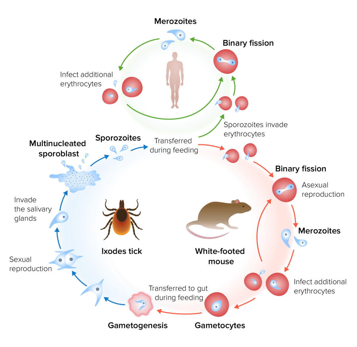
  • Tick carrying sporozoites Sporozoites The product of meiotic division of zygotes in parasitic protozoa comprising haploid cells. These infective cells invade the host and undergo asexual reproduction producing merozoites (or other forms) and ultimately gametocytes. Antimalarial Drugs → attaches to a mouse → transfers sporozoites Sporozoites The product of meiotic division of zygotes in parasitic protozoa comprising haploid cells. These infective cells invade the host and undergo asexual reproduction producing merozoites (or other forms) and ultimately gametocytes. Antimalarial Drugs
  • Sporozoites Sporozoites The product of meiotic division of zygotes in parasitic protozoa comprising haploid cells. These infective cells invade the host and undergo asexual reproduction producing merozoites (or other forms) and ultimately gametocytes. Antimalarial Drugs enter RBCs RBCs Erythrocytes, or red blood cells (RBCs), are the most abundant cells in the blood. While erythrocytes in the fetus are initially produced in the yolk sac then the liver, the bone marrow eventually becomes the main site of production. Erythrocytes: Histology.
  • Once inside the RBC, sporozoites Sporozoites The product of meiotic division of zygotes in parasitic protozoa comprising haploid cells. These infective cells invade the host and undergo asexual reproduction producing merozoites (or other forms) and ultimately gametocytes. Antimalarial Drugs differentiate into trophozoites Trophozoites Cells or feeding stage in the life cycle of sporozoan protozoa. In the malarial parasite, the trophozoite develops from the merozoite and then splits into the schizont. Trophozoites that are left over from cell division can go on to form gametocytes. Amebicides.
  • Trophozoites Trophozoites Cells or feeding stage in the life cycle of sporozoan protozoa. In the malarial parasite, the trophozoite develops from the merozoite and then splits into the schizont. Trophozoites that are left over from cell division can go on to form gametocytes. Amebicides undergo asexual replication ( binary fission Binary fission Cell Types: Eukaryotic versus Prokaryotic) → merozoites Merozoites Uninuclear cells or a stage in the life cycle of sporozoan protozoa. Merozoites, released from ruptured multinucleate schizonts, enter the bloodstream and infect the erythrocytes. Plasmodium/Malaria
  • Merozoites Merozoites Uninuclear cells or a stage in the life cycle of sporozoan protozoa. Merozoites, released from ruptured multinucleate schizonts, enter the bloodstream and infect the erythrocytes. Plasmodium/Malaria produce gametocytes Gametocytes Plasmodium/Malaria.
  • Tick takes a blood meal → gametes are transferred to the tick
  • Gametes are fertilized in the tick’s gut → sexual replication 
  • Invasion into the salivary gland Salivary gland Glands that secrete saliva in the mouth. There are three pairs of salivary glands (parotid gland; sublingual gland; submandibular gland). Diseases of the Salivary Glands of the tick → development into sporozoites Sporozoites The product of meiotic division of zygotes in parasitic protozoa comprising haploid cells. These infective cells invade the host and undergo asexual reproduction producing merozoites (or other forms) and ultimately gametocytes. Antimalarial Drugs

Inside a human host:

  • Tick carrying sporozoites Sporozoites The product of meiotic division of zygotes in parasitic protozoa comprising haploid cells. These infective cells invade the host and undergo asexual reproduction producing merozoites (or other forms) and ultimately gametocytes. Antimalarial Drugs → attaches to a host → transfer sporozoites Sporozoites The product of meiotic division of zygotes in parasitic protozoa comprising haploid cells. These infective cells invade the host and undergo asexual reproduction producing merozoites (or other forms) and ultimately gametocytes. Antimalarial Drugs into the dermis Dermis A layer of vascularized connective tissue underneath the epidermis. The surface of the dermis contains innervated papillae. Embedded in or beneath the dermis are sweat glands; hair follicles; and sebaceous glands. Skin: Structure and Functions (typically the 2nd or 3rd day of attachment)
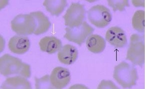
  • Sporozoites Sporozoites The product of meiotic division of zygotes in parasitic protozoa comprising haploid cells. These infective cells invade the host and undergo asexual reproduction producing merozoites (or other forms) and ultimately gametocytes. Antimalarial Drugs enter RBCs RBCs Erythrocytes, or red blood cells (RBCs), are the most abundant cells in the blood. While erythrocytes in the fetus are initially produced in the yolk sac then the liver, the bone marrow eventually becomes the main site of production. Erythrocytes: Histology → differentiate into trophozoites Trophozoites Cells or feeding stage in the life cycle of sporozoan protozoa. In the malarial parasite, the trophozoite develops from the merozoite and then splits into the schizont. Trophozoites that are left over from cell division can go on to form gametocytes. Amebicides (appear as multiple delicate rings)
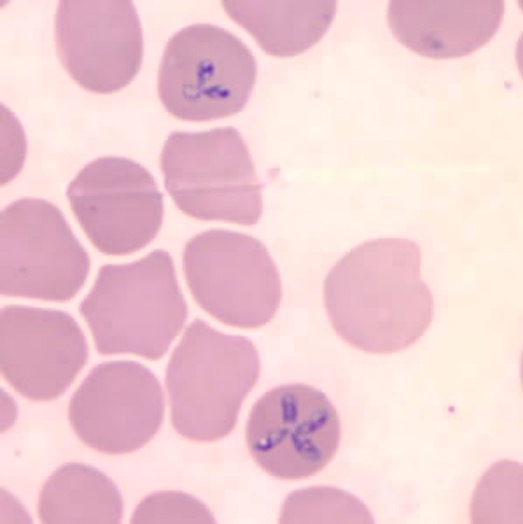
  • Trophozoites Trophozoites Cells or feeding stage in the life cycle of sporozoan protozoa. In the malarial parasite, the trophozoite develops from the merozoite and then splits into the schizont. Trophozoites that are left over from cell division can go on to form gametocytes. Amebicides undergo binary fission Binary fission Cell Types: Eukaryotic versus Prokaryotic merozoites Merozoites Uninuclear cells or a stage in the life cycle of sporozoan protozoa. Merozoites, released from ruptured multinucleate schizonts, enter the bloodstream and infect the erythrocytes. Plasmodium/Malaria (appear as tetrad structures or “ Maltese Cross Maltese Cross Babesia/Babesiosis”)
  • Merozoites Merozoites Uninuclear cells or a stage in the life cycle of sporozoan protozoa. Merozoites, released from ruptured multinucleate schizonts, enter the bloodstream and infect the erythrocytes. Plasmodium/Malaria escape Escape With constant immune mechanisms holding unstable tumor cells in equilibrium, tumor-cell variants may emerge. These cancer cells may express fewer antigens on their surfaces or lose their MHC class I expression.Variants may also protect themselves from T-cell attack via expression of IC molecules on their surfaces, like normal cells.Creation of an immunosuppressive state in the microenvironment is another way to grow without immunologic interference. Cancer Immunotherapy → rupture the RBCs RBCs Erythrocytes, or red blood cells (RBCs), are the most abundant cells in the blood. While erythrocytes in the fetus are initially produced in the yolk sac then the liver, the bone marrow eventually becomes the main site of production. Erythrocytes: Histology (hemolysis) → invasion of other nearby RBCs RBCs Erythrocytes, or red blood cells (RBCs), are the most abundant cells in the blood. While erythrocytes in the fetus are initially produced in the yolk sac then the liver, the bone marrow eventually becomes the main site of production. Erythrocytes: Histology → cycle repeats
  • Infected RBCs RBCs Erythrocytes, or red blood cells (RBCs), are the most abundant cells in the blood. While erythrocytes in the fetus are initially produced in the yolk sac then the liver, the bone marrow eventually becomes the main site of production. Erythrocytes: Histology are recognized as abnormal in the spleen Spleen The spleen is the largest lymphoid organ in the body, located in the LUQ of the abdomen, superior to the left kidney and posterior to the stomach at the level of the 9th-11th ribs just below the diaphragm. The spleen is highly vascular and acts as an important blood filter, cleansing the blood of pathogens and damaged erythrocytes. Spleen: Anatomy → targeted for destruction by macrophages Macrophages The relatively long-lived phagocytic cell of mammalian tissues that are derived from blood monocytes. Main types are peritoneal macrophages; alveolar macrophages; histiocytes; kupffer cells of the liver; and osteoclasts. They may further differentiate within chronic inflammatory lesions to epithelioid cells or may fuse to form foreign body giant cells or langhans giant cells. Innate Immunity: Phagocytes and Antigen Presentation
  • Hemolysis and host immune response → clinical manifestations
The life cycle and transmission of babesia

Life cycle and transmission of Babesia

Image by Lecturio.

Clinical Presentation

Symptoms

The incubation Incubation The amount time between exposure to an infectious agent and becoming symptomatic. Rabies Virus period for babesiosis Babesiosis Babesiosis is an infection caused by a protozoa belonging to the genus, Babesia. The most common Babesia seen in the United States is B. microti, which is transmitted by the Ixodes tick. The protozoa thrive and replicate within host erythrocytes. Lysis of erythrocytes and the body’s immune response result in clinical symptoms. Babesia/Babesiosis is 1–4 weeks.

Mild-to-moderate disease:[2,4‒6]

  • Flu-like symptoms Flu-Like Symptoms Babesia/Babesiosis:
    • Fatigue Fatigue The state of weariness following a period of exertion, mental or physical, characterized by a decreased capacity for work and reduced efficiency to respond to stimuli. Fibromyalgia
    • Fever Fever Fever is defined as a measured body temperature of at least 38°C (100.4°F). Fever is caused by circulating endogenous and/or exogenous pyrogens that increase levels of prostaglandin E2 in the hypothalamus. Fever is commonly associated with chills, rigors, sweating, and flushing of the skin. Fever 
    • Chills Chills The sudden sensation of being cold. It may be accompanied by shivering. Fever
    • Diaphoresis
    • Malaise Malaise Tick-borne Encephalitis Virus
    • Myalgia Myalgia Painful sensation in the muscles. Ion Channel Myopathy
    • Arthralgia Arthralgia Pain in the joint. Rheumatic Fever
    • Headache Headache The symptom of pain in the cranial region. It may be an isolated benign occurrence or manifestation of a wide variety of headache disorders. Brain Abscess
  • Evidence of hemolysis:
    • Yellow skin Skin The skin, also referred to as the integumentary system, is the largest organ of the body. The skin is primarily composed of the epidermis (outer layer) and dermis (deep layer). The epidermis is primarily composed of keratinocytes that undergo rapid turnover, while the dermis contains dense layers of connective tissue. Skin: Structure and Functions
    • Dark urine
  • Less common symptoms:
    • Anorexia Anorexia The lack or loss of appetite accompanied by an aversion to food and the inability to eat. It is the defining characteristic of the disorder anorexia nervosa. Anorexia Nervosa
    • Nausea Nausea An unpleasant sensation in the stomach usually accompanied by the urge to vomit. Common causes are early pregnancy, sea and motion sickness, emotional stress, intense pain, food poisoning, and various enteroviruses. Antiemetics
    • Sore throat Sore throat Pharyngitis is an inflammation of the back of the throat (pharynx). Pharyngitis is usually caused by an upper respiratory tract infection, which is viral in most cases. It typically results in a sore throat and fever. Other symptoms may include a runny nose, cough, headache, and hoarseness. Pharyngitis
    • Dry cough Dry Cough Strongyloidiasis
    • Conjunctival injection

Severe disease:[2,4‒6]

  • Infected individuals tend to have more intense symptoms.
  • In addition, those infected may experience:
    • Altered mental status Altered Mental Status Sepsis in Children
    • Abdominal pain Abdominal Pain Acute Abdomen
    • Vomiting Vomiting The forcible expulsion of the contents of the stomach through the mouth. Hypokalemia
    • Diarrhea Diarrhea Diarrhea is defined as ≥ 3 watery or loose stools in a 24-hour period. There are a multitude of etiologies, which can be classified based on the underlying mechanism of disease. The duration of symptoms (acute or chronic) and characteristics of the stools (e.g., watery, bloody, steatorrheic, mucoid) can help guide further diagnostic evaluation. Diarrhea
    • Shortness of breath Shortness of breath Dyspnea is the subjective sensation of breathing discomfort. Dyspnea is a normal manifestation of heavy physical or psychological exertion, but also may be caused by underlying conditions (both pulmonary and extrapulmonary). Dyspnea
    • Neck stiffness Neck Stiffness Meningitis
    • Photophobia Photophobia Abnormal sensitivity to light. This may occur as a manifestation of eye diseases; migraine; subarachnoid hemorrhage; meningitis; and other disorders. Photophobia may also occur in association with depression and other mental disorders. Migraine Headache
    • Hyperesthesia
  • Complications:
    • ARDS
    • Severe anemia Anemia Anemia is a condition in which individuals have low Hb levels, which can arise from various causes. Anemia is accompanied by a reduced number of RBCs and may manifest with fatigue, shortness of breath, pallor, and weakness. Subtypes are classified by the size of RBCs, chronicity, and etiology. Anemia: Overview and Types
    • Heart failure Heart Failure A heterogeneous condition in which the heart is unable to pump out sufficient blood to meet the metabolic need of the body. Heart failure can be caused by structural defects, functional abnormalities (ventricular dysfunction), or a sudden overload beyond its capacity. Chronic heart failure is more common than acute heart failure which results from sudden insult to cardiac function, such as myocardial infarction. Total Anomalous Pulmonary Venous Return (TAPVR)
    • Splenic rupture Splenic rupture Splenic rupture is a medical emergency that carries a significant risk of hypovolemic shock and death. Injury to the spleen accounts for nearly half of all injuries to intra-abdominal organs. The most common reason for a rupture of the spleen is blunt abdominal trauma, specifically, motor vehicle accidents. Rupture of the Spleen
    • Hepatitis
    • Renal failure Renal failure Conditions in which the kidneys perform below the normal level in the ability to remove wastes, concentrate urine, and maintain electrolyte balance; blood pressure; and calcium metabolism. Renal insufficiency can be classified by the degree of kidney damage (as measured by the level of proteinuria) and reduction in glomerular filtration rate. Crush Syndrome
    • Disseminated intravascular coagulation Disseminated intravascular coagulation Disseminated intravascular coagulation (DIC) is a condition characterized by systemic bodywide activation of the coagulation cascade. This cascade results in both widespread microvascular thrombi contributing to multiple organ dysfunction and consumption of clotting factors and platelets, leading to hemorrhage. Disseminated Intravascular Coagulation
    • Coma Coma Coma is defined as a deep state of unarousable unresponsiveness, characterized by a score of 3 points on the GCS. A comatose state can be caused by a multitude of conditions, making the precise epidemiology and prognosis of coma difficult to determine. Coma
    • Shock Shock Shock is a life-threatening condition associated with impaired circulation that results in tissue hypoxia. The different types of shock are based on the underlying cause: distributive (↑ cardiac output (CO), ↓ systemic vascular resistance (SVR)), cardiogenic (↓ CO, ↑ SVR), hypovolemic (↓ CO, ↑ SVR), obstructive (↓ CO), and mixed. Types of Shock
    • Death

Physical exam[2,5]

  • General appearance:
    • Fever Fever Fever is defined as a measured body temperature of at least 38°C (100.4°F). Fever is caused by circulating endogenous and/or exogenous pyrogens that increase levels of prostaglandin E2 in the hypothalamus. Fever is commonly associated with chills, rigors, sweating, and flushing of the skin. Fever
    • Jaundice Jaundice Jaundice is the abnormal yellowing of the skin and/or sclera caused by the accumulation of bilirubin. Hyperbilirubinemia is caused by either an increase in bilirubin production or a decrease in the hepatic uptake, conjugation, or excretion of bilirubin. Jaundice
  • Eyes, ears, nose Nose The nose is the human body’s primary organ of smell and functions as part of the upper respiratory system. The nose may be best known for inhaling oxygen and exhaling carbon dioxide, but it also contributes to other important functions, such as tasting. The anatomy of the nose can be divided into the external nose and the nasal cavity. Nose Anatomy (External & Internal), and throat Throat The pharynx is a component of the digestive system that lies posterior to the nasal cavity, oral cavity, and larynx. The pharynx can be divided into the oropharynx, nasopharynx, and laryngopharynx. Pharyngeal muscles play an integral role in vital processes such as breathing, swallowing, and speaking. Pharynx: Anatomy:
  • Abdominal exam:
    • Splenomegaly Splenomegaly Splenomegaly is pathologic enlargement of the spleen that is attributable to numerous causes, including infections, hemoglobinopathies, infiltrative processes, and outflow obstruction of the portal vein. Splenomegaly 
    • Hepatomegaly
  • Skin Skin The skin, also referred to as the integumentary system, is the largest organ of the body. The skin is primarily composed of the epidermis (outer layer) and dermis (deep layer). The epidermis is primarily composed of keratinocytes that undergo rapid turnover, while the dermis contains dense layers of connective tissue. Skin: Structure and Functions:
    • Petechiae Petechiae Primary Skin Lesions may be present.
    • A rash Rash Rocky Mountain Spotted Fever may represent a concurrent Lyme disease Lyme disease Lyme disease is a tick-borne infection caused by the gram-negative spirochete Borrelia burgdorferi. Lyme disease is transmitted by the black-legged Ixodes tick (known as a deer tick), which is only found in specific geographic regions. Patient presentation can vary depending on the stage of the disease and may include a characteristic erythema migrans rash. Lyme Disease infection.

Diagnosis and Management

Epidemiology of infectious diseases is affected by location; therefore, diagnostic and treatment approaches may vary. The following information is based on sources from the US and Europe.
Diagnosis

  • Diagnostic testing options:[4,5]
    • Blood smear Blood smear Myeloperoxidase Deficiency:
      • Maltese cross Maltese Cross Babesia/Babesiosis” within RBCs RBCs Erythrocytes, or red blood cells (RBCs), are the most abundant cells in the blood. While erythrocytes in the fetus are initially produced in the yolk sac then the liver, the bone marrow eventually becomes the main site of production. Erythrocytes: Histology
      • Ring forms within RBCs RBCs Erythrocytes, or red blood cells (RBCs), are the most abundant cells in the blood. While erythrocytes in the fetus are initially produced in the yolk sac then the liver, the bone marrow eventually becomes the main site of production. Erythrocytes: Histology may also be present.
    • Indirect fluorescent antibody Indirect fluorescent antibody A form of fluorescent antibody technique commonly used to detect serum antibodies and immune complexes in tissues and microorganisms in specimens from patients with infectious diseases. The technique involves formation of an antigen-antibody complex which is labeled with fluorescein-conjugated anti-immunoglobulin antibody. Chikungunya Virus (IFA) testing for Babesia Babesia Babesiosis is an infection caused by a protozoa belonging to the genus, Babesia. The most common Babesia seen in the United States is B. microti, which is transmitted by the Ixodes tick. The protozoa thrive and replicate within host erythrocytes. Lysis of erythrocytes and the body’s immune response result in clinical symptoms. Babesia/Babesiosis antibodies Antibodies Immunoglobulins (Igs), also known as antibodies, are glycoprotein molecules produced by plasma cells that act in immune responses by recognizing and binding particular antigens. The various Ig classes are IgG (the most abundant), IgM, IgE, IgD, and IgA, which differ in their biologic features, structure, target specificity, and distribution. Immunoglobulins: Types and Functions
    • PCR PCR Polymerase chain reaction (PCR) is a technique that amplifies DNA fragments exponentially for analysis. The process is highly specific, allowing for the targeting of specific genomic sequences, even with minuscule sample amounts. The PCR cycles multiple times through 3 phases: denaturation of the template DNA, annealing of a specific primer to the individual DNA strands, and synthesis/elongation of new DNA molecules. Polymerase Chain Reaction (PCR) for Babesia Babesia Babesiosis is an infection caused by a protozoa belonging to the genus, Babesia. The most common Babesia seen in the United States is B. microti, which is transmitted by the Ixodes tick. The protozoa thrive and replicate within host erythrocytes. Lysis of erythrocytes and the body’s immune response result in clinical symptoms. Babesia/Babesiosis DNA DNA A deoxyribonucleotide polymer that is the primary genetic material of all cells. Eukaryotic and prokaryotic organisms normally contain DNA in a double-stranded state, yet several important biological processes transiently involve single-stranded regions. DNA, which consists of a polysugar-phosphate backbone possessing projections of purines (adenine and guanine) and pyrimidines (thymine and cytosine), forms a double helix that is held together by hydrogen bonds between these purines and pyrimidines (adenine to thymine and guanine to cytosine). DNA Types and Structure:
      • Remains positive for months (at least 2 years in immunosuppressed individuals) even after treatment. 
      • Not used to determine duration of treatment
  • Diagnostic approach:[46]
    • Preferred diagnostic tests Diagnostic tests Diagnostic tests are important aspects in making a diagnosis. Some of the most important epidemiological values of diagnostic tests include sensitivity and specificity, false positives and false negatives, positive and negative predictive values, likelihood ratios, and pre-test and post-test probabilities. Epidemiological Values of Diagnostic Tests:
      • Peripheral blood smear Peripheral Blood Smear Anemia: Overview and Types
      • PCR PCR Polymerase chain reaction (PCR) is a technique that amplifies DNA fragments exponentially for analysis. The process is highly specific, allowing for the targeting of specific genomic sequences, even with minuscule sample amounts. The PCR cycles multiple times through 3 phases: denaturation of the template DNA, annealing of a specific primer to the individual DNA strands, and synthesis/elongation of new DNA molecules. Polymerase Chain Reaction (PCR)
    • For those with a positive serology Serology The study of serum, especially of antigen-antibody reactions in vitro. Yellow Fever Virus (4-fold increase in Babesia Babesia Babesiosis is an infection caused by a protozoa belonging to the genus, Babesia. The most common Babesia seen in the United States is B. microti, which is transmitted by the Ixodes tick. The protozoa thrive and replicate within host erythrocytes. Lysis of erythrocytes and the body’s immune response result in clinical symptoms. Babesia/Babesiosis IgG IgG The major immunoglobulin isotype class in normal human serum. There are several isotype subclasses of igg, for example, igg1, igg2a, and igg2b. Hypersensitivity Pneumonitis):[2]
      • Confirmation via peripheral smear or PCR PCR Polymerase chain reaction (PCR) is a technique that amplifies DNA fragments exponentially for analysis. The process is highly specific, allowing for the targeting of specific genomic sequences, even with minuscule sample amounts. The PCR cycles multiple times through 3 phases: denaturation of the template DNA, annealing of a specific primer to the individual DNA strands, and synthesis/elongation of new DNA molecules. Polymerase Chain Reaction (PCR) is recommended prior to treatment.
      • Antibodies Antibodies Immunoglobulins (Igs), also known as antibodies, are glycoprotein molecules produced by plasma cells that act in immune responses by recognizing and binding particular antigens. The various Ig classes are IgG (the most abundant), IgM, IgE, IgD, and IgA, which differ in their biologic features, structure, target specificity, and distribution. Immunoglobulins: Types and Functions can remain positive for ≥ 1 year, even with clearance of infection (treated or not treated).
  • Supporting evaluation:[2,4]
    • CBC:
      • Hemolytic anemia Hemolytic Anemia Hemolytic anemia (HA) is the term given to a large group of anemias that are caused by the premature destruction/hemolysis of circulating red blood cells (RBCs). Hemolysis can occur within (intravascular hemolysis) or outside the blood vessels (extravascular hemolysis). Hemolytic Anemia (↓ hemoglobin, ↑ LDH LDH Osteosarcoma, ↓ haptoglobin)
      • Neutropenia Neutropenia Neutrophils are an important component of the immune system and play a significant role in the eradication of infections. Low numbers of circulating neutrophils, referred to as neutropenia, predispose the body to recurrent infections or sepsis, though patients can also be asymptomatic. Neutropenia
      • Thrombocytopenia Thrombocytopenia Thrombocytopenia occurs when the platelet count is < 150,000 per microliter. The normal range for platelets is usually 150,000-450,000/µL of whole blood. Thrombocytopenia can be a result of decreased production, increased destruction, or splenic sequestration of platelets. Patients are often asymptomatic until platelet counts are < 50,000/µL. Thrombocytopenia
    • Liver function tests Liver function tests Liver function tests, also known as hepatic function panels, are one of the most commonly performed screening blood tests. Such tests are also used to detect, evaluate, and monitor acute and chronic liver diseases. Liver Function Tests:
    • Basic metabolic panel Basic Metabolic Panel Primary vs Secondary Headaches:
      • ↑ BUN
      • ↑ Creatinine
  • Consider evaluating for coinfections:[2]
    • Borrelia burgdorferi Borrelia burgdorferi A specific species of bacteria, part of the borrelia burgdorferi group, whose common name is lyme disease spirochete. Borrelia ( Lyme disease Lyme disease Lyme disease is a tick-borne infection caused by the gram-negative spirochete Borrelia burgdorferi. Lyme disease is transmitted by the black-legged Ixodes tick (known as a deer tick), which is only found in specific geographic regions. Patient presentation can vary depending on the stage of the disease and may include a characteristic erythema migrans rash. Lyme Disease)
    • Anaplasma phagocytophilum Anaplasma phagocytophilum A species of gram-negative bacteria in the genus anaplasma, family anaplasmataceae, formerly called ehrlichia phagocytophila or ehrlichia equi. This organism is tick-borne (ixodes) and causes disease in horses and sheep. In humans, it causes human granulocytic ehrlichiosis. Ehrlichiosis and Anaplasmosis ( anaplasmosis Anaplasmosis Anaplasmosis is a tick-borne bacterial infection. The most common causative species include Anaplasma phagocytophilum, which infect and multiply within granulocytes. The clinical presentation can vary widely, but often includes fever, malaise, headache, myalgia, and arthralgias. Ehrlichiosis and Anaplasmosis)

Management

The mainstay of treatment is antibiotics and educating individuals on preventive methods to avoid tick bites. Since babesiosis Babesiosis Babesiosis is an infection caused by a protozoa belonging to the genus, Babesia. The most common Babesia seen in the United States is B. microti, which is transmitted by the Ixodes tick. The protozoa thrive and replicate within host erythrocytes. Lysis of erythrocytes and the body’s immune response result in clinical symptoms. Babesia/Babesiosis is relatively rare in the United Kingdom, the following are based on U.S. recommendations:

Babesiosis Babesiosis Babesiosis is an infection caused by a protozoa belonging to the genus, Babesia. The most common Babesia seen in the United States is B. microti, which is transmitted by the Ixodes tick. The protozoa thrive and replicate within host erythrocytes. Lysis of erythrocytes and the body’s immune response result in clinical symptoms. Babesia/Babesiosis secondary to B. microti B. microti A species of protozoa infecting humans via the intermediate tick vector ixodes scapularis. The other hosts are the mouse peromyscus leucopus and meadow vole microtus pennsylvanicus, which are fed on by the tick. Other primates can be experimentally infected with babesia microti. Babesia/Babesiosis

  • Antimicrobial therapy:[4,5]
    • 1st-line treatment: azithromycin Azithromycin A semi-synthetic macrolide antibiotic structurally related to erythromycin. It has been used in the treatment of Mycobacterium avium intracellulare infections, toxoplasmosis, and cryptosporidiosis. Macrolides and Ketolides and atovaquone Atovaquone A hydroxynaphthoquinone that has antimicrobial activity and is being used in antimalarial protocols. Antimalarial Drugs 
    • Alternative: quinine Quinine An alkaloid derived from the bark of the cinchona tree. It is used as an antimalarial drug, and is the active ingredient in extracts of the cinchona that have been used for that purpose since before 1633. Quinine is also a mild antipyretic and analgesic and has been used in common cold preparations for that purpose. It was used commonly and as a bitter and flavoring agent, and is still useful for the treatment of babesiosis. Quinine is also useful in some muscular disorders, especially nocturnal leg cramps and myotonia congenita, because of its direct effects on muscle membrane and sodium channels. The mechanisms of its antimalarial effects are not well understood. Antimalarial Drugs and clindamycin Clindamycin An antibacterial agent that is a semisynthetic analog of lincomycin. Lincosamides (typical treatment in Europe)[6]
  • Considerations:[4]
    • Clindamycin Clindamycin An antibacterial agent that is a semisynthetic analog of lincomycin. Lincosamides and quinine Quinine An alkaloid derived from the bark of the cinchona tree. It is used as an antimalarial drug, and is the active ingredient in extracts of the cinchona that have been used for that purpose since before 1633. Quinine is also a mild antipyretic and analgesic and has been used in common cold preparations for that purpose. It was used commonly and as a bitter and flavoring agent, and is still useful for the treatment of babesiosis. Quinine is also useful in some muscular disorders, especially nocturnal leg cramps and myotonia congenita, because of its direct effects on muscle membrane and sodium channels. The mechanisms of its antimalarial effects are not well understood. Antimalarial Drugs are the 1st choice for pregnant women.
    • Typical duration of treatment is 7–10 days (especially in those with a low risk of relapse Relapse Relapsing Fever).
    • For those at high risk of relapse Relapse Relapsing Fever, antibiotics are given for ≥ 6 weeks.
    • The same antibiotic options are used for relapses.
  • Exchange transfusion is indicated if:[3,5]
  • Monitoring:[3,5]
    • During treatment:
      • Monitor CBC
      • Monitor renal and hepatic panel
      • Check peripheral blood smears (every 2–3 days) to monitor parasitemia.
    • In immunocompetent individuals, peripheral blood smears are no longer checked after symptoms resolve.
    • In immunocompromised immunocompromised A human or animal whose immunologic mechanism is deficient because of an immunodeficiency disorder or other disease or as the result of the administration of immunosuppressive drugs or radiation. Gastroenteritis individuals:
      • Continue checking peripheral blood smears until symptoms resolve and smears are negative for 2 weeks.
      • Check PCR PCR Polymerase chain reaction (PCR) is a technique that amplifies DNA fragments exponentially for analysis. The process is highly specific, allowing for the targeting of specific genomic sequences, even with minuscule sample amounts. The PCR cycles multiple times through 3 phases: denaturation of the template DNA, annealing of a specific primer to the individual DNA strands, and synthesis/elongation of new DNA molecules. Polymerase Chain Reaction (PCR) if symptoms continue even though smears are negative.
  • Splenic rupture Splenic rupture Splenic rupture is a medical emergency that carries a significant risk of hypovolemic shock and death. Injury to the spleen accounts for nearly half of all injuries to intra-abdominal organs. The most common reason for a rupture of the spleen is blunt abdominal trauma, specifically, motor vehicle accidents. Rupture of the Spleen:[3]
Table: Summary of antibiotic treatment recommendations for babesiosis Babesiosis Babesiosis is an infection caused by a protozoa belonging to the genus, Babesia. The most common Babesia seen in the United States is B. microti, which is transmitted by the Ixodes tick. The protozoa thrive and replicate within host erythrocytes. Lysis of erythrocytes and the body’s immune response result in clinical symptoms. Babesia/Babesiosis[3,5]
Treatment indications Adult dose Pediatric dose
1st-line therapy for mild-moderate diseasea
  • Azithromycin Azithromycin A semi-synthetic macrolide antibiotic structurally related to erythromycin. It has been used in the treatment of Mycobacterium avium intracellulare infections, toxoplasmosis, and cryptosporidiosis. Macrolides and Ketolides: 500 mg orally on day 1, then 250 mg daily (1 g daily for immunocompromised immunocompromised A human or animal whose immunologic mechanism is deficient because of an immunodeficiency disorder or other disease or as the result of the administration of immunosuppressive drugs or radiation. Gastroenteritis individuals)
PLUS
  • Atovaquone Atovaquone A hydroxynaphthoquinone that has antimicrobial activity and is being used in antimalarial protocols. Antimalarial Drugsb: 750 mg oral every 12 hours for 7–10 days
  • Azithromycin Azithromycin A semi-synthetic macrolide antibiotic structurally related to erythromycin. It has been used in the treatment of Mycobacterium avium intracellulare infections, toxoplasmosis, and cryptosporidiosis. Macrolides and Ketolides: 10 mg/kg (up to 500 mg/dose) orally on day 1, then 5 mg/kg (up to 250 mg/ dose) orally daily
PLUS
  • Atovaquone Atovaquone A hydroxynaphthoquinone that has antimicrobial activity and is being used in antimalarial protocols. Antimalarial Drugsb: 20 mg/kg (up to 750 mg/dose) orally every 12 hours
1st-line therapy for severe diseasec
  • Azithromycin Azithromycin A semi-synthetic macrolide antibiotic structurally related to erythromycin. It has been used in the treatment of Mycobacterium avium intracellulare infections, toxoplasmosis, and cryptosporidiosis. Macrolides and Ketolides: 500 mg IV daily (step-down therapy [oral] once improved)
PLUS
  • Atovaquone Atovaquone A hydroxynaphthoquinone that has antimicrobial activity and is being used in antimalarial protocols. Antimalarial Drugsb:750 mg orally every 12 hours
  • Azithromycin Azithromycin A semi-synthetic macrolide antibiotic structurally related to erythromycin. It has been used in the treatment of Mycobacterium avium intracellulare infections, toxoplasmosis, and cryptosporidiosis. Macrolides and Ketolides: 10 mg/kg (up to 500 mg/dose) IV daily (step-down therapy (oral) once improved)
PLUS
  • Atovaquone Atovaquone A hydroxynaphthoquinone that has antimicrobial activity and is being used in antimalarial protocols. Antimalarial Drugs 20 mg/kg (up to 750 mg/dose) orally every 12 hours
Alternative therapy for mild-moderate diseasea
  • Clindamycin Clindamycin An antibacterial agent that is a semisynthetic analog of lincomycin. Lincosamides 600 mg orally every 8 hours
PLUS
  • Quinine Quinine An alkaloid derived from the bark of the cinchona tree. It is used as an antimalarial drug, and is the active ingredient in extracts of the cinchona that have been used for that purpose since before 1633. Quinine is also a mild antipyretic and analgesic and has been used in common cold preparations for that purpose. It was used commonly and as a bitter and flavoring agent, and is still useful for the treatment of babesiosis. Quinine is also useful in some muscular disorders, especially nocturnal leg cramps and myotonia congenita, because of its direct effects on muscle membrane and sodium channels. The mechanisms of its antimalarial effects are not well understood. Antimalarial Drugs sulfate 650 mg orally every 8 hours for 7–10 days
  • Clindamycin Clindamycin An antibacterial agent that is a semisynthetic analog of lincomycin. Lincosamides: 7–10 mg/kg (up to 600 mg/dose) orally every 6–8 hours
PLUS
  • Quinine Quinine An alkaloid derived from the bark of the cinchona tree. It is used as an antimalarial drug, and is the active ingredient in extracts of the cinchona that have been used for that purpose since before 1633. Quinine is also a mild antipyretic and analgesic and has been used in common cold preparations for that purpose. It was used commonly and as a bitter and flavoring agent, and is still useful for the treatment of babesiosis. Quinine is also useful in some muscular disorders, especially nocturnal leg cramps and myotonia congenita, because of its direct effects on muscle membrane and sodium channels. The mechanisms of its antimalarial effects are not well understood. Antimalarial Drugs sulfate 8 mg/kg (up to 650 mg/dose) orally every 8 hours for 7–10 days
Alternative therapy for severe diseasec
  • Clindamycin Clindamycin An antibacterial agent that is a semisynthetic analog of lincomycin. Lincosamides 600 mg IV every 6 hours (step-down therapy (oral) once improved)
PLUS
  • Quinine Quinine An alkaloid derived from the bark of the cinchona tree. It is used as an antimalarial drug, and is the active ingredient in extracts of the cinchona that have been used for that purpose since before 1633. Quinine is also a mild antipyretic and analgesic and has been used in common cold preparations for that purpose. It was used commonly and as a bitter and flavoring agent, and is still useful for the treatment of babesiosis. Quinine is also useful in some muscular disorders, especially nocturnal leg cramps and myotonia congenita, because of its direct effects on muscle membrane and sodium channels. The mechanisms of its antimalarial effects are not well understood. Antimalarial Drugs sulfate 650 mg orally every 8 hours for 7–10 days
  • Clindamycin Clindamycin An antibacterial agent that is a semisynthetic analog of lincomycin. Lincosamides: 7–10 mg/kg (up to 600 mg/dose) IV every 6–8 hours
PLUS
  • Quinine Quinine An alkaloid derived from the bark of the cinchona tree. It is used as an antimalarial drug, and is the active ingredient in extracts of the cinchona that have been used for that purpose since before 1633. Quinine is also a mild antipyretic and analgesic and has been used in common cold preparations for that purpose. It was used commonly and as a bitter and flavoring agent, and is still useful for the treatment of babesiosis. Quinine is also useful in some muscular disorders, especially nocturnal leg cramps and myotonia congenita, because of its direct effects on muscle membrane and sodium channels. The mechanisms of its antimalarial effects are not well understood. Antimalarial Drugs sulfate: 8 mg/kg (up to 650 mg/dose) orally every 8 hours
a: Mild-moderate disease: Treatment usually lasts for 7–10 days (longer for immunocompromised individuals).
b: Taken with a fatty meal
c: Severe disease: Treatment of acute disease plus step-down therapy usually lasts 7–10 days (longer for immunocompromised individuals).

Non– B. microti B. microti A species of protozoa infecting humans via the intermediate tick vector ixodes scapularis. The other hosts are the mouse peromyscus leucopus and meadow vole microtus pennsylvanicus, which are fed on by the tick. Other primates can be experimentally infected with babesia microti. Babesia/Babesiosis babesiosis Babesiosis Babesiosis is an infection caused by a protozoa belonging to the genus, Babesia. The most common Babesia seen in the United States is B. microti, which is transmitted by the Ixodes tick. The protozoa thrive and replicate within host erythrocytes. Lysis of erythrocytes and the body’s immune response result in clinical symptoms. Babesia/Babesiosis[4‒6]

  • B. duncani B. duncani Babesia/Babesiosis:
    • Recommended treatment:  IV clindamycin Clindamycin An antibacterial agent that is a semisynthetic analog of lincomycin. Lincosamides plus oral quinine Quinine An alkaloid derived from the bark of the cinchona tree. It is used as an antimalarial drug, and is the active ingredient in extracts of the cinchona that have been used for that purpose since before 1633. Quinine is also a mild antipyretic and analgesic and has been used in common cold preparations for that purpose. It was used commonly and as a bitter and flavoring agent, and is still useful for the treatment of babesiosis. Quinine is also useful in some muscular disorders, especially nocturnal leg cramps and myotonia congenita, because of its direct effects on muscle membrane and sodium channels. The mechanisms of its antimalarial effects are not well understood. Antimalarial Drugs for 7–10 days
    • Azithromycin Azithromycin A semi-synthetic macrolide antibiotic structurally related to erythromycin. It has been used in the treatment of Mycobacterium avium intracellulare infections, toxoplasmosis, and cryptosporidiosis. Macrolides and Ketolides plus atovaquone Atovaquone A hydroxynaphthoquinone that has antimicrobial activity and is being used in antimalarial protocols. Antimalarial Drugs not fully evaluated
  • B. divergens B. divergens Babesia/Babesiosis:
    • Associated with fulminant illness
    • Antibiotics plus exchange transfusion (early in the illness)
    • Recommended treatment: clindamycin Clindamycin An antibacterial agent that is a semisynthetic analog of lincomycin. Lincosamides plus quinine Quinine An alkaloid derived from the bark of the cinchona tree. It is used as an antimalarial drug, and is the active ingredient in extracts of the cinchona that have been used for that purpose since before 1633. Quinine is also a mild antipyretic and analgesic and has been used in common cold preparations for that purpose. It was used commonly and as a bitter and flavoring agent, and is still useful for the treatment of babesiosis. Quinine is also useful in some muscular disorders, especially nocturnal leg cramps and myotonia congenita, because of its direct effects on muscle membrane and sodium channels. The mechanisms of its antimalarial effects are not well understood. Antimalarial Drugs or clindamycin Clindamycin An antibacterial agent that is a semisynthetic analog of lincomycin. Lincosamides alone

Prevention[4‒6]

Precautions should be taken in endemic areas, particularly in individuals at risk for severe disease and complications.

  • Tick prevention Tick prevention Chemical, biological, or medical measures designed to prevent the spread of ticks or the concomitant infestations which result in tick-borne diseases. It includes the veterinary as well as the public health aspects of tick and mite control. Babesia/Babesiosis:
    • Protective clothing 
    • Use of tick repellents Tick Repellents Ehrlichiosis and Anaplasmosis
    • Check for and remove ticks Ticks Blood-sucking acarid parasites of the order ixodida comprising two families: the softbacked ticks (argasidae) and hardbacked ticks (ixodidae). Ticks are larger than their relatives, the mites. They penetrate the skin of their host by means of highly specialized, hooked mouth parts and feed on its blood. Ticks attack all groups of terrestrial vertebrates. In humans they are responsible for many tick-borne diseases, including the transmission of rocky mountain spotted fever; tularemia; babesiosis; african swine fever; and relapsing fever. Coxiella/Q Fever.
  • Screening Screening Preoperative Care of donated blood and organs
  • There is no prophylactic therapy or vaccination Vaccination Vaccination is the administration of a substance to induce the immune system to develop protection against a disease. Unlike passive immunization, which involves the administration of pre-performed antibodies, active immunization constitutes the administration of a vaccine to stimulate the body to produce its own antibodies. Vaccination.

Comparison of Intraerythrocytic Parasites

The table below summarizes the characteristics of parasites that infect RBCs RBCs Erythrocytes, or red blood cells (RBCs), are the most abundant cells in the blood. While erythrocytes in the fetus are initially produced in the yolk sac then the liver, the bone marrow eventually becomes the main site of production. Erythrocytes: Histology.

Table: Comparison of intraerythrocytic parasites
Organism Babesia Babesia Babesiosis is an infection caused by a protozoa belonging to the genus, Babesia. The most common Babesia seen in the United States is B. microti, which is transmitted by the Ixodes tick. The protozoa thrive and replicate within host erythrocytes. Lysis of erythrocytes and the body’s immune response result in clinical symptoms. Babesia/Babesiosis Plasmodium Plasmodium A genus of protozoa that comprise the malaria parasites of mammals. Four species infect humans (although occasional infections with primate malarias may occur). These are plasmodium falciparum; plasmodium malariae; plasmodium ovale, and plasmodium vivax. Species causing infection in vertebrates other than man include: plasmodium berghei; plasmodium chabaudi; p. Vinckei, and plasmodium yoelii in rodents; p. Brasilianum, plasmodium cynomolgi; and plasmodium knowlesi in monkeys; and plasmodium gallinaceum in chickens. Antimalarial Drugs
Disease Babesiosis Babesiosis Babesiosis is an infection caused by a protozoa belonging to the genus, Babesia. The most common Babesia seen in the United States is B. microti, which is transmitted by the Ixodes tick. The protozoa thrive and replicate within host erythrocytes. Lysis of erythrocytes and the body’s immune response result in clinical symptoms. Babesia/Babesiosis Malaria Malaria Malaria is an infectious parasitic disease affecting humans and other animals. Most commonly transmitted via the bite of a female Anopheles mosquito infected with microorganisms of the Plasmodium genus. Patients present with fever, chills, myalgia, headache, and diaphoresis. Plasmodium/Malaria
Microscopic appearance
  • Sporozoa
  • Pear shaped
  • Sporozoa
  • Thin, elongated
Reservoir Reservoir Animate or inanimate sources which normally harbor disease-causing organisms and thus serve as potential sources of disease outbreaks. Reservoirs are distinguished from vectors (disease vectors) and carriers, which are agents of disease transmission rather than continuing sources of potential disease outbreaks. Humans may serve both as disease reservoirs and carriers. Escherichia coli White-footed mouse
  • Monkeys
  • Humans
Transmission Ixodes tick Anopheles Anopheles A genus of mosquitoes (culicidae) that are known vectors of malaria. Plasmodium/Malaria mosquito
Common regions
  • North America
  • Europe
Clinical
  • Flu-like symptoms Flu-Like Symptoms Babesia/Babesiosis
  • Abdominal symptoms
  • Hepatosplenomegaly Hepatosplenomegaly Cytomegalovirus
  • Hemolytic anemia Hemolytic Anemia Hemolytic anemia (HA) is the term given to a large group of anemias that are caused by the premature destruction/hemolysis of circulating red blood cells (RBCs). Hemolysis can occur within (intravascular hemolysis) or outside the blood vessels (extravascular hemolysis). Hemolytic Anemia
  • Renal failure Renal failure Conditions in which the kidneys perform below the normal level in the ability to remove wastes, concentrate urine, and maintain electrolyte balance; blood pressure; and calcium metabolism. Renal insufficiency can be classified by the degree of kidney damage (as measured by the level of proteinuria) and reduction in glomerular filtration rate. Crush Syndrome
  • ARDS
  • HF
  • DIC DIC Disseminated intravascular coagulation (DIC) is a condition characterized by systemic bodywide activation of the coagulation cascade. This cascade results in both widespread microvascular thrombi contributing to multiple organ dysfunction and consumption of clotting factors and platelets, leading to hemorrhage. Disseminated Intravascular Coagulation
  • Flu-like symptoms Flu-Like Symptoms Babesia/Babesiosis
  • Abdominal symptoms
  • Hepatosplenomegaly Hepatosplenomegaly Cytomegalovirus
  • Hemolytic anemia Hemolytic Anemia Hemolytic anemia (HA) is the term given to a large group of anemias that are caused by the premature destruction/hemolysis of circulating red blood cells (RBCs). Hemolysis can occur within (intravascular hemolysis) or outside the blood vessels (extravascular hemolysis). Hemolytic Anemia (more severe)
  • Renal failure Renal failure Conditions in which the kidneys perform below the normal level in the ability to remove wastes, concentrate urine, and maintain electrolyte balance; blood pressure; and calcium metabolism. Renal insufficiency can be classified by the degree of kidney damage (as measured by the level of proteinuria) and reduction in glomerular filtration rate. Crush Syndrome
  • ARDS
  • DIC DIC Disseminated intravascular coagulation (DIC) is a condition characterized by systemic bodywide activation of the coagulation cascade. This cascade results in both widespread microvascular thrombi contributing to multiple organ dysfunction and consumption of clotting factors and platelets, leading to hemorrhage. Disseminated Intravascular Coagulation
  • Hypoglycemia Hypoglycemia Hypoglycemia is an emergency condition defined as a serum glucose level ≤ 70 mg/dL (≤ 3.9 mmol/L) in diabetic patients. In nondiabetic patients, there is no specific or defined limit for normal serum glucose levels, and hypoglycemia is defined mainly by its clinical features. Hypoglycemia
Diagnosis
  • Blood smear Blood smear Myeloperoxidase Deficiency (preferred)
  • PCR PCR Polymerase chain reaction (PCR) is a technique that amplifies DNA fragments exponentially for analysis. The process is highly specific, allowing for the targeting of specific genomic sequences, even with minuscule sample amounts. The PCR cycles multiple times through 3 phases: denaturation of the template DNA, annealing of a specific primer to the individual DNA strands, and synthesis/elongation of new DNA molecules. Polymerase Chain Reaction (PCR) (preferred)
  • IFA
  • Blood smear Blood smear Myeloperoxidase Deficiency
  • Antigen Antigen Substances that are recognized by the immune system and induce an immune reaction. Vaccination testing
  • PCR PCR Polymerase chain reaction (PCR) is a technique that amplifies DNA fragments exponentially for analysis. The process is highly specific, allowing for the targeting of specific genomic sequences, even with minuscule sample amounts. The PCR cycles multiple times through 3 phases: denaturation of the template DNA, annealing of a specific primer to the individual DNA strands, and synthesis/elongation of new DNA molecules. Polymerase Chain Reaction (PCR) (not widely available)
Management
  • Azithromycin Azithromycin A semi-synthetic macrolide antibiotic structurally related to erythromycin. It has been used in the treatment of Mycobacterium avium intracellulare infections, toxoplasmosis, and cryptosporidiosis. Macrolides and Ketolides and atovaquone Atovaquone A hydroxynaphthoquinone that has antimicrobial activity and is being used in antimalarial protocols. Antimalarial Drugs
  • Clindamycin Clindamycin An antibacterial agent that is a semisynthetic analog of lincomycin. Lincosamides and quinine Quinine An alkaloid derived from the bark of the cinchona tree. It is used as an antimalarial drug, and is the active ingredient in extracts of the cinchona that have been used for that purpose since before 1633. Quinine is also a mild antipyretic and analgesic and has been used in common cold preparations for that purpose. It was used commonly and as a bitter and flavoring agent, and is still useful for the treatment of babesiosis. Quinine is also useful in some muscular disorders, especially nocturnal leg cramps and myotonia congenita, because of its direct effects on muscle membrane and sodium channels. The mechanisms of its antimalarial effects are not well understood. Antimalarial Drugs
Depends on species, severity, and resistance Resistance Physiologically, the opposition to flow of air caused by the forces of friction. As a part of pulmonary function testing, it is the ratio of driving pressure to the rate of air flow. Ventilation: Mechanics of Breathing patterns, but may include a combination of:
  • Atovaquone Atovaquone A hydroxynaphthoquinone that has antimicrobial activity and is being used in antimalarial protocols. Antimalarial Drugs
  • Proguanil Proguanil A biguanide compound which metabolizes in the body to form cycloguanil, an anti-malaria agent. Antimalarial Drugs
  • Quinine Quinine An alkaloid derived from the bark of the cinchona tree. It is used as an antimalarial drug, and is the active ingredient in extracts of the cinchona that have been used for that purpose since before 1633. Quinine is also a mild antipyretic and analgesic and has been used in common cold preparations for that purpose. It was used commonly and as a bitter and flavoring agent, and is still useful for the treatment of babesiosis. Quinine is also useful in some muscular disorders, especially nocturnal leg cramps and myotonia congenita, because of its direct effects on muscle membrane and sodium channels. The mechanisms of its antimalarial effects are not well understood. Antimalarial Drugs
  • Tetracyclines Tetracyclines Tetracyclines are a class of broad-spectrum antibiotics indicated for a wide variety of bacterial infections. These medications bind the 30S ribosomal subunit to inhibit protein synthesis of bacteria. Tetracyclines cover gram-positive and gram-negative organisms, as well as atypical bacteria such as chlamydia, mycoplasma, spirochetes, and even protozoa. Tetracyclines
  • Mefloquine Mefloquine A phospholipid-interacting antimalarial drug (antimalarials). It is very effective against plasmodium falciparum with very few side effects. Antimalarial Drugs
  • Chloroquine Chloroquine The prototypical antimalarial agent with a mechanism that is not well understood. It has also been used to treat rheumatoid arthritis, systemic lupus erythematosus, and in the systemic therapy of amebic liver abscesses. Antimalarial Drugs
HF: heart failure
DIC: disseminated intravascular coagulation
IFA: indirect fluorescent antibody

Differential Diagnosis

  • Malaria Malaria Malaria is an infectious parasitic disease affecting humans and other animals. Most commonly transmitted via the bite of a female Anopheles mosquito infected with microorganisms of the Plasmodium genus. Patients present with fever, chills, myalgia, headache, and diaphoresis. Plasmodium/Malaria: a mosquito-borne infectious disease caused by Plasmodium Plasmodium A genus of protozoa that comprise the malaria parasites of mammals. Four species infect humans (although occasional infections with primate malarias may occur). These are plasmodium falciparum; plasmodium malariae; plasmodium ovale, and plasmodium vivax. Species causing infection in vertebrates other than man include: plasmodium berghei; plasmodium chabaudi; p. Vinckei, and plasmodium yoelii in rodents; p. Brasilianum, plasmodium cynomolgi; and plasmodium knowlesi in monkeys; and plasmodium gallinaceum in chickens. Antimalarial Drugs species. Malaria Malaria Malaria is an infectious parasitic disease affecting humans and other animals. Most commonly transmitted via the bite of a female Anopheles mosquito infected with microorganisms of the Plasmodium genus. Patients present with fever, chills, myalgia, headache, and diaphoresis. Plasmodium/Malaria often presents with fever Fever Fever is defined as a measured body temperature of at least 38°C (100.4°F). Fever is caused by circulating endogenous and/or exogenous pyrogens that increase levels of prostaglandin E2 in the hypothalamus. Fever is commonly associated with chills, rigors, sweating, and flushing of the skin. Fever, rigors Rigors Fever, diaphoresis, jaundice Jaundice Jaundice is the abnormal yellowing of the skin and/or sclera caused by the accumulation of bilirubin. Hyperbilirubinemia is caused by either an increase in bilirubin production or a decrease in the hepatic uptake, conjugation, or excretion of bilirubin. Jaundice, abdominal pain Abdominal Pain Acute Abdomen, hemolytic anemia Hemolytic Anemia Hemolytic anemia (HA) is the term given to a large group of anemias that are caused by the premature destruction/hemolysis of circulating red blood cells (RBCs). Hemolysis can occur within (intravascular hemolysis) or outside the blood vessels (extravascular hemolysis). Hemolytic Anemia, hepatosplenomegaly Hepatosplenomegaly Cytomegalovirus, and renal impairment. A blood smear Blood smear Myeloperoxidase Deficiency in malaria Malaria Malaria is an infectious parasitic disease affecting humans and other animals. Most commonly transmitted via the bite of a female Anopheles mosquito infected with microorganisms of the Plasmodium genus. Patients present with fever, chills, myalgia, headache, and diaphoresis. Plasmodium/Malaria shows a single pleomorphic Pleomorphic Bacteroides ring. The “ Maltese Cross Maltese Cross Babesia/Babesiosis” finding is not seen. Rapid testing for Plasmodium Plasmodium A genus of protozoa that comprise the malaria parasites of mammals. Four species infect humans (although occasional infections with primate malarias may occur). These are plasmodium falciparum; plasmodium malariae; plasmodium ovale, and plasmodium vivax. Species causing infection in vertebrates other than man include: plasmodium berghei; plasmodium chabaudi; p. Vinckei, and plasmodium yoelii in rodents; p. Brasilianum, plasmodium cynomolgi; and plasmodium knowlesi in monkeys; and plasmodium gallinaceum in chickens. Antimalarial Drugs antigens can also be performed. Management requires a prolonged course of multiple antimalarial drugs Antimalarial drugs Malaria, a vector-borne parasitic disease caused by Plasmodium spp., is transmitted via injection of sporozoites or immature forms of the parasite into a person’s bloodstream. Sporozoites then infect the hepatocytes and differentiate into schizonts, which subsequently rupture, and merozoites invade red blood cells. Antimalarial Drugs.
  • Lyme disease Lyme disease Lyme disease is a tick-borne infection caused by the gram-negative spirochete Borrelia burgdorferi. Lyme disease is transmitted by the black-legged Ixodes tick (known as a deer tick), which is only found in specific geographic regions. Patient presentation can vary depending on the stage of the disease and may include a characteristic erythema migrans rash. Lyme Disease: a tick-borne infection caused by the gram-negative spirochete Spirochete Treponema is a gram-negative, microaerophilic spirochete. Owing to its very thin structure, it is not easily seen on Gram stain, but can be visualized using dark-field microscopy. This spirochete contains endoflagella, which allow for a characteristic corkscrew movement. Treponema, Borrelia burgdorferi Borrelia burgdorferi A specific species of bacteria, part of the borrelia burgdorferi group, whose common name is lyme disease spirochete. Borrelia. Lyme disease Lyme disease Lyme disease is a tick-borne infection caused by the gram-negative spirochete Borrelia burgdorferi. Lyme disease is transmitted by the black-legged Ixodes tick (known as a deer tick), which is only found in specific geographic regions. Patient presentation can vary depending on the stage of the disease and may include a characteristic erythema migrans rash. Lyme Disease is also transmitted by the Ixodes tick. The presentation of Lyme disease Lyme disease Lyme disease is a tick-borne infection caused by the gram-negative spirochete Borrelia burgdorferi. Lyme disease is transmitted by the black-legged Ixodes tick (known as a deer tick), which is only found in specific geographic regions. Patient presentation can vary depending on the stage of the disease and may include a characteristic erythema migrans rash. Lyme Disease can vary depending on the stage of the disease and may include the characteristic rash Rash Rocky Mountain Spotted Fever known as erythema Erythema Redness of the skin produced by congestion of the capillaries. This condition may result from a variety of disease processes. Chalazion migrans (not present in babesiosis Babesiosis Babesiosis is an infection caused by a protozoa belonging to the genus, Babesia. The most common Babesia seen in the United States is B. microti, which is transmitted by the Ixodes tick. The protozoa thrive and replicate within host erythrocytes. Lysis of erythrocytes and the body’s immune response result in clinical symptoms. Babesia/Babesiosis). Neurological, cardiac, ocular, and joint manifestations are also common in later stages. Diagnosis of Lyme disease Lyme disease Lyme disease is a tick-borne infection caused by the gram-negative spirochete Borrelia burgdorferi. Lyme disease is transmitted by the black-legged Ixodes tick (known as a deer tick), which is only found in specific geographic regions. Patient presentation can vary depending on the stage of the disease and may include a characteristic erythema migrans rash. Lyme Disease relies on clinical findings and tick exposure, and is supported by serological testing. Antibiotics are used for treatment. 
  • AntibioticsViral hepatitis: liver Liver The liver is the largest gland in the human body. The liver is found in the superior right quadrant of the abdomen and weighs approximately 1.5 kilograms. Its main functions are detoxification, metabolism, nutrient storage (e.g., iron and vitamins), synthesis of coagulation factors, formation of bile, filtration, and storage of blood. Liver: Anatomy inflammation Inflammation Inflammation is a complex set of responses to infection and injury involving leukocytes as the principal cellular mediators in the body’s defense against pathogenic organisms. Inflammation is also seen as a response to tissue injury in the process of wound healing. The 5 cardinal signs of inflammation are pain, heat, redness, swelling, and loss of function. Inflammation due to infection with the hepatitis virus Virus Viruses are infectious, obligate intracellular parasites composed of a nucleic acid core surrounded by a protein capsid. Viruses can be either naked (non-enveloped) or enveloped. The classification of viruses is complex and based on many factors, including type and structure of the nucleoid and capsid, the presence of an envelope, the replication cycle, and the host range. Virology. Patients Patients Individuals participating in the health care system for the purpose of receiving therapeutic, diagnostic, or preventive procedures. Clinician–Patient Relationship present with a prodromal flu-like illness, followed by jaundice Jaundice Jaundice is the abnormal yellowing of the skin and/or sclera caused by the accumulation of bilirubin. Hyperbilirubinemia is caused by either an increase in bilirubin production or a decrease in the hepatic uptake, conjugation, or excretion of bilirubin. Jaundice, hepatosplenomegaly Hepatosplenomegaly Cytomegalovirus, and elevated transaminases Transaminases A subclass of enzymes of the transferase class that catalyze the transfer of an amino group from a donor (generally an amino acid) to an acceptor (generally a 2-keto acid). Most of these enzymes are pyridoxyl phosphate proteins. Autoimmune Hepatitis. Hepatitis serologies are used in the diagnosis and help differentiate hepatitis from babesiosis Babesiosis Babesiosis is an infection caused by a protozoa belonging to the genus, Babesia. The most common Babesia seen in the United States is B. microti, which is transmitted by the Ixodes tick. The protozoa thrive and replicate within host erythrocytes. Lysis of erythrocytes and the body’s immune response result in clinical symptoms. Babesia/Babesiosis. Management of acute hepatitis Acute Hepatitis Autoimmune Hepatitis is supportive. 
  • Rocky Mountain spotted fever Fever Fever is defined as a measured body temperature of at least 38°C (100.4°F). Fever is caused by circulating endogenous and/or exogenous pyrogens that increase levels of prostaglandin E2 in the hypothalamus. Fever is commonly associated with chills, rigors, sweating, and flushing of the skin. Fever: a disease caused by Rickettsia rickettsii Rickettsia rickettsii A species of gram-negative, aerobic bacteria that is the etiologic agent of rocky mountain spotted fever. Its cells are slightly smaller and more uniform in size than those of rickettsia prowazekii. Rickettsia that presents with fever Fever Fever is defined as a measured body temperature of at least 38°C (100.4°F). Fever is caused by circulating endogenous and/or exogenous pyrogens that increase levels of prostaglandin E2 in the hypothalamus. Fever is commonly associated with chills, rigors, sweating, and flushing of the skin. Fever, fatigue Fatigue The state of weariness following a period of exertion, mental or physical, characterized by a decreased capacity for work and reduced efficiency to respond to stimuli. Fibromyalgia, headache Headache The symptom of pain in the cranial region. It may be an isolated benign occurrence or manifestation of a wide variety of headache disorders. Brain Abscess, and a rash Rash Rocky Mountain Spotted Fever following a tick bite. However, Rocky Mountain spotted fever Fever Fever is defined as a measured body temperature of at least 38°C (100.4°F). Fever is caused by circulating endogenous and/or exogenous pyrogens that increase levels of prostaglandin E2 in the hypothalamus. Fever is commonly associated with chills, rigors, sweating, and flushing of the skin. Fever is associated with the Dermacentor Dermacentor A widely distributed genus of ticks, in the family ixodidae, including a number that infest humans and other mammals. Several are vectors of diseases such as tularemia; rocky mountain spotted fever; colorado tick fever; and anaplasmosis. Rickettsia tick. Diagnosis is made based on the clinical features, biopsy Biopsy Removal and pathologic examination of specimens from the living body. Ewing Sarcoma of the rash Rash Rocky Mountain Spotted Fever, and serological testing. Treatment is with antibiotics including doxycycline.
  • Ehrlichiosis Ehrlichiosis Ehrlichiosis is a tick-borne bacterial infection. The most common causative species include Ehrlichia chaffeensis, which infect and multiply within monocytes. The clinical presentation can vary widely, but often includes fever, malaise, headache, myalgia, and arthralgias. Ehrlichiosis and Anaplasmosis and anaplasmosis Anaplasmosis Anaplasmosis is a tick-borne bacterial infection. The most common causative species include Anaplasma phagocytophilum, which infect and multiply within granulocytes. The clinical presentation can vary widely, but often includes fever, malaise, headache, myalgia, and arthralgias. Ehrlichiosis and Anaplasmosis: tick-borne infections Infections Invasion of the host organism by microorganisms or their toxins or by parasites that can cause pathological conditions or diseases. Chronic Granulomatous Disease caused by Ehrlichia chaffeensis Ehrlichia chaffeensis A species of gram-negative bacteria that is the causative agent of human ehrlichiosis. This organism was first discovered at fort chaffee, arkansas, when blood samples from suspected human ehrlichiosis patients were studied. Ehrlichiosis and Anaplasmosis and Anaplasmosis Anaplasmosis Anaplasmosis is a tick-borne bacterial infection. The most common causative species include Anaplasma phagocytophilum, which infect and multiply within granulocytes. The clinical presentation can vary widely, but often includes fever, malaise, headache, myalgia, and arthralgias. Ehrlichiosis and Anaplasmosis phagocytophilum, respectively. Symptoms of ehrlichiosis Ehrlichiosis Ehrlichiosis is a tick-borne bacterial infection. The most common causative species include Ehrlichia chaffeensis, which infect and multiply within monocytes. The clinical presentation can vary widely, but often includes fever, malaise, headache, myalgia, and arthralgias. Ehrlichiosis and Anaplasmosis and anaplasmosis Anaplasmosis Anaplasmosis is a tick-borne bacterial infection. The most common causative species include Anaplasma phagocytophilum, which infect and multiply within granulocytes. The clinical presentation can vary widely, but often includes fever, malaise, headache, myalgia, and arthralgias. Ehrlichiosis and Anaplasmosis include fever Fever Fever is defined as a measured body temperature of at least 38°C (100.4°F). Fever is caused by circulating endogenous and/or exogenous pyrogens that increase levels of prostaglandin E2 in the hypothalamus. Fever is commonly associated with chills, rigors, sweating, and flushing of the skin. Fever, headache Headache The symptom of pain in the cranial region. It may be an isolated benign occurrence or manifestation of a wide variety of headache disorders. Brain Abscess, and malaise Malaise Tick-borne Encephalitis Virus. Disseminated intravascular coagulation Disseminated intravascular coagulation Disseminated intravascular coagulation (DIC) is a condition characterized by systemic bodywide activation of the coagulation cascade. This cascade results in both widespread microvascular thrombi contributing to multiple organ dysfunction and consumption of clotting factors and platelets, leading to hemorrhage. Disseminated Intravascular Coagulation, multiorgan failure, and coma Coma Coma is defined as a deep state of unarousable unresponsiveness, characterized by a score of 3 points on the GCS. A comatose state can be caused by a multitude of conditions, making the precise epidemiology and prognosis of coma difficult to determine. Coma can also occur with severe disease. The diagnosis is made using PCR PCR Polymerase chain reaction (PCR) is a technique that amplifies DNA fragments exponentially for analysis. The process is highly specific, allowing for the targeting of specific genomic sequences, even with minuscule sample amounts. The PCR cycles multiple times through 3 phases: denaturation of the template DNA, annealing of a specific primer to the individual DNA strands, and synthesis/elongation of new DNA molecules. Polymerase Chain Reaction (PCR). Treatment of both diseases is with doxycycline.

References

  1. Krause, P., Vannier, E. (2019). Babesiosis: Microbiology, epidemiology, and pathogenesis. Retrieved on March 09, 2021, from https://www.uptodate.com/contents/babesiosis-microbiology-epidemiology-and-pathogenesis
  2. Krause, P., Vannier, E. (2019). Babesiosis: Clinical manifestations and diagnosis. Retrieved on March 09, 2021, from https://www.uptodate.com/contents/babesiosis-clinical-manifestations-and-diagnosis
  3. Krause, P., Vannier, E. (2021). Babesiosis: Treatment and prevention. Retrieved on March 09, 2021, from https://www.uptodate.com/contents/babesiosis-treatment-and-prevention
  4. Centers for Disease Control and Prevention. (2020). Babesiosis. Retrieved June 18, 2022, from https://www.cdc.gov/parasites/babesiosis/index.html
  5. Krause, P., et al. (2021). Clinical practice guidelines by the Infectious Diseases Society of America (IDSA): 2020 guideline on diagnosis and management of babesiosis. Clinical Infectious Diseases 72(2):e49–e64. https://doi.org/10.1093/cid/ciaa1216
  6. European Centre for Disease Prevention and Control. (n.d.). Facts about babesiosis. Retrieved June 19, 2022,from https://www.ecdc.europa.eu/en/all-topics-z/babesiosis/facts-about-babesiosis

Create your free account or log in to continue reading!

Sign up now and get free access to Lecturio with concept pages, medical videos, and questions for your medical education.

User Reviews

Details