Advertisement

Advertisement

Advertisement

Advertisement

Aortic Dissection

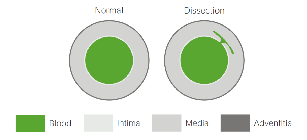
Aortic dissection occurs due to shearing stress from pulsatile pressure causing a tear in the tunica intima of the aortic wall. This tear allows blood to flow into the media, creating a “false lumen.” Aortic dissection is most commonly caused by uncontrolled hypertension. Complications arise due to partial occlusion of vital branches off the aorta and reduced blood flow to the brain, visceral organs, and extremities. Patients often present with acute, tearing chest or back pain. Computed tomography is the diagnostic modality of choice. All type A dissections (ascending aorta) are a surgical emergency due to the risk of imminent rupture. Type B dissections (descending aorta) can be managed medically with impulse control using beta-blockers and calcium channel blockers. If there is evidence of malperfusion to visceral organs or extremities, aneurysm dilation to > 5 cm, retrograde extension into the ascending aorta, or intractable pain, the patient will need evaluation for endovascular or open repair.

Last updated: May 16, 2024
Editorial responsibility: Stanley Oiseth, Lindsay Jones, Evelin Maza

Advertisement

Advertisement

Advertisement

Advertisement

Advertisement

Advertisement

Overview

Epidemiology

  • 5–30 cases per 1 million people per year
  • More common in men than women (3:1)
  • More common in African Americans than Caucasians
  • Peak incidence Incidence The number of new cases of a given disease during a given period in a specified population. It also is used for the rate at which new events occur in a defined population. It is differentiated from prevalence, which refers to all cases in the population at a given time. Measures of Disease Frequency at age 50–65 years

Etiology

  • Hypertension Hypertension Hypertension, or high blood pressure, is a common disease that manifests as elevated systemic arterial pressures. Hypertension is most often asymptomatic and is found incidentally as part of a routine physical examination or during triage for an unrelated medical encounter. Hypertension (70% of dissection patients Patients Individuals participating in the health care system for the purpose of receiving therapeutic, diagnostic, or preventive procedures. Clinician–Patient Relationship)
  • Atherosclerosis Atherosclerosis Atherosclerosis is a common form of arterial disease in which lipid deposition forms a plaque in the blood vessel walls. Atherosclerosis is an incurable disease, for which there are clearly defined risk factors that often can be reduced through a change in lifestyle and behavior of the patient. Atherosclerosis
  • Connective tissue Connective tissue Connective tissues originate from embryonic mesenchyme and are present throughout the body except inside the brain and spinal cord. The main function of connective tissues is to provide structural support to organs. Connective tissues consist of cells and an extracellular matrix. Connective Tissue: Histology disorders
    • Marfan syndrome Marfan syndrome Marfan syndrome is a genetic condition with autosomal dominant inheritance. Marfan syndrome affects the elasticity of connective tissues throughout the body, most notably in the cardiovascular, ocular, and musculoskeletal systems. Marfan Syndrome
    • Ehlers-Danlos syndrome Ehlers-Danlos syndrome Ehlers-Danlos syndrome (EDS) is a heterogeneous group of inherited connective tissue disorders that are characterized by hyperextensible skin, hypermobile joints, and fragility of the skin and connective tissue. Ehlers-Danlos Syndrome
    • Loeys-Dietz syndrome
    • Cystic Cystic Fibrocystic Change medial necrosis Necrosis The death of cells in an organ or tissue due to disease, injury or failure of the blood supply. Ischemic Cell Damage 
  • Vasculitis Vasculitis Inflammation of any one of the blood vessels, including the arteries; veins; and rest of the vasculature system in the body. Systemic Lupus Erythematosus
    • Giant cell arteritis Giant Cell Arteritis Giant cell arteritis (GCA), also known as temporal arteritis, is a type of large-vessel vasculitis that predominantly affects the aorta and its major branches, with a predilection for the branches of the carotid (including the temporal artery). Giant cell arteritis is defined by inflammatory leukocytes in the vessel walls leading to reactive damage, ischemia, and necrosis. Giant Cell Arteritis
    • Takayasu arteritis
    • Behcet’s disease
  • Anatomic abnormalities
  • Trauma involving rapid deceleration Deceleration A decrease in the rate of speed. Blunt Chest Trauma
  • Iatrogenic Iatrogenic Any adverse condition in a patient occurring as the result of treatment by a physician, surgeon, or other health professional, especially infections acquired by a patient during the course of treatment. Anterior Cord Syndrome
    • Prior cardiac or aortic surgery
    • Cardiac catheterization Cardiac Catheterization Procedures in which placement of cardiac catheters is performed for therapeutic or diagnostic procedures. Cardiac Surgery
  • Drugs
    • Amphetamines Amphetamines Analogs or derivatives of amphetamine. Many are sympathomimetics and central nervous system stimulators causing excitation, vasopressin, bronchodilation, and to varying degrees, anorexia, analepsis, nasal decongestion, and some smooth muscle relaxation. Stimulants
    • Cocaine Cocaine An alkaloid ester extracted from the leaves of plants including coca. It is a local anesthetic and vasoconstrictor and is clinically used for that purpose, particularly in the eye, ear, nose, and throat. It also has powerful central nervous system effects similar to the amphetamines and is a drug of abuse. Cocaine, like amphetamines, acts by multiple mechanisms on brain catecholaminergic neurons; the mechanism of its reinforcing effects is thought to involve inhibition of dopamine uptake. Local Anesthetics

Classification

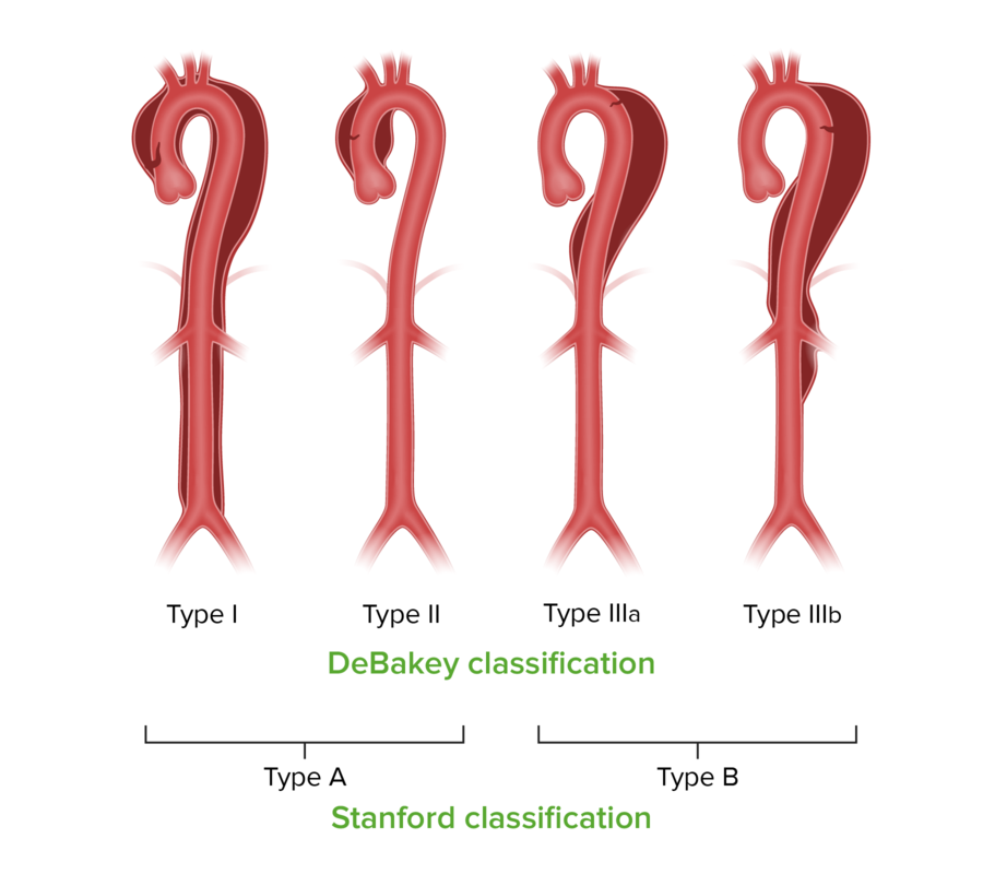
Stanford and debakey classifications for aortic dissection

Stanford and DeBakey classifications for aortic dissection

Image by Lecturio.
Pathophysiology

  • The aorta Aorta The main trunk of the systemic arteries. Mediastinum and Great Vessels: Anatomy is more likely to rupture than any other vessel in the body.
    • 90% of dissections occur within 10 cm of the aortic valve Aortic valve The valve between the left ventricle and the ascending aorta which prevents backflow into the left ventricle. Heart: Anatomy.
    • 2nd-most common location is just distal to the take-off of the left subclavian.
  • Contributing factors to aortic injury:
    • Continuously high pulsatile pressure and shear stress
    • Connective tissue Connective tissue Connective tissues originate from embryonic mesenchyme and are present throughout the body except inside the brain and spinal cord. The main function of connective tissues is to provide structural support to organs. Connective tissues consist of cells and an extracellular matrix. Connective Tissue: Histology disorders are characterized by cystic Cystic Fibrocystic Change medial necrosis Necrosis The death of cells in an organ or tissue due to disease, injury or failure of the blood supply. Ischemic Cell Damage.
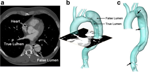
  • Intimal tear develops → blood flows into the media → formation of a false lumen
    • True lumen
      • Lined by intima
      • Typically the smaller lumen
    • False lumen
      • Within the media
      • Larger lumen, slower flow Flow Blood flows through the heart, arteries, capillaries, and veins in a closed, continuous circuit. Flow is the movement of volume per unit of time. Flow is affected by the pressure gradient and the resistance fluid encounters between 2 points. Vascular resistance is the opposition to flow, which is caused primarily by blood friction against vessel walls. Vascular Resistance, Flow, and Mean Arterial Pressure, and elevated mean pressures
      • Elevated pressures can cause compression Compression Blunt Chest Trauma of the true lumen.
  • Dissections can extend proximally and/or distally for variable Variable Variables represent information about something that can change. The design of the measurement scales, or of the methods for obtaining information, will determine the data gathered and the characteristics of that data. As a result, a variable can be qualitative or quantitative, and may be further classified into subgroups. Types of Variables distances.
    • Antegrade toward the iliac bifurcation
    • Retrograde toward the aortic root and heart
  • Consequences:
    • Compromised blood supply to arterial branches off the aorta Aorta The main trunk of the systemic arteries. Mediastinum and Great Vessels: Anatomy
      • Ischemic stroke Ischemic Stroke An ischemic stroke (also known as cerebrovascular accident) is an acute neurologic injury that occurs as a result of brain ischemia; this condition may be due to cerebral blood vessel occlusion by thrombosis or embolism, or rarely due to systemic hypoperfusion. Ischemic Stroke
      • Spinal cord Spinal cord The spinal cord is the major conduction pathway connecting the brain to the body; it is part of the CNS. In cross section, the spinal cord is divided into an H-shaped area of gray matter (consisting of synapsing neuronal cell bodies) and a surrounding area of white matter (consisting of ascending and descending tracts of myelinated axons). Spinal Cord: Anatomy ischemia Ischemia A hypoperfusion of the blood through an organ or tissue caused by a pathologic constriction or obstruction of its blood vessels, or an absence of blood circulation. Ischemic Cell Damage
      • Mesenteric ischemia Ischemia A hypoperfusion of the blood through an organ or tissue caused by a pathologic constriction or obstruction of its blood vessels, or an absence of blood circulation. Ischemic Cell Damage
      • Renal ischemia Ischemia A hypoperfusion of the blood through an organ or tissue caused by a pathologic constriction or obstruction of its blood vessels, or an absence of blood circulation. Ischemic Cell Damage
    • If the dissection spreads through the adventitia, there is a significant risk for aortic rupture.
      • Increased risk in ascending aortic dissections
      • Leads to hemorrhagic shock Hemorrhagic shock Acute hemorrhage or excessive fluid loss resulting in hypovolemia. Hemothorax and death
    • Compression Compression Blunt Chest Trauma symptoms from an enlarged false lumen
    • Ascending aortic dissections:
      • Hemopericardium → cardiac tamponade Tamponade Pericardial effusion, usually of rapid onset, exceeding ventricular filling pressures and causing collapse of the heart with a markedly reduced cardiac output. Pericarditis 
      • Occlusion of the coronary sinus Coronary Sinus A short vein that collects about two thirds of the venous blood from the myocardium and drains into the right atrium. Coronary sinus, normally located between the left atrium and left ventricle on the posterior surface of the heart, can serve as an anatomical reference for cardiac procedures. Atrial Septal Defect (ASD) → coronary ischemia Ischemia A hypoperfusion of the blood through an organ or tissue caused by a pathologic constriction or obstruction of its blood vessels, or an absence of blood circulation. Ischemic Cell Damage
      • Severe aortic regurgitation Regurgitation Gastroesophageal Reflux Disease (GERD) ( AR AR Aortic regurgitation (AR) is a cardiac condition characterized by the backflow of blood from the aorta to the left ventricle during diastole. Aortic regurgitation is associated with an abnormal aortic valve and/or aortic root stemming from multiple causes, commonly rheumatic heart disease as well as congenital and degenerative valvular disorders. Aortic Regurgitation) heart failure Heart Failure A heterogeneous condition in which the heart is unable to pump out sufficient blood to meet the metabolic need of the body. Heart failure can be caused by structural defects, functional abnormalities (ventricular dysfunction), or a sudden overload beyond its capacity. Chronic heart failure is more common than acute heart failure which results from sudden insult to cardiac function, such as myocardial infarction. Total Anomalous Pulmonary Venous Return (TAPVR)
    • Descending aortic dissections: extension Extension Examination of the Upper Limbs into the iliac arteries Arteries Arteries are tubular collections of cells that transport oxygenated blood and nutrients from the heart to the tissues of the body. The blood passes through the arteries in order of decreasing luminal diameter, starting in the largest artery (the aorta) and ending in the small arterioles. Arteries are classified into 3 types: large elastic arteries, medium muscular arteries, and small arteries and arterioles. Arteries: Histology → claudication and limb ischemia Ischemia A hypoperfusion of the blood through an organ or tissue caused by a pathologic constriction or obstruction of its blood vessels, or an absence of blood circulation. Ischemic Cell Damage
  • Other mechanisms of dissection:

Clinical Presentation

Symptoms

  • Sudden-onset, severe, tearing chest pain Tearing Chest Pain Chest Pain (90% of patients Patients Individuals participating in the health care system for the purpose of receiving therapeutic, diagnostic, or preventive procedures. Clinician–Patient Relationship)
    • Anterior chest pain Pain An unpleasant sensation induced by noxious stimuli which are detected by nerve endings of nociceptive neurons. Pain: Types and Pathways (Stanford type A)
    • Back pain Pain An unpleasant sensation induced by noxious stimuli which are detected by nerve endings of nociceptive neurons. Pain: Types and Pathways between the shoulder blades (Stanford type B) 
    • Radiation Radiation Emission or propagation of acoustic waves (sound), electromagnetic energy waves (such as light; radio waves; gamma rays; or x-rays), or a stream of subatomic particles (such as electrons; neutrons; protons; or alpha particles). Osteosarcoma to the neck Neck The part of a human or animal body connecting the head to the rest of the body. Peritonsillar Abscess or jaw Jaw The jaw is made up of the mandible, which comprises the lower jaw, and the maxilla, which comprises the upper jaw. The mandible articulates with the temporal bone via the temporomandibular joint (TMJ). The 4 muscles of mastication produce the movements of the TMJ to ensure the efficient chewing of food. Jaw and Temporomandibular Joint: Anatomy, particularly if dissection extends into the branches of the aortic arch Aortic arch Mediastinum and Great Vessels: Anatomy
  • Painless dissection occurs in 10% of individuals, most commonly those with connective tissue Connective tissue Connective tissues originate from embryonic mesenchyme and are present throughout the body except inside the brain and spinal cord. The main function of connective tissues is to provide structural support to organs. Connective tissues consist of cells and an extracellular matrix. Connective Tissue: Histology disorder.
  • Neurologic symptoms:
    • Syncope Syncope Syncope is a short-term loss of consciousness and loss of postural stability followed by spontaneous return of consciousness to the previous neurologic baseline without the need for resuscitation. The condition is caused by transient interruption of cerebral blood flow that may be benign or related to a underlying life-threatening condition. Syncope
    • Altered mental status Altered Mental Status Sepsis in Children
    • Stroke
    • Hemiplegia/ hemiparesis Hemiparesis The term hemiparesis refers to mild to moderate weakness involving one side of the body. Epidural Hemorrhage
    • Hemianesthesia 
  • Compression Compression Blunt Chest Trauma symptoms:

Physical exam

  • Cardiovascular:
    • Hypertension Hypertension Hypertension, or high blood pressure, is a common disease that manifests as elevated systemic arterial pressures. Hypertension is most often asymptomatic and is found incidentally as part of a routine physical examination or during triage for an unrelated medical encounter. Hypertension due to:
      • Baseline essential hypertension Hypertension Hypertension, or high blood pressure, is a common disease that manifests as elevated systemic arterial pressures. Hypertension is most often asymptomatic and is found incidentally as part of a routine physical examination or during triage for an unrelated medical encounter. Hypertension
      • ↑ In circulating catecholamines Catecholamines A general class of ortho-dihydroxyphenylalkylamines derived from tyrosine. Adrenal Hormones
    • Hypotension Hypotension Hypotension is defined as low blood pressure, specifically < 90/60 mm Hg, and is most commonly a physiologic response. Hypotension may be mild, serious, or life threatening, depending on the cause. Hypotension
      • Poor prognostic sign
      • May be due to cardiac tamponade Tamponade Pericardial effusion, usually of rapid onset, exceeding ventricular filling pressures and causing collapse of the heart with a markedly reduced cardiac output. Pericarditis, severe AR AR Aortic regurgitation (AR) is a cardiac condition characterized by the backflow of blood from the aorta to the left ventricle during diastole. Aortic regurgitation is associated with an abnormal aortic valve and/or aortic root stemming from multiple causes, commonly rheumatic heart disease as well as congenital and degenerative valvular disorders. Aortic Regurgitation, or aortic rupture
    • > 20-point difference in blood pressure between arms
    • Asymmetric pulses (pulse deficit)
    • Tachycardia Tachycardia Abnormally rapid heartbeat, usually with a heart rate above 100 beats per minute for adults. Tachycardia accompanied by disturbance in the cardiac depolarization (cardiac arrhythmia) is called tachyarrhythmia. Sepsis in Children
    • AR AR Aortic regurgitation (AR) is a cardiac condition characterized by the backflow of blood from the aorta to the left ventricle during diastole. Aortic regurgitation is associated with an abnormal aortic valve and/or aortic root stemming from multiple causes, commonly rheumatic heart disease as well as congenital and degenerative valvular disorders. Aortic Regurgitation murmur
      • Auscultated to the left of the sternal border
      • Soft, high-pitched, early diastolic decrescendo 
      • Wide pulse pressure
    • Evidence of cardiac tamponade Tamponade Pericardial effusion, usually of rapid onset, exceeding ventricular filling pressures and causing collapse of the heart with a markedly reduced cardiac output. Pericarditis:
  • Musculoskeletal:

Diagnosis

Initial imaging

  • Chest X-ray X-ray Penetrating electromagnetic radiation emitted when the inner orbital electrons of an atom are excited and release radiant energy. X-ray wavelengths range from 1 pm to 10 nm. Hard x-rays are the higher energy, shorter wavelength x-rays. Soft x-rays or grenz rays are less energetic and longer in wavelength. The short wavelength end of the x-ray spectrum overlaps the gamma rays wavelength range. The distinction between gamma rays and x-rays is based on their radiation source. Pulmonary Function Tests may show:
  • Electrocardiogram Electrocardiogram An electrocardiogram (ECG) is a graphic representation of the electrical activity of the heart plotted against time. Adhesive electrodes are affixed to the skin surface allowing measurement of cardiac impulses from many angles. The ECG provides 3-dimensional information about the conduction system of the heart, the myocardium, and other cardiac structures. Electrocardiogram (ECG) ( ECG ECG An electrocardiogram (ECG) is a graphic representation of the electrical activity of the heart plotted against time. Adhesive electrodes are affixed to the skin surface allowing measurement of cardiac impulses from many angles. The ECG provides 3-dimensional information about the conduction system of the heart, the myocardium, and other cardiac structures. Electrocardiogram (ECG)) to evaluate for signs of myocardial infarction Myocardial infarction MI is ischemia and death of an area of myocardial tissue due to insufficient blood flow and oxygenation, usually from thrombus formation on a ruptured atherosclerotic plaque in the epicardial arteries. Clinical presentation is most commonly with chest pain, but women and patients with diabetes may have atypical symptoms. Myocardial Infarction

Laboratory workup

  • D-dimer D-dimer Deep Vein Thrombosis
    • Negative values can be used to rule out dissection.
    • Elevated values are nonspecific.
  • Complete blood count
    • Anemia Anemia Anemia is a condition in which individuals have low Hb levels, which can arise from various causes. Anemia is accompanied by a reduced number of RBCs and may manifest with fatigue, shortness of breath, pallor, and weakness. Subtypes are classified by the size of RBCs, chronicity, and etiology. Anemia: Overview and Types → poor prognosis Prognosis A prediction of the probable outcome of a disease based on a individual’s condition and the usual course of the disease as seen in similar situations. Non-Hodgkin Lymphomas, possible rupture
    • Leukocytosis Leukocytosis A transient increase in the number of leukocytes in a body fluid. West Nile Virus → stress state  
  • Basic metabolic panel Basic Metabolic Panel Primary vs Secondary Headaches
    • ↑ Creatinine → renal artery Renal artery A branch of the abdominal aorta which supplies the kidneys, adrenal glands and ureters. Glomerular Filtration ischemia Ischemia A hypoperfusion of the blood through an organ or tissue caused by a pathologic constriction or obstruction of its blood vessels, or an absence of blood circulation. Ischemic Cell Damage or prerenal azotemia Azotemia A biochemical abnormality referring to an elevation of blood urea nitrogen and creatinine. Azotemia can be produced by kidney diseases or other extrarenal disorders. When azotemia becomes associated with a constellation of clinical signs, it is termed uremia. Acute Kidney Injury
  • ↑ Troponin → coronary artery Coronary Artery Truncus Arteriosus involvement and myocardial ischemia Myocardial ischemia A disorder of cardiac function caused by insufficient blood flow to the muscle tissue of the heart. The decreased blood flow may be due to narrowing of the coronary arteries (coronary artery disease), to obstruction by a thrombus (coronary thrombosis), or less commonly, to diffuse narrowing of arterioles and other small vessels within the heart. Coronary Heart Disease
  • ↑ Lactic acid → poor perfusion
  • Type and screen → if blood products are needed
  • Prothrombin time Prothrombin time Clotting time of plasma recalcified in the presence of excess tissue thromboplastin. Factors measured are fibrinogen; prothrombin; factor V; factor VII; and factor X. Hemostasis and partial prothromboplastin time → evaluate for coagulopathy

Definitive diagnosis

  • CT angiogram CT angiogram A non-invasive method that uses a ct scanner for capturing images of blood vessels and tissues. A contrast material is injected, which helps produce detailed images that aid in diagnosing vascular diseases. Pulmonary Function Tests of the chest, abdomen, and pelvis Pelvis The pelvis consists of the bony pelvic girdle, the muscular and ligamentous pelvic floor, and the pelvic cavity, which contains viscera, vessels, and multiple nerves and muscles. The pelvic girdle, composed of 2 “hip” bones and the sacrum, is a ring-like bony structure of the axial skeleton that links the vertebral column with the lower extremities. Pelvis: Anatomy
    • Used in stable patients Stable Patients Blunt Chest Trauma
    • Use caution if the patient has abnormal renal function.
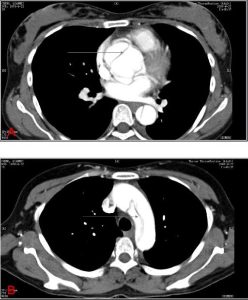
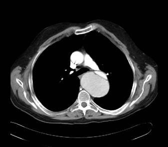
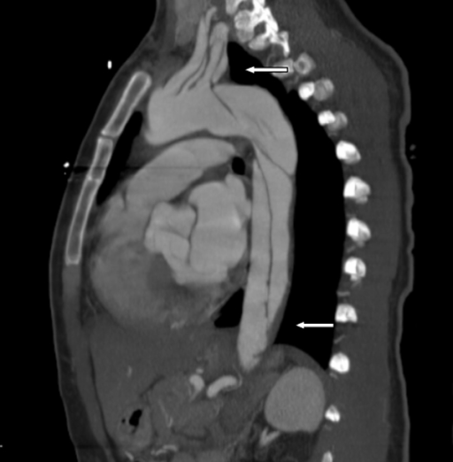
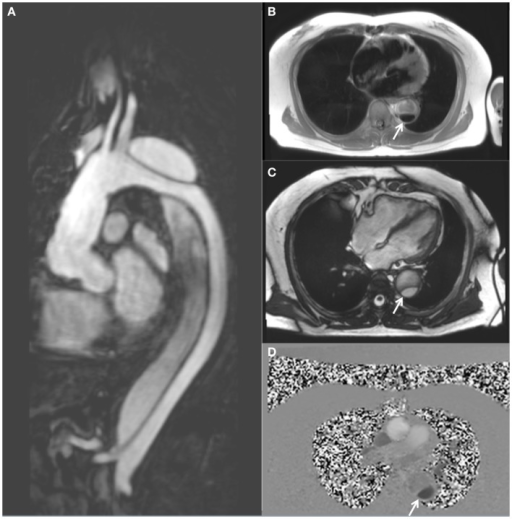
    • Findings: true and false lumen, intimal flap, intraluminal thrombus
  • Transesophageal echocardiography Echocardiography Ultrasonic recording of the size, motion, and composition of the heart and surrounding tissues. The standard approach is transthoracic. Tricuspid Valve Atresia (TVA) ( TEE TEE Ultrasonic recording of the size, motion, and composition of the heart and surrounding tissues using a transducer placed in the esophagus. Imaging of the Heart and Great Vessels)
  • Magnetic resonance angiography Angiography Radiography of blood vessels after injection of a contrast medium. Cardiac Surgery ( MRA MRA Imaging of the Heart and Great Vessels): an alternative to CT angiogram CT angiogram A non-invasive method that uses a ct scanner for capturing images of blood vessels and tissues. A contrast material is injected, which helps produce detailed images that aid in diagnosing vascular diseases. Pulmonary Function Tests

Management

Acute management

  • 2 large-bore intravenous (IV) lines
  • Cardiac and arterial blood pressure monitoring
  • Supplemental oxygen Supplemental Oxygen Respiratory Failure
  • Analgesia Analgesia Methods of pain relief that may be used with or in place of analgesics. Anesthesiology: History and Basic Concepts
  • Anti-impulse therapy (minimize shear stress):
    • Target systolic blood pressure < 120 mm Hg 
    • Target heart rate Heart rate The number of times the heart ventricles contract per unit of time, usually per minute. Cardiac Physiology < 60/min
    • 1st line: IV beta-blockers Beta-blockers Drugs that bind to but do not activate beta-adrenergic receptors thereby blocking the actions of beta-adrenergic agonists. Adrenergic beta-antagonists are used for treatment of hypertension, cardiac arrhythmias, angina pectoris, glaucoma, migraine headaches, and anxiety. Class 2 Antiarrhythmic Drugs (Beta Blockers) ( esmolol Esmolol Antiadrenergic Drugs, labetalol Labetalol A salicylamide derivative that is a non-cardioselective blocker of beta-adrenergic receptors and alpha-1 adrenergic receptors. Subarachnoid Hemorrhage) to prevent reflex tachycardia Tachycardia Abnormally rapid heartbeat, usually with a heart rate above 100 beats per minute for adults. Tachycardia accompanied by disturbance in the cardiac depolarization (cardiac arrhythmia) is called tachyarrhythmia. Sepsis in Children
    • Alternatives: IV nitroprusside Nitroprusside A powerful vasodilator used in emergencies to lower blood pressure or to improve cardiac function. It is also an indicator for free sulfhydryl groups in proteins. Nitrates or calcium Calcium A basic element found in nearly all tissues. It is a member of the alkaline earth family of metals with the atomic symbol ca, atomic number 20, and atomic weight 40. Calcium is the most abundant mineral in the body and combines with phosphorus to form calcium phosphate in the bones and teeth. It is essential for the normal functioning of nerves and muscles and plays a role in blood coagulation (as factor IV) and in many enzymatic processes. Electrolytes channel blockers 
  • For hypotension Hypotension Hypotension is defined as low blood pressure, specifically < 90/60 mm Hg, and is most commonly a physiologic response. Hypotension may be mild, serious, or life threatening, depending on the cause. Hypotension:

Type A dissections

Type A dissections require emergent surgical intervention for replacement of the involved ascending aorta Ascending aorta Mediastinum and Great Vessels: Anatomy or the entire ascending aorta Ascending aorta Mediastinum and Great Vessels: Anatomy with Dacron graft Graft A piece of living tissue that is surgically transplanted Organ Transplantation.

Type B dissections

  • Can be managed with anti-impulse therapy
  • May require surgical intervention with open versus endovascular repair for the following scenarios:

Long-term management

  • Oral antihypertensive therapy (lifelong)
  • Avoid strenuous physical activity.
  • Young patients Patients Individuals participating in the health care system for the purpose of receiving therapeutic, diagnostic, or preventive procedures. Clinician–Patient Relationship may be screened for genetic conditions.
  • Serial follow-up imaging to monitor for:
    • Dissection extension Extension Examination of the Upper Limbs or recurrence
    • Aneurysm Aneurysm An aneurysm is a bulging, weakened area of a blood vessel that causes an abnormal widening of its diameter > 1.5 times the size of the native vessel. Aneurysms occur more often in arteries than in veins and are at risk of dissection and rupture, which can be life-threatening. Thoracic Aortic Aneurysms formation
    • Surgical anastamotic leakage

Differential Diagnosis

  • Myocardial infarction Myocardial infarction MI is ischemia and death of an area of myocardial tissue due to insufficient blood flow and oxygenation, usually from thrombus formation on a ruptured atherosclerotic plaque in the epicardial arteries. Clinical presentation is most commonly with chest pain, but women and patients with diabetes may have atypical symptoms. Myocardial Infarction: ischemia Ischemia A hypoperfusion of the blood through an organ or tissue caused by a pathologic constriction or obstruction of its blood vessels, or an absence of blood circulation. Ischemic Cell Damage of the myocardium Myocardium The muscle tissue of the heart. It is composed of striated, involuntary muscle cells connected to form the contractile pump to generate blood flow. Heart: Anatomy due to partial or complete obstruction of the coronary arteries Arteries Arteries are tubular collections of cells that transport oxygenated blood and nutrients from the heart to the tissues of the body. The blood passes through the arteries in order of decreasing luminal diameter, starting in the largest artery (the aorta) and ending in the small arterioles. Arteries are classified into 3 types: large elastic arteries, medium muscular arteries, and small arteries and arterioles. Arteries: Histology. Most often due to atherosclerosis Atherosclerosis Atherosclerosis is a common form of arterial disease in which lipid deposition forms a plaque in the blood vessel walls. Atherosclerosis is an incurable disease, for which there are clearly defined risk factors that often can be reduced through a change in lifestyle and behavior of the patient. Atherosclerosis of the coronary arteries Arteries Arteries are tubular collections of cells that transport oxygenated blood and nutrients from the heart to the tissues of the body. The blood passes through the arteries in order of decreasing luminal diameter, starting in the largest artery (the aorta) and ending in the small arterioles. Arteries are classified into 3 types: large elastic arteries, medium muscular arteries, and small arteries and arterioles. Arteries: Histology. Presentation can mimic a dissection, with acute-onset chest pain Pain An unpleasant sensation induced by noxious stimuli which are detected by nerve endings of nociceptive neurons. Pain: Types and Pathways. However, the pain Pain An unpleasant sensation induced by noxious stimuli which are detected by nerve endings of nociceptive neurons. Pain: Types and Pathways does not typically radiate to the back and gradually increases in severity, and is usually accompanied by elevated troponins, typical ischemic ECG ECG An electrocardiogram (ECG) is a graphic representation of the electrical activity of the heart plotted against time. Adhesive electrodes are affixed to the skin surface allowing measurement of cardiac impulses from many angles. The ECG provides 3-dimensional information about the conduction system of the heart, the myocardium, and other cardiac structures. Electrocardiogram (ECG) changes (ST elevations or depressions), and wall motion abnormalities on transthoracic echo.
  • Horner’s syndrome: interruption to the sympathetic chain innervating the eye due to compression Compression Blunt Chest Trauma, lesion of the neuron, or stroke. Characterized by a constricted pupil Pupil The pupil is the space within the eye that permits light to project onto the retina. Anatomically located in front of the lens, the pupil’s size is controlled by the surrounding iris. The pupil provides insight into the function of the central and autonomic nervous systems. Pupil: Physiology and Abnormalities ( miosis Miosis Pupil: Physiology and Abnormalities), drooping of the upper eyelid ( ptosis Ptosis Cranial Nerve Palsies), and absence of sweating of the face (anhidrosis). A chest X-ray X-ray Penetrating electromagnetic radiation emitted when the inner orbital electrons of an atom are excited and release radiant energy. X-ray wavelengths range from 1 pm to 10 nm. Hard x-rays are the higher energy, shorter wavelength x-rays. Soft x-rays or grenz rays are less energetic and longer in wavelength. The short wavelength end of the x-ray spectrum overlaps the gamma rays wavelength range. The distinction between gamma rays and x-rays is based on their radiation source. Pulmonary Function Tests should be ordered to evaluate for an apical lung mass Mass Three-dimensional lesion that occupies a space within the breast Imaging of the Breast.
  • Aortic regurgitation Regurgitation Gastroesophageal Reflux Disease (GERD): insufficiency of the aortic valve Aortic valve The valve between the left ventricle and the ascending aorta which prevents backflow into the left ventricle. Heart: Anatomy, causing the backflow of blood into the left ventricle following systole Systole Period of contraction of the heart, especially of the heart ventricles. Cardiac Cycle when the valve is closed. Characterized by soft, high-pitched, early diastolic, decrescendo murmur heard best at the 3rd intercostal space left of the sternal border. Transthoracic echocardiography Transthoracic Echocardiography Imaging of the Heart and Great Vessels is used to confirm the diagnosis.
  • Pericardial effusion Pericardial effusion Fluid accumulation within the pericardium. Serous effusions are associated with pericardial diseases. Hemopericardium is associated with trauma. Lipid-containing effusion (chylopericardium) results from leakage of thoracic duct. Severe cases can lead to cardiac tamponade. Pericardial Effusion and Cardiac Tamponade and tamponade Tamponade Pericardial effusion, usually of rapid onset, exceeding ventricular filling pressures and causing collapse of the heart with a markedly reduced cardiac output. Pericarditis: fluid in the pericardial sac, which can cause compression Compression Blunt Chest Trauma of the heart and lead to tamponade Tamponade Pericardial effusion, usually of rapid onset, exceeding ventricular filling pressures and causing collapse of the heart with a markedly reduced cardiac output. Pericarditis physiology. Tamponade Tamponade Pericardial effusion, usually of rapid onset, exceeding ventricular filling pressures and causing collapse of the heart with a markedly reduced cardiac output. Pericarditis physiology prevents the heart from filling, resulting in hemodynamic collapse. Beck’s triad Beck’s triad The triad describes the classic findings in cardiac tamponade: hypotension, jugular venous distension, muffled heart sounds on auscultation. Pericardial Effusion and Cardiac Tamponade of distended neck Neck The part of a human or animal body connecting the head to the rest of the body. Peritonsillar Abscess veins Veins Veins are tubular collections of cells, which transport deoxygenated blood and waste from the capillary beds back to the heart. Veins are classified into 3 types: small veins/venules, medium veins, and large veins. Each type contains 3 primary layers: tunica intima, tunica media, and tunica adventitia. Veins: Histology, hypotension Hypotension Hypotension is defined as low blood pressure, specifically < 90/60 mm Hg, and is most commonly a physiologic response. Hypotension may be mild, serious, or life threatening, depending on the cause. Hypotension, and muffled heart sounds Muffled Heart Sounds Pericardial Effusion and Cardiac Tamponade is often seen on physical exam. Diagnosis is confirmed with transthoracic echocardiography Transthoracic Echocardiography Imaging of the Heart and Great Vessels, and management involves drainage of the pericardium Pericardium A conical fibroserous sac surrounding the heart and the roots of the great vessels (aorta; venae cavae; pulmonary artery). Pericardium consists of two sacs: the outer fibrous pericardium and the inner serous pericardium. The latter consists of an outer parietal layer facing the fibrous pericardium, and an inner visceral layer (epicardium) resting next to the heart, and a pericardial cavity between these two layers. Heart: Anatomy.
  • Pneumothorax Pneumothorax A pneumothorax is a life-threatening condition in which air collects in the pleural space, causing partial or full collapse of the lung. A pneumothorax can be traumatic or spontaneous. Patients present with a sudden onset of sharp chest pain, dyspnea, and diminished breath sounds on exam. Pneumothorax: a life-threatening condition in which air collects in the pleural space Pleural space The thin serous membrane enveloping the lungs (lung) and lining the thoracic cavity. Pleura consist of two layers, the inner visceral pleura lying next to the pulmonary parenchyma and the outer parietal pleura. Between the two layers is the pleural cavity which contains a thin film of liquid. Pleuritis, causing partial or full collapse of the lung. Pneumothorax Pneumothorax A pneumothorax is a life-threatening condition in which air collects in the pleural space, causing partial or full collapse of the lung. A pneumothorax can be traumatic or spontaneous. Patients present with a sudden onset of sharp chest pain, dyspnea, and diminished breath sounds on exam. Pneumothorax can be traumatic or spontaneous. Patients Patients Individuals participating in the health care system for the purpose of receiving therapeutic, diagnostic, or preventive procedures. Clinician–Patient Relationship present with a sudden onset of sharp chest pain Sharp Chest Pain Chest Pain, dyspnea Dyspnea Dyspnea is the subjective sensation of breathing discomfort. Dyspnea is a normal manifestation of heavy physical or psychological exertion, but also may be caused by underlying conditions (both pulmonary and extrapulmonary). Dyspnea, and diminished breath sounds on exam. A large or tension pneumothorax Tension Pneumothorax Pneumothorax can result in cardiopulmonary collapse. Diagnosis is made with imaging. Management includes needle decompression Needle Decompression Pneumothorax and chest tube ( thoracostomy Thoracostomy Surgical procedure involving the creation of an opening (stoma) into the chest cavity for drainage; used in the treatment of pleural effusion; pneumothorax; hemothorax; and empyema. Hemothorax) placement.
  • Pulmonary embolism Pulmonary Embolism Pulmonary embolism (PE) is a potentially fatal condition that occurs as a result of intraluminal obstruction of the main pulmonary artery or its branches. The causative factors include thrombi, air, amniotic fluid, and fat. In PE, gas exchange is impaired due to the decreased return of deoxygenated blood to the lungs. Pulmonary Embolism: obstruction of the pulmonary arteries Arteries Arteries are tubular collections of cells that transport oxygenated blood and nutrients from the heart to the tissues of the body. The blood passes through the arteries in order of decreasing luminal diameter, starting in the largest artery (the aorta) and ending in the small arterioles. Arteries are classified into 3 types: large elastic arteries, medium muscular arteries, and small arteries and arterioles. Arteries: Histology, most often due to thrombus migration from the deep venous system. Signs and symptoms include pleuritic chest pain Pain An unpleasant sensation induced by noxious stimuli which are detected by nerve endings of nociceptive neurons. Pain: Types and Pathways, dyspnea Dyspnea Dyspnea is the subjective sensation of breathing discomfort. Dyspnea is a normal manifestation of heavy physical or psychological exertion, but also may be caused by underlying conditions (both pulmonary and extrapulmonary). Dyspnea, tachypnea Tachypnea Increased respiratory rate. Pulmonary Examination, and tachycardia Tachycardia Abnormally rapid heartbeat, usually with a heart rate above 100 beats per minute for adults. Tachycardia accompanied by disturbance in the cardiac depolarization (cardiac arrhythmia) is called tachyarrhythmia. Sepsis in Children. Severe cases can result in hemodynamic instability or cardiopulmonary arrest Cardiopulmonary arrest Cardiac arrest is the sudden, complete cessation of cardiac output with hemodynamic collapse. Patients present as pulseless, unresponsive, and apneic. Rhythms associated with cardiac arrest are ventricular fibrillation/tachycardia, asystole, or pulseless electrical activity. Cardiac Arrest. A chest CT with angiography Angiography Radiography of blood vessels after injection of a contrast medium. Cardiac Surgery is the primary method of diagnosis. Management includes oxygenation, anticoagulation Anticoagulation Pulmonary Hypertension Drugs, and thrombolytic therapy for unstable patients Unstable Patients Blunt Chest Trauma.

References

  1. Mancini, M.C. (2020). Aortic dissection. In Geibel, J. (Ed.), Medscape. Retrieved January 17, 2021, from https://emedicine.medscape.com/article/2062452-overview
  2. Black, III, J.H., and Manning, W.J. (2020). Overview of acute aortic dissection and other acute aortic syndromes. In Collins, K.A. (Ed.), UpToDate. Retrieved January 17, 2021, from https://www.uptodate.com/contents/overview-of-acute-aortic-dissection-and-other-acute-aortic-syndromes
  3. Black, III, J.H., and Manning W.J. (2020). Clinical features of acute aortic dissection. In Collins, K.A. (Ed.), UpToDate. Retrieved January 19, 2021, from https://www.uptodate.com/contents/clinical-features-and-diagnosis-of-acute-aortic-dissection
  4. Black, III, J.H., and Manning, W.J. (2019). Management of acute aortic dissection. In Collins, K.A. (Ed.), UpToDate. Retrieved January 19, 2021, from https://www.uptodate.com/contents/management-of-acute-aortic-dissection
  5. Farber, M.A. (2020). Aortic dissection. [online] MSD Manual Professional Version. Retrieved January 19, 2021, from https://www.merckmanuals.com/professional/cardiovascular-disorders/diseases-of-the-aorta-and-its-branches/aortic-dissection
  6. Wang, S.S. (2018). Aortic regurgitation workup. In O’Brien, T. X. (Ed.), Medscape. Retrieved January 18, 2021, from https://emedicine.medscape.com/article/150490-workup
  7. Yarlagadda, C. (2018). Cardiac tamponade. In O’Brien, T. X. (Ed.), Medscape. Retrieved January 18, 2021, from https://emedicine.medscape.com/article/152083-overview

Create your free account or log in to continue reading!

Sign up now and get free access to Lecturio with concept pages, medical videos, and questions for your medical education.

User Reviews

Details