Advertisement

Advertisement

Advertisement

Advertisement

Anticoagulants

Anticoagulants are drugs that retard or interrupt the coagulation cascade Coagulation cascade The coagulation cascade is a series of reactions that ultimately generates a strong, cross-linked fibrin clot. Hemostasis. The primary classes of available anticoagulants include heparins, vitamin K-dependent antagonists (e.g., warfarin), direct thrombin Thrombin An enzyme formed from prothrombin that converts fibrinogen to fibrin. Hemostasis inhibitors, and factor Xa inhibitors. Anticoagulants are used in the treatment and prevention of thrombotic and embolic diseases including cardioembolic ischemic stroke Ischemic Stroke An ischemic stroke (also known as cerebrovascular accident) is an acute neurologic injury that occurs as a result of brain ischemia; this condition may be due to cerebral blood vessel occlusion by thrombosis or embolism, or rarely due to systemic hypoperfusion. Ischemic Stroke, acute coronary syndrome, and venous thromboembolism Thromboembolism Obstruction of a blood vessel (embolism) by a blood clot (thrombus) in the blood stream. Systemic Lupus Erythematosus, among other conditions. Patients Patients Individuals participating in the health care system for the purpose of receiving therapeutic, diagnostic, or preventive procedures. Clinician–Patient Relationship with atrial fibrillation Atrial fibrillation Atrial fibrillation (AF or Afib) is a supraventricular tachyarrhythmia and the most common kind of arrhythmia. It is caused by rapid, uncontrolled atrial contractions and uncoordinated ventricular responses. Atrial Fibrillation or thrombophilias Thrombophilias Hypercoagulable states (also referred to as thrombophilias) are a group of hematologic diseases defined by an increased risk of clot formation (i.e., thrombosis) due to either an increase in procoagulants, a decrease in anticoagulants, or a decrease in fibrinolysis. Hypercoagulable States may require indefinite or lifelong anticoagulation Anticoagulation Pulmonary Hypertension Drugs. Accordingly, the route of administration, drug interactions, pharmacokinetics Pharmacokinetics Pharmacokinetics is the science that analyzes how the human body interacts with a drug. Pharmacokinetics examines how the drug is absorbed, distributed, metabolized, and excreted by the body. Pharmacokinetics and Pharmacodynamics, and availability of reversal factors should be considered while selecting the anticoagulant therapy.

Last updated: Jan 17, 2024

Editorial responsibility: Stanley Oiseth, Lindsay Jones, Evelin Maza

Advertisement

Advertisement

Advertisement

Advertisement

Advertisement

Advertisement

Overview

Definition

Anticoagulants are a category of drugs that inhibit the coagulation cascade Coagulation cascade The coagulation cascade is a series of reactions that ultimately generates a strong, cross-linked fibrin clot. Hemostasis.

General indications

Anticoagulants are indicated in the treatment and prophylaxis Prophylaxis Cephalosporins of thrombotic events including:

  • Venous thromboembolism Thromboembolism Obstruction of a blood vessel (embolism) by a blood clot (thrombus) in the blood stream. Systemic Lupus Erythematosus ( VTE VTE Obstruction of a vein or veins (embolism) by a blood clot (thrombus) in the bloodstream. Hypercoagulable States)
  • Arterial thrombosis Arterial Thrombosis Hypercoagulable States
    • MI MI MI is ischemia and death of an area of myocardial tissue due to insufficient blood flow and oxygenation, usually from thrombus formation on a ruptured atherosclerotic plaque in the epicardial arteries. Clinical presentation is most commonly with chest pain, but women and patients with diabetes may have atypical symptoms. Myocardial Infarction
    • Stroke 
  • Atrial fibrillation Atrial fibrillation Atrial fibrillation (AF or Afib) is a supraventricular tachyarrhythmia and the most common kind of arrhythmia. It is caused by rapid, uncontrolled atrial contractions and uncoordinated ventricular responses. Atrial Fibrillation ( AFib Afib Atrial fibrillation (AF or Afib) is a supraventricular tachyarrhythmia and the most common kind of arrhythmia. It is caused by rapid, uncontrolled atrial contractions and uncoordinated ventricular responses. Atrial Fibrillation)
  • After a heart valve replacement
  • Thrombophilias Thrombophilias Hypercoagulable states (also referred to as thrombophilias) are a group of hematologic diseases defined by an increased risk of clot formation (i.e., thrombosis) due to either an increase in procoagulants, a decrease in anticoagulants, or a decrease in fibrinolysis. Hypercoagulable States

Classification

There are several primary classes of anticoagulants:

  • Heparins:
    • Unfractionated heparin
    • Low-molecular-weight heparins (LMWHs)
  • Vitamin K Vitamin K A lipid cofactor that is required for normal blood clotting. Several forms of vitamin K have been identified: vitamin K 1 (phytomenadione) derived from plants, vitamin K 2 (menaquinone) from bacteria, and synthetic naphthoquinone provitamins, vitamin K 3 (menadione). Vitamin k 3 provitamins, after being alkylated in vivo, exhibit the antifibrinolytic activity of vitamin k. Green leafy vegetables, liver, cheese, butter, and egg yolk are good sources of vitamin k. Fat-soluble Vitamins and their Deficiencies antagonists
  • Direct thrombin Thrombin An enzyme formed from prothrombin that converts fibrinogen to fibrin. Hemostasis inhibitors
  • Factor Xa inhibitors:
    • Direct factor Xa inhibitors
    • Indirect factor Xa inhibitors

Physiology: Overview of the coagulation cascade Coagulation cascade The coagulation cascade is a series of reactions that ultimately generates a strong, cross-linked fibrin clot. Hemostasis

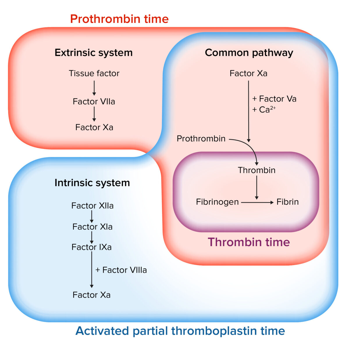
The coagulation cascade Coagulation cascade The coagulation cascade is a series of reactions that ultimately generates a strong, cross-linked fibrin clot. Hemostasis is a series of reactions that ultimately generates a strong, cross-linked fibrin Fibrin A protein derived from fibrinogen in the presence of thrombin, which forms part of the blood clot. Rapidly Progressive Glomerulonephritis clot. This cascade is also known as secondary hemostasis Secondary hemostasis The coagulation cascade is a series of reactions that ultimately generates a strong, cross-linked fibrin clot. Hemostasis.

Coagulation factors Coagulation factors Endogenous substances, usually proteins, that are involved in the blood coagulation process. Hemostasis:

A number of coagulation factors Coagulation factors Endogenous substances, usually proteins, that are involved in the blood coagulation process. Hemostasis undergo sequential Sequential Computed Tomography (CT) activation down 1 of 2 pathways:

  • Extrinsic pathway Extrinsic pathway The extrinsic pathway is the primary physiological mechanism by which clotting is initiated Hemostasis:
    • Primarily responsible for initiation of the cascade
    • Involves tissue factor and factor VII Factor VII Heat- and storage-stable plasma protein that is activated by tissue thromboplastin to form factor viia in the extrinsic pathway of blood coagulation. The activated form then catalyzes the activation of factor X to factor Xa. Hemostasis
    • Assessed by the PT
  • Intrinsic pathway Intrinsic pathway The intrinsic pathway is mainly responsible for the amplification of factor X activation Hemostasis:
    • Primarily involved in amplification of the cascade
    • Involves factors XII, XI, IX, and VIII
    • Assessed by the aPTT

Common pathway Common pathway Hemostasis:
  • The extrinsic and intrinsic pathways join together when factor X Factor X Storage-stable glycoprotein blood coagulation factor that can be activated to factor Xa by both the intrinsic and extrinsic pathways. A deficiency of factor X, sometimes called stuart-prower factor deficiency, may lead to a systemic coagulation disorder. Hemostasis is activated to factor Xa at the beginning of the final common pathway Common pathway Hemostasis.
  • Involves factors X, V, II ( prothrombin Prothrombin A plasma protein that is the inactive precursor of thrombin. It is converted to thrombin by a prothrombin activator complex consisting of factor Xa, factor V, phospholipid, and calcium ions. Hemostasis), and I ( fibrinogen Fibrinogen Plasma glycoprotein clotted by thrombin, composed of a dimer of three non-identical pairs of polypeptide chains (alpha, beta, gamma) held together by disulfide bonds. Fibrinogen clotting is a sol-gel change involving complex molecular arrangements: whereas fibrinogen is cleaved by thrombin to form polypeptides a and b, the proteolytic action of other enzymes yields different fibrinogen degradation products. Hemostasis)
  • Prothrombinase Prothrombinase Hemostasis complex: 
    • A procoagulant, multi-component enzyme complex involving factor Xa (the protease Protease Enzyme of the human immunodeficiency virus that is required for post-translational cleavage of gag and gag-pol precursor polyproteins into functional products needed for viral assembly. HIV protease is an aspartic protease encoded by the amino terminus of the pol gene. HIV Infection and AIDS), factor Va VA Ventilation: Mechanics of Breathing (the cofactor), and prothrombin Prothrombin A plasma protein that is the inactive precursor of thrombin. It is converted to thrombin by a prothrombin activator complex consisting of factor Xa, factor V, phospholipid, and calcium ions. Hemostasis (the substrate Substrate A substance upon which the enzyme acts. Basics of Enzymes)
    • Activation of prothrombin Prothrombin A plasma protein that is the inactive precursor of thrombin. It is converted to thrombin by a prothrombin activator complex consisting of factor Xa, factor V, phospholipid, and calcium ions. Hemostasis (factor II) → thrombin Thrombin An enzyme formed from prothrombin that converts fibrinogen to fibrin. Hemostasis (factor IIa)
  • Thrombin Thrombin An enzyme formed from prothrombin that converts fibrinogen to fibrin. Hemostasis converts fibrinogen Fibrinogen Plasma glycoprotein clotted by thrombin, composed of a dimer of three non-identical pairs of polypeptide chains (alpha, beta, gamma) held together by disulfide bonds. Fibrinogen clotting is a sol-gel change involving complex molecular arrangements: whereas fibrinogen is cleaved by thrombin to form polypeptides a and b, the proteolytic action of other enzymes yields different fibrinogen degradation products. Hemostasis fibrin Fibrin A protein derived from fibrinogen in the presence of thrombin, which forms part of the blood clot. Rapidly Progressive Glomerulonephritis, which is able to form a stable clot
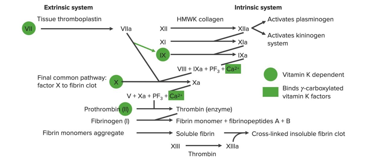
Overview of the coagulation cascade

Overview of the coagulation cascade
a: activated form
PF3: platelet factor 3 (phospholipids)
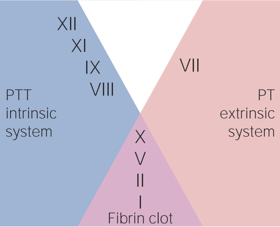
Image by Lecturio.

Related videos

Heparins

Natural heparins are a group of large, endogenously produced polysaccharides Polysaccharides Basics of Carbohydrates of varying sizes that are not completely understood. Heparins have anticoagulant, anti-inflammatory, and possibly anti-angiogenic effects.

Table: Heparins
UFH LMWH
Agents UFH
  • Enoxaparin (Lovenox®)
  • Dalteparin (Fragmin®)
Mechanism of action Binds to and potentiates antithrombin
  • Heparin induces a conformational change in antithrombin, which ↑ its activity 1,000‒4,000 fold
  • Antithrombin inactivates:
    • Factor Xa
    • Thrombin Thrombin An enzyme formed from prothrombin that converts fibrinogen to fibrin. Hemostasis (Factor IIa)
  • Inactivation of thrombin Thrombin An enzyme formed from prothrombin that converts fibrinogen to fibrin. Hemostasis requires larger heparin molecules → UFH has ↑↑ thrombin-inactivation effects compared to LMWHs
Physiologic effects Inactivation of Factor Xa and thrombin Thrombin An enzyme formed from prothrombin that converts fibrinogen to fibrin. Hemostasis Inactivation of Factor Xa and, to a much lesser extent, thrombin Thrombin An enzyme formed from prothrombin that converts fibrinogen to fibrin. Hemostasis
Absorption Absorption Absorption involves the uptake of nutrient molecules and their transfer from the lumen of the GI tract across the enterocytes and into the interstitial space, where they can be taken up in the venous or lymphatic circulation. Digestion and Absorption Administered IV (rarely SC):
  • IV absorption Absorption Absorption involves the uptake of nutrient molecules and their transfer from the lumen of the GI tract across the enterocytes and into the interstitial space, where they can be taken up in the venous or lymphatic circulation. Digestion and Absorption is immediate
  • SC absorption Absorption Absorption involves the uptake of nutrient molecules and their transfer from the lumen of the GI tract across the enterocytes and into the interstitial space, where they can be taken up in the venous or lymphatic circulation. Digestion and Absorption is variable Variable Variables represent information about something that can change. The design of the measurement scales, or of the methods for obtaining information, will determine the data gathered and the characteristics of that data. As a result, a variable can be qualitative or quantitative, and may be further classified into subgroups. Types of Variables: peak activity at 2‒4 hours
Distribution Vd = ~ 35 mL/kg Vd = 4.3 L
Metabolism
  • Liver Liver The liver is the largest gland in the human body. The liver is found in the superior right quadrant of the abdomen and weighs approximately 1.5 kilograms. Its main functions are detoxification, metabolism, nutrient storage (e.g., iron and vitamins), synthesis of coagulation factors, formation of bile, filtration, and storage of blood. Liver: Anatomy
  • Reticuloendothelial system
Liver Liver The liver is the largest gland in the human body. The liver is found in the superior right quadrant of the abdomen and weighs approximately 1.5 kilograms. Its main functions are detoxification, metabolism, nutrient storage (e.g., iron and vitamins), synthesis of coagulation factors, formation of bile, filtration, and storage of blood. Liver: Anatomy
Elimination Elimination The initial damage and destruction of tumor cells by innate and adaptive immunity. Completion of the phase means no cancer growth. Cancer Immunotherapy
  • Renally excreted
  • Elimination Elimination The initial damage and destruction of tumor cells by innate and adaptive immunity. Completion of the phase means no cancer growth. Cancer Immunotherapy may be ↓ in patients Patients Individuals participating in the health care system for the purpose of receiving therapeutic, diagnostic, or preventive procedures. Clinician–Patient Relationship with kidney disease
  • Longer half-life Half-Life The time it takes for a substance (drug, radioactive nuclide, or other) to lose half of its pharmacologic, physiologic, or radiologic activity. Pharmacokinetics and Pharmacodynamics than UFH: Enoxaparin: 4.5‒7 hours
Monitoring
  • aPTT
  • Anti-factor Xa activity
  • Activated clotting time
  • Factor Xa levels
  • Note: aPTT is not reliable
Reversal agent Protamine sulfate Protamine sulfate
Complications
  • Transient thrombocytopenia Thrombocytopenia Thrombocytopenia occurs when the platelet count is < 150,000 per microliter. The normal range for platelets is usually 150,000-450,000/µL of whole blood. Thrombocytopenia can be a result of decreased production, increased destruction, or splenic sequestration of platelets. Patients are often asymptomatic until platelet counts are < 50,000/µL. Thrombocytopenia
  • HITT
  • Osteoporosis Osteoporosis Osteoporosis refers to a decrease in bone mass and density leading to an increased number of fractures. There are 2 forms of osteoporosis: primary, which is commonly postmenopausal or senile; and secondary, which is a manifestation of immobilization, underlying medical disorders, or long-term use of certain medications. Osteoporosis with chronic use
Lower risks of HITT and osteoporosis Osteoporosis Osteoporosis refers to a decrease in bone mass and density leading to an increased number of fractures. There are 2 forms of osteoporosis: primary, which is commonly postmenopausal or senile; and secondary, which is a manifestation of immobilization, underlying medical disorders, or long-term use of certain medications. Osteoporosis than UFH
Specific contraindications Contraindications A condition or factor associated with a recipient that makes the use of a drug, procedure, or physical agent improper or inadvisable. Contraindications may be absolute (life threatening) or relative (higher risk of complications in which benefits may outweigh risks). Noninvasive Ventilation
Notes
  • Highly acidic → will be neutralized by a base (e.g., protamine sulfate)
  • A mixture of large molecules with varying sizes
  • Isolated from porcine or bovine intestines
  • Unlike direct thrombin Thrombin An enzyme formed from prothrombin that converts fibrinogen to fibrin. Hemostasis inhibitors, does not affect thrombin Thrombin An enzyme formed from prothrombin that converts fibrinogen to fibrin. Hemostasis already within clots
  • Very predictable effects → monitoring is rarely needed
  • Derived from UFH
UFH: unfractionated heparin
LMWH: low-molecular-weight heparin
HITT: heparin-induced thrombocytopenia and thrombosis

Vitamin K-Dependent Antagonists: Warfarin

Table: Vitamin K-dependant antagonists: Warfarin (Coumadin®)
Mechanism of action
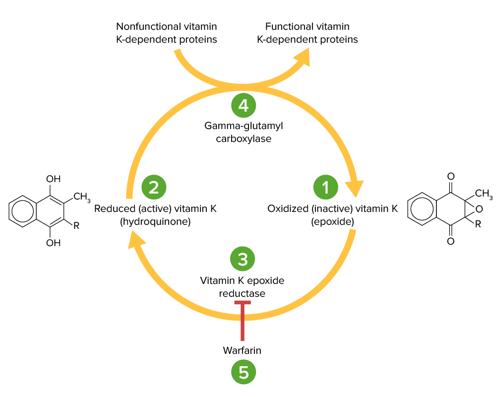
  • Competitively inhibits vitamin K Vitamin K A lipid cofactor that is required for normal blood clotting. Several forms of vitamin K have been identified: vitamin K 1 (phytomenadione) derived from plants, vitamin K 2 (menaquinone) from bacteria, and synthetic naphthoquinone provitamins, vitamin K 3 (menadione). Vitamin k 3 provitamins, after being alkylated in vivo, exhibit the antifibrinolytic activity of vitamin k. Green leafy vegetables, liver, cheese, butter, and egg yolk are good sources of vitamin k. Fat-soluble Vitamins and their Deficiencies epoxide reductase → depletes “active” vitamin K Vitamin K A lipid cofactor that is required for normal blood clotting. Several forms of vitamin K have been identified: vitamin K 1 (phytomenadione) derived from plants, vitamin K 2 (menaquinone) from bacteria, and synthetic naphthoquinone provitamins, vitamin K 3 (menadione). Vitamin k 3 provitamins, after being alkylated in vivo, exhibit the antifibrinolytic activity of vitamin k. Green leafy vegetables, liver, cheese, butter, and egg yolk are good sources of vitamin k. Fat-soluble Vitamins and their Deficiencies reserves required for the formation of vitamin K-dependent factors:
    • Procoagulant factors II, VII, IX, X
    • Anticoagulant proteins Proteins Linear polypeptides that are synthesized on ribosomes and may be further modified, crosslinked, cleaved, or assembled into complex proteins with several subunits. The specific sequence of amino acids determines the shape the polypeptide will take, during protein folding, and the function of the protein. Energy Homeostasis C and S
  • Note: Because Proteins Proteins Linear polypeptides that are synthesized on ribosomes and may be further modified, crosslinked, cleaved, or assembled into complex proteins with several subunits. The specific sequence of amino acids determines the shape the polypeptide will take, during protein folding, and the function of the protein. Energy Homeostasis C and S have a shorter half-life Half-Life The time it takes for a substance (drug, radioactive nuclide, or other) to lose half of its pharmacologic, physiologic, or radiologic activity. Pharmacokinetics and Pharmacodynamics than the procoagulant factors, patients Patients Individuals participating in the health care system for the purpose of receiving therapeutic, diagnostic, or preventive procedures. Clinician–Patient Relationship develop a transient hypercoagulable Hypercoagulable Hypercoagulable states (also referred to as thrombophilias) are a group of hematologic diseases defined by an increased risk of clot formation (i.e., thrombosis) due to either an increase in procoagulants, a decrease in anticoagulants, or a decrease in fibrinolysis. Hypercoagulable States state for several days after warfarin therapy.
    • Patients Patients Individuals participating in the health care system for the purpose of receiving therapeutic, diagnostic, or preventive procedures. Clinician–Patient Relationship typically require coadministration of an additional anticoagulant until a therapeutic INR is achieved.
Absorption Absorption Absorption involves the uptake of nutrient molecules and their transfer from the lumen of the GI tract across the enterocytes and into the interstitial space, where they can be taken up in the venous or lymphatic circulation. Digestion and Absorption
  • Completely absorbed orally
  • Peak activity approximately 4 hours
Metabolism Metabolized in liver Liver The liver is the largest gland in the human body. The liver is found in the superior right quadrant of the abdomen and weighs approximately 1.5 kilograms. Its main functions are detoxification, metabolism, nutrient storage (e.g., iron and vitamins), synthesis of coagulation factors, formation of bile, filtration, and storage of blood. Liver: Anatomy:
  • Primarily via CYP2C9
  • Minor pathways via CYP2C8 CYP2C8 A liver microsomal cytochrome p450 hydroxylase that oxidizes a broad spectrum of substrates including steroids, fatty acids, and xenobiotics. Examples of pharmaceutical substrates for cyp2c8 include; paclitaxol; torsemide; and; amodiaquine. Class 3 Antiarrhythmic Drugs (Potassium Channel Blockers), 2C18, 2C19, 1A2, and 3A4
Distribution
  • Vd = 0.14 L/kg
  • Protein binding: 99%
Elimination Elimination The initial damage and destruction of tumor cells by innate and adaptive immunity. Completion of the phase means no cancer growth. Cancer Immunotherapy
  • Renally excreted as metabolites
  • Half-life Half-Life The time it takes for a substance (drug, radioactive nuclide, or other) to lose half of its pharmacologic, physiologic, or radiologic activity. Pharmacokinetics and Pharmacodynamics: 20‒60 hours (highly variable Variable Variables represent information about something that can change. The design of the measurement scales, or of the methods for obtaining information, will determine the data gathered and the characteristics of that data. As a result, a variable can be qualitative or quantitative, and may be further classified into subgroups. Types of Variables)
Monitoring
Reversal agent/ antidote Antidote An antidote is a substance that counteracts poisoning or toxicity. Substances that can cause poisoning include heavy metals (from occupation, treatments, or diet), alcohols, environmental toxins, and medications. Antidotes of Common Poisonings Vitamin K Vitamin K A lipid cofactor that is required for normal blood clotting. Several forms of vitamin K have been identified: vitamin K 1 (phytomenadione) derived from plants, vitamin K 2 (menaquinone) from bacteria, and synthetic naphthoquinone provitamins, vitamin K 3 (menadione). Vitamin k 3 provitamins, after being alkylated in vivo, exhibit the antifibrinolytic activity of vitamin k. Green leafy vegetables, liver, cheese, butter, and egg yolk are good sources of vitamin k. Fat-soluble Vitamins and their Deficiencies (takes several hours for effect)
Interactions Warfarin has numerous drug, herbal, and dietary interactions:
  • CYP450 inducers (e.g., carbamazepine Carbamazepine A dibenzazepine that acts as a sodium channel blocker. It is used as an anticonvulsant for the treatment of grand mal and psychomotor or focal seizures. It may also be used in the management of bipolar disorder, and has analgesic properties. First-Generation Anticonvulsant Drugs, phenytoin Phenytoin An anticonvulsant that is used to treat a wide variety of seizures. The mechanism of therapeutic action is not clear, although several cellular actions have been described including effects on ion channels, active transport, and general membrane stabilization. Phenytoin has been proposed for several other therapeutic uses, but its use has been limited by its many adverse effects and interactions with other drugs. First-Generation Anticonvulsant Drugs, barbiturates Barbiturates A class of chemicals derived from barbituric acid or thiobarbituric acid. Many of these are gaba modulators used as hypnotics and sedatives, as anesthetics, or as anticonvulsants. Intravenous Anesthetics, rifampin Rifampin A semisynthetic antibiotic produced from streptomyces mediterranei. It has a broad antibacterial spectrum, including activity against several forms of Mycobacterium. In susceptible organisms it inhibits dna-dependent RNA polymerase activity by forming a stable complex with the enzyme. It thus suppresses the initiation of RNA synthesis. Rifampin is bactericidal, and acts on both intracellular and extracellular organisms. Epiglottitis) → ↑ clearance → ↓ INR
  • CYP450 inhibitors (e.g., amiodarone Amiodarone An antianginal and class III antiarrhythmic drug. It increases the duration of ventricular and atrial muscle action by inhibiting potassium channels and voltage-gated sodium channels. There is a resulting decrease in heart rate and in vascular resistance. Pulmonary Fibrosis, selective-serotonin reuptake inhibitors) → ↓ clearance → ↑ INR
  • Broad-spectrum Broad-Spectrum Fluoroquinolones antibiotics: kill normal gut flora Normal gut flora All of the microbial organisms that naturally exist within the gastrointestinal tract. Escherichia coli that biosynthesize vitamin K Vitamin K A lipid cofactor that is required for normal blood clotting. Several forms of vitamin K have been identified: vitamin K 1 (phytomenadione) derived from plants, vitamin K 2 (menaquinone) from bacteria, and synthetic naphthoquinone provitamins, vitamin K 3 (menadione). Vitamin k 3 provitamins, after being alkylated in vivo, exhibit the antifibrinolytic activity of vitamin k. Green leafy vegetables, liver, cheese, butter, and egg yolk are good sources of vitamin k. Fat-soluble Vitamins and their Deficiencies vitamin K deficiency Vitamin K Deficiency A nutritional condition produced by a deficiency of vitamin K in the diet, characterized by an increased tendency to hemorrhage (hemorrhagic disorders). Such bleeding episodes may be particularly severe in newborn infants. Fat-soluble Vitamins and their Deficiencies → ↑ INR
Specific contraindications Contraindications A condition or factor associated with a recipient that makes the use of a drug, procedure, or physical agent improper or inadvisable. Contraindications may be absolute (life threatening) or relative (higher risk of complications in which benefits may outweigh risks). Noninvasive Ventilation Pregnancy Pregnancy The status during which female mammals carry their developing young (embryos or fetuses) in utero before birth, beginning from fertilization to birth. Pregnancy: Diagnosis, Physiology, and Care (warfarin is teratogenic)
Complications
Notes Patients Patients Individuals participating in the health care system for the purpose of receiving therapeutic, diagnostic, or preventive procedures. Clinician–Patient Relationship with CYP2C9 variants have enzyme activity → require dose

Vitamin k cycle

Vitamin K cycle:
Vitamin K epoxide (1) is inactive and converted to its active, reduced form, vitamin K hydroquinone (2), by vitamin K epoxide reductase (VKOR; 3). Vitamin K hydroquinone is a cofactor in the carboxylation of specific glutamate residues within the vitamin K-dependent proteins (factors II, VII, IX, X, protein C and S), a process which is necessary to activate them. The carboxylation reaction is catalyzed by gamma-glutamyl carboxylase (4). Vitamin K hydroquinone is oxidized to the epoxide form when it acts as a cofactor, but is then recycled back to the hydroquinone form by VKOR. Warfarin inhibits VKOR (5) so that vitamin K cannot be recycled from its oxidized form to the reduced form. Thus, vitamin K-dependent proteins cannot be activated.

Image by Lecturio.

Related videos

Direct Thrombin Inhibitors

Table: Direct thrombin Thrombin An enzyme formed from prothrombin that converts fibrinogen to fibrin. Hemostasis inhibitors
Oral agents (DOAC) Parenteral agents
Agents Dabigatran (Pradaxa®)
  • Bivalirudin (Angiomax®)
  • Argatroban (Argatra®)
  • Lepirudin (discontinued)
Mechanism of action Binds to and functionally inhibits thrombin Thrombin An enzyme formed from prothrombin that converts fibrinogen to fibrin. Hemostasis in both serum and clots
Absorption Absorption Absorption involves the uptake of nutrient molecules and their transfer from the lumen of the GI tract across the enterocytes and into the interstitial space, where they can be taken up in the venous or lymphatic circulation. Digestion and Absorption Onset of action: immediate
Distribution
  • Vd = 50‒70 L
  • Protein binding: 35%
Bivalirudin:
  • Vd = 240 mL/kg
  • Protein binding 0%
Argatroban:
  • Vd = 174 mL/kg
  • Protein binding: 54%
Metabolism Liver Liver The liver is the largest gland in the human body. The liver is found in the superior right quadrant of the abdomen and weighs approximately 1.5 kilograms. Its main functions are detoxification, metabolism, nutrient storage (e.g., iron and vitamins), synthesis of coagulation factors, formation of bile, filtration, and storage of blood. Liver: Anatomy
  • Bivalirudin: kidney, liver Liver The liver is the largest gland in the human body. The liver is found in the superior right quadrant of the abdomen and weighs approximately 1.5 kilograms. Its main functions are detoxification, metabolism, nutrient storage (e.g., iron and vitamins), synthesis of coagulation factors, formation of bile, filtration, and storage of blood. Liver: Anatomy, and other sites (proteolytic cleavage)
  • Argatroban: liver Liver The liver is the largest gland in the human body. The liver is found in the superior right quadrant of the abdomen and weighs approximately 1.5 kilograms. Its main functions are detoxification, metabolism, nutrient storage (e.g., iron and vitamins), synthesis of coagulation factors, formation of bile, filtration, and storage of blood. Liver: Anatomy
Elimination Elimination The initial damage and destruction of tumor cells by innate and adaptive immunity. Completion of the phase means no cancer growth. Cancer Immunotherapy
  • Renally excreted
  • Half-life Half-Life The time it takes for a substance (drug, radioactive nuclide, or other) to lose half of its pharmacologic, physiologic, or radiologic activity. Pharmacokinetics and Pharmacodynamics: 12‒17 hours
  • Bivalirudin:
    • Renally excreted
    • Half-life Half-Life The time it takes for a substance (drug, radioactive nuclide, or other) to lose half of its pharmacologic, physiologic, or radiologic activity. Pharmacokinetics and Pharmacodynamics: 25 minutes
  • Argatroban:
    • Fecal excretion
    • Half-life Half-Life The time it takes for a substance (drug, radioactive nuclide, or other) to lose half of its pharmacologic, physiologic, or radiologic activity. Pharmacokinetics and Pharmacodynamics: approximately 45 minutes
Monitoring aPTT ACT
Antidote Antidote An antidote is a substance that counteracts poisoning or toxicity. Substances that can cause poisoning include heavy metals (from occupation, treatments, or diet), alcohols, environmental toxins, and medications. Antidotes of Common Poisonings Idarucizumab (Praxbind®) None
Specific contraindications Contraindications A condition or factor associated with a recipient that makes the use of a drug, procedure, or physical agent improper or inadvisable. Contraindications may be absolute (life threatening) or relative (higher risk of complications in which benefits may outweigh risks). Noninvasive Ventilation None (beyond general contraindications Contraindications A condition or factor associated with a recipient that makes the use of a drug, procedure, or physical agent improper or inadvisable. Contraindications may be absolute (life threatening) or relative (higher risk of complications in which benefits may outweigh risks). Noninvasive Ventilation listed above)
Notes Often used as an alternative in patients Patients Individuals participating in the health care system for the purpose of receiving therapeutic, diagnostic, or preventive procedures. Clinician–Patient Relationship with a history of HITT
DOAC: direct oral anticoagulant
ACT: activated clotting time
HITT: heparin-induced thrombocytopenia and thrombosis

Factor Xa Inhibitors

Table: Factor Xa inhibitors
Direct factor Xa inhibitors (DOACs) Indirect factor Xa inhibitors
Agents
  • Rivaroxaban (Xarelto®)
  • Apixaban (Eliquis®)
  • Edoxaban (Lixiana®, Savaysa®)
  • Fondaparinux (Arixtra®)
  • Note: Heparins can be considered indirect factor Xa inhibitors.
Mechanism of action Directly binds to and inhibits factor Xa
  • Binds to antithrombin (similar to heparin) → inactivates factor Xa → inhibits thrombin Thrombin An enzyme formed from prothrombin that converts fibrinogen to fibrin. Hemostasis formation
  • Does not inhibit thrombin Thrombin An enzyme formed from prothrombin that converts fibrinogen to fibrin. Hemostasis (too small)
Absorption Absorption Absorption involves the uptake of nutrient molecules and their transfer from the lumen of the GI tract across the enterocytes and into the interstitial space, where they can be taken up in the venous or lymphatic circulation. Digestion and Absorption
  • Oral
  • Rapidly absorbed
Distribution Rivaroxaban:
  • Vd = ~ 50 L
  • Protein binding: ~ 95%
Apixaban:
  • Vc = ~ 21 L
  • Protein binding: ~ 87%
  • Vd = 7‒11 L
  • Protein binding: ~ 95% (to antithrombin)
Metabolism Primarily metabolized in the liver Liver The liver is the largest gland in the human body. The liver is found in the superior right quadrant of the abdomen and weighs approximately 1.5 kilograms. Its main functions are detoxification, metabolism, nutrient storage (e.g., iron and vitamins), synthesis of coagulation factors, formation of bile, filtration, and storage of blood. Liver: Anatomy by CYP3A4 CYP3A4 Class 3 Antiarrhythmic Drugs (Potassium Channel Blockers) Eliminated unchanged
Elimination Elimination The initial damage and destruction of tumor cells by innate and adaptive immunity. Completion of the phase means no cancer growth. Cancer Immunotherapy
  • Renal and fecal/ bile Bile An emulsifying agent produced in the liver and secreted into the duodenum. Its composition includes bile acids and salts; cholesterol; and electrolytes. It aids digestion of fats in the duodenum. Gallbladder and Biliary Tract: Anatomy excretion
  • Half-life Half-Life The time it takes for a substance (drug, radioactive nuclide, or other) to lose half of its pharmacologic, physiologic, or radiologic activity. Pharmacokinetics and Pharmacodynamics:
    • Rivaroxaban: 5‒9 hours
    • Apixaban: approximately 12 hours
  • Renally excreted
  • Half-life Half-Life The time it takes for a substance (drug, radioactive nuclide, or other) to lose half of its pharmacologic, physiologic, or radiologic activity. Pharmacokinetics and Pharmacodynamics: 17‒21 hours, prolonged in renal impairment
Monitoring
  • Generally not required
  • Anti-Xa activity calibrated specifically for the medication
  • Generally not required
  • Anti-Xa activity calibrated specifically for fondaparinux
Antidote Antidote An antidote is a substance that counteracts poisoning or toxicity. Substances that can cause poisoning include heavy metals (from occupation, treatments, or diet), alcohols, environmental toxins, and medications. Antidotes of Common Poisonings Andexanet alfa None
Specific contraindications Contraindications A condition or factor associated with a recipient that makes the use of a drug, procedure, or physical agent improper or inadvisable. Contraindications may be absolute (life threatening) or relative (higher risk of complications in which benefits may outweigh risks). Noninvasive Ventilation None (beyond general contraindications Contraindications A condition or factor associated with a recipient that makes the use of a drug, procedure, or physical agent improper or inadvisable. Contraindications may be absolute (life threatening) or relative (higher risk of complications in which benefits may outweigh risks). Noninvasive Ventilation listed above) Thrombocytopenia Thrombocytopenia Thrombocytopenia occurs when the platelet count is < 150,000 per microliter. The normal range for platelets is usually 150,000-450,000/µL of whole blood. Thrombocytopenia can be a result of decreased production, increased destruction, or splenic sequestration of platelets. Patients are often asymptomatic until platelet counts are < 50,000/µL. Thrombocytopenia associated with a positive antiplatelet antibody test
Notes A synthetic pentasaccharide with a functional site similar to heparin
DOAC: direct oral anticoagulant

Reversal and Cessation of Anticoagulation

Considerations

Factors to consider prior to reversing an anticoagulant:

  • Indication for anticoagulation Anticoagulation Pulmonary Hypertension Drugs (assess risk of bleeding versus risk of thrombosis Thrombosis Formation and development of a thrombus or blood clot in the blood vessel. Epidemic Typhus)
  • Type of anticoagulant, dose, and timing of last dose
  • Reasons for reversal:
    • Accidental or intentional overdose
    • Emergent versus elective procedure/surgery
    • Acute bleeding event

Elective procedures and surgery

  • Determine if cessation of the anticoagulant is required:
    • Estimate bleeding and thromboembolic risks
    • If possible, avoid reversing anticoagulation Anticoagulation Pulmonary Hypertension Drugs during the initial phases immediately after a thrombotic event.
    • Can the procedure be delayed if the thromboembolic risk is transient?
  • Determine the appropriate timing of anticoagulation Anticoagulation Pulmonary Hypertension Drugs interruption:
    • When to discontinue the anticoagulant?
    • How long should it be held?
    • Determine whether bridging therapy is required either before or after the procedure:
      • Example: High-risk patients Patients Individuals participating in the health care system for the purpose of receiving therapeutic, diagnostic, or preventive procedures. Clinician–Patient Relationship on warfarin may be started on LMWH while off warfarin, because LMWH can be stopped closer to the procedure.
  • Consider placement of an inferior vena cava filter Inferior vena cava filter Mechanical devices inserted in the inferior vena cava that prevent the migration of blood clots from deep venous thrombosis of the leg. Hypercoagulable States.
  • Various guidelines and protocols exist based on the specific:
    • Anticoagulant
    • Procedure
    • Thrombotic risk to the patient
Table: General guidelines for stopping anticoagulation Anticoagulation Pulmonary Hypertension Drugs therapy prior to invasive procedures
Medication Time prior to the procedure for which the medication should be stopped
Warfarin 5 days
Heparin 4 hours
LMWH
  • 12 hours for prophylactic dosing
  • 24 hours for therapeutic dosing
DOACs: Dabigatran, rivaroxaban, apixaban, edoxaban
  • 24 hours for patients Patients Individuals participating in the health care system for the purpose of receiving therapeutic, diagnostic, or preventive procedures. Clinician–Patient Relationship at low risk
  • 48 hours for patients Patients Individuals participating in the health care system for the purpose of receiving therapeutic, diagnostic, or preventive procedures. Clinician–Patient Relationship at high risk
DOAC: direct oral anticoagulant
LMWH: low-molecular-weight heparin

Bleeding while on anticoagulants

  • Stop the anticoagulant.
  • Supportive treatment including blood components and local hemostasis Hemostasis Hemostasis refers to the innate, stepwise body processes that occur following vessel injury, resulting in clot formation and cessation of bleeding. Hemostasis occurs in 2 phases, namely, primary and secondary. Primary hemostasis involves forming a plug that stops the bleeding temporarily. Secondary hemostasis involves the activation of the coagulation cascade. Hemostasis measures 
  • Hemodialysis Hemodialysis Procedures which temporarily or permanently remedy insufficient cleansing of body fluids by the kidneys. Crush Syndrome is of little use in anticoagulant reversal.
  • Use reversal agents to allow clotting to occur.
Table: Reversal agents
Medication Reversal agent
Warfarin
  • Vitamin K Vitamin K A lipid cofactor that is required for normal blood clotting. Several forms of vitamin K have been identified: vitamin K 1 (phytomenadione) derived from plants, vitamin K 2 (menaquinone) from bacteria, and synthetic naphthoquinone provitamins, vitamin K 3 (menadione). Vitamin k 3 provitamins, after being alkylated in vivo, exhibit the antifibrinolytic activity of vitamin k. Green leafy vegetables, liver, cheese, butter, and egg yolk are good sources of vitamin k. Fat-soluble Vitamins and their Deficiencies
  • PCC
Heparin and LMWHs Protamine sulfate
Dabigatran Idarucizumab (Praxbind®)
Argatroban, bivalirudin None
Apixaban, rivaroxaban Andexanet alfa
Fondaparinux None, consider recombinant activated factor VII Factor VII Heat- and storage-stable plasma protein that is activated by tissue thromboplastin to form factor viia in the extrinsic pathway of blood coagulation. The activated form then catalyzes the activation of factor X to factor Xa. Hemostasis
LMWH: low-molecular-weight heparin
PCC: prothrombin complex concentrate

Clinical Relevance

Some of the most common therapeutic uses of anticoagulants include:

  • Deep vein thrombosis Thrombosis Formation and development of a thrombus or blood clot in the blood vessel. Epidemic Typhus ( DVT DVT Deep vein thrombosis (DVT) usually occurs in the deep veins of the lower extremities. The affected veins include the femoral, popliteal, iliofemoral, and pelvic veins. Proximal DVT is more likely to cause a pulmonary embolism (PE) and is generally considered more serious. Deep Vein Thrombosis): a clot that has formed in the deep veins Veins Veins are tubular collections of cells, which transport deoxygenated blood and waste from the capillary beds back to the heart. Veins are classified into 3 types: small veins/venules, medium veins, and large veins. Each type contains 3 primary layers: tunica intima, tunica media, and tunica adventitia. Veins: Histology, most commonly in the calf. Patients Patients Individuals participating in the health care system for the purpose of receiving therapeutic, diagnostic, or preventive procedures. Clinician–Patient Relationship with DVT DVT Deep vein thrombosis (DVT) usually occurs in the deep veins of the lower extremities. The affected veins include the femoral, popliteal, iliofemoral, and pelvic veins. Proximal DVT is more likely to cause a pulmonary embolism (PE) and is generally considered more serious. Deep Vein Thrombosis may present with pain Pain An unpleasant sensation induced by noxious stimuli which are detected by nerve endings of nociceptive neurons. Pain: Types and Pathways, redness Redness Inflammation, and swelling Swelling Inflammation distal to the thrombus. Proximal DVT DVT Deep vein thrombosis (DVT) usually occurs in the deep veins of the lower extremities. The affected veins include the femoral, popliteal, iliofemoral, and pelvic veins. Proximal DVT is more likely to cause a pulmonary embolism (PE) and is generally considered more serious. Deep Vein Thrombosis is more likely to cause a pulmonary embolism Pulmonary Embolism Pulmonary embolism (PE) is a potentially fatal condition that occurs as a result of intraluminal obstruction of the main pulmonary artery or its branches. The causative factors include thrombi, air, amniotic fluid, and fat. In PE, gas exchange is impaired due to the decreased return of deoxygenated blood to the lungs. Pulmonary Embolism (PE) and is generally considered more serious. Ultrasound can be used to visualize the thrombus. Anticoagulation Anticoagulation Pulmonary Hypertension Drugs is the primary mode of treatment.
  • Thrombotic PE: a potentially fatal condition that occurs as a result of vascular obstruction of the main pulmonary artery Pulmonary artery The short wide vessel arising from the conus arteriosus of the right ventricle and conveying unaerated blood to the lungs. Lungs: Anatomy or its branches due to a thrombus. Thrombotic PEs PES Removal of plasma and replacement with various fluids, e.g., fresh frozen plasma, plasma protein fractions (ppf), albumin preparations, dextran solutions, saline. Used in treatment of autoimmune diseases, immune complex diseases, diseases of excess plasma factors, and other conditions. Thrombotic Thrombocytopenic Purpura commonly arise from a DVT DVT Deep vein thrombosis (DVT) usually occurs in the deep veins of the lower extremities. The affected veins include the femoral, popliteal, iliofemoral, and pelvic veins. Proximal DVT is more likely to cause a pulmonary embolism (PE) and is generally considered more serious. Deep Vein Thrombosis in the leg Leg The lower leg, or just “leg” in anatomical terms, is the part of the lower limb between the knee and the ankle joint. The bony structure is composed of the tibia and fibula bones, and the muscles of the leg are grouped into the anterior, lateral, and posterior compartments by extensions of fascia. Leg: Anatomy; thus, patients Patients Individuals participating in the health care system for the purpose of receiving therapeutic, diagnostic, or preventive procedures. Clinician–Patient Relationship may present with unilateral lower extremity edema Edema Edema is a condition in which excess serous fluid accumulates in the body cavity or interstitial space of connective tissues. Edema is a symptom observed in several medical conditions. It can be categorized into 2 types, namely, peripheral (in the extremities) and internal (in an organ or body cavity). Edema and/or calf pain Pain An unpleasant sensation induced by noxious stimuli which are detected by nerve endings of nociceptive neurons. Pain: Types and Pathways, in addition to dyspnea Dyspnea Dyspnea is the subjective sensation of breathing discomfort. Dyspnea is a normal manifestation of heavy physical or psychological exertion, but also may be caused by underlying conditions (both pulmonary and extrapulmonary). Dyspnea and/or chest pain Pain An unpleasant sensation induced by noxious stimuli which are detected by nerve endings of nociceptive neurons. Pain: Types and Pathways. The diagnosis is usually made based on a chest CT. Management is aimed at stabilizing unstable patients Unstable Patients Blunt Chest Trauma. Anticoagulation Anticoagulation Pulmonary Hypertension Drugs is indicated in patients Patients Individuals participating in the health care system for the purpose of receiving therapeutic, diagnostic, or preventive procedures. Clinician–Patient Relationship with a thrombotic PE.
  • Atrial fibrillation Atrial fibrillation Atrial fibrillation (AF or Afib) is a supraventricular tachyarrhythmia and the most common kind of arrhythmia. It is caused by rapid, uncontrolled atrial contractions and uncoordinated ventricular responses. Atrial Fibrillation: a supraventricular tachyarrhythmia Tachyarrhythmia A tachyarrhythmia is a rapid heart rhythm, regular or irregular, with a rate > 100 beats/min. Tachyarrhythmia may or may not be accompanied by symptoms of hemodynamic change. Tachyarrhythmias and the most common kind of arrhythmia. Atrial fibrillation Atrial fibrillation Atrial fibrillation (AF or Afib) is a supraventricular tachyarrhythmia and the most common kind of arrhythmia. It is caused by rapid, uncontrolled atrial contractions and uncoordinated ventricular responses. Atrial Fibrillation is caused by rapid, uncontrolled atrial contractions and uncoordinated ventricular responses. Diagnosis is confirmed based on an ECG ECG An electrocardiogram (ECG) is a graphic representation of the electrical activity of the heart plotted against time. Adhesive electrodes are affixed to the skin surface allowing measurement of cardiac impulses from many angles. The ECG provides 3-dimensional information about the conduction system of the heart, the myocardium, and other cardiac structures. Electrocardiogram (ECG) that will show an “irregularly irregular” heartbeat without distinct P waves and with narrow QRS complexes. Atrial fibrillation Atrial fibrillation Atrial fibrillation (AF or Afib) is a supraventricular tachyarrhythmia and the most common kind of arrhythmia. It is caused by rapid, uncontrolled atrial contractions and uncoordinated ventricular responses. Atrial Fibrillation increases the risk of thromboembolic events and anticoagulation Anticoagulation Pulmonary Hypertension Drugs is often indicated. Treatment is primarily based on ventricular rate and rhythm control, which can be achieved through drug therapy and/or cardioversion Cardioversion Atrial Fibrillation
  • Myocardial infarction Myocardial infarction MI is ischemia and death of an area of myocardial tissue due to insufficient blood flow and oxygenation, usually from thrombus formation on a ruptured atherosclerotic plaque in the epicardial arteries. Clinical presentation is most commonly with chest pain, but women and patients with diabetes may have atypical symptoms. Myocardial Infarction: ischemia Ischemia A hypoperfusion of the blood through an organ or tissue caused by a pathologic constriction or obstruction of its blood vessels, or an absence of blood circulation. Ischemic Cell Damage of the myocardial tissue due to a complete obstruction or drastic constriction of the coronary artery Coronary Artery Truncus Arteriosus. Myocardial infarction Myocardial infarction MI is ischemia and death of an area of myocardial tissue due to insufficient blood flow and oxygenation, usually from thrombus formation on a ruptured atherosclerotic plaque in the epicardial arteries. Clinical presentation is most commonly with chest pain, but women and patients with diabetes may have atypical symptoms. Myocardial Infarction is usually accompanied by an increase in cardiac enzymes Enzymes Enzymes are complex protein biocatalysts that accelerate chemical reactions without being consumed by them. Due to the body’s constant metabolic needs, the absence of enzymes would make life unsustainable, as reactions would occur too slowly without these molecules. Basics of Enzymes, typical ECG ECG An electrocardiogram (ECG) is a graphic representation of the electrical activity of the heart plotted against time. Adhesive electrodes are affixed to the skin surface allowing measurement of cardiac impulses from many angles. The ECG provides 3-dimensional information about the conduction system of the heart, the myocardium, and other cardiac structures. Electrocardiogram (ECG) changes, and chest pain Pain An unpleasant sensation induced by noxious stimuli which are detected by nerve endings of nociceptive neurons. Pain: Types and Pathways. Treatment depends on the timing of presentation and available resources, but most patients Patients Individuals participating in the health care system for the purpose of receiving therapeutic, diagnostic, or preventive procedures. Clinician–Patient Relationship initially receive anticoagulation Anticoagulation Pulmonary Hypertension Drugs therapy, antiplatelet therapy, and medications that decrease the oxygen demand of the heart.
  • Thromboembolic ischemic stroke Ischemic Stroke An ischemic stroke (also known as cerebrovascular accident) is an acute neurologic injury that occurs as a result of brain ischemia; this condition may be due to cerebral blood vessel occlusion by thrombosis or embolism, or rarely due to systemic hypoperfusion. Ischemic Stroke: ischemia Ischemia A hypoperfusion of the blood through an organ or tissue caused by a pathologic constriction or obstruction of its blood vessels, or an absence of blood circulation. Ischemic Cell Damage of the brain Brain The part of central nervous system that is contained within the skull (cranium). Arising from the neural tube, the embryonic brain is comprised of three major parts including prosencephalon (the forebrain); mesencephalon (the midbrain); and rhombencephalon (the hindbrain). The developed brain consists of cerebrum; cerebellum; and other structures in the brain stem. Nervous System: Anatomy, Structure, and Classification due to a thrombotic or embolic obstruction of blood flow Blood flow Blood flow refers to the movement of a certain volume of blood through the vasculature over a given unit of time (e.g., mL per minute). Vascular Resistance, Flow, and Mean Arterial Pressure. Thrombotic strokes are caused by clots within the large or small vessels in the brain Brain The part of central nervous system that is contained within the skull (cranium). Arising from the neural tube, the embryonic brain is comprised of three major parts including prosencephalon (the forebrain); mesencephalon (the midbrain); and rhombencephalon (the hindbrain). The developed brain consists of cerebrum; cerebellum; and other structures in the brain stem. Nervous System: Anatomy, Structure, and Classification. Embolic strokes are due to clots that break off from elsewhere and ultimately become lodged in the brain Brain The part of central nervous system that is contained within the skull (cranium). Arising from the neural tube, the embryonic brain is comprised of three major parts including prosencephalon (the forebrain); mesencephalon (the midbrain); and rhombencephalon (the hindbrain). The developed brain consists of cerebrum; cerebellum; and other structures in the brain stem. Nervous System: Anatomy, Structure, and Classification; these clots often arise from cardiac or carotid sources. Patients Patients Individuals participating in the health care system for the purpose of receiving therapeutic, diagnostic, or preventive procedures. Clinician–Patient Relationship present with neurologic deficits Neurologic Deficits High-Risk Headaches, and the diagnosis is made using a CT scan. Management is complex, but the initial treatment often involves the use of anticoagulants.
  • Thrombophilias Thrombophilias Hypercoagulable states (also referred to as thrombophilias) are a group of hematologic diseases defined by an increased risk of clot formation (i.e., thrombosis) due to either an increase in procoagulants, a decrease in anticoagulants, or a decrease in fibrinolysis. Hypercoagulable States/ hypercoagulable Hypercoagulable Hypercoagulable states (also referred to as thrombophilias) are a group of hematologic diseases defined by an increased risk of clot formation (i.e., thrombosis) due to either an increase in procoagulants, a decrease in anticoagulants, or a decrease in fibrinolysis. Hypercoagulable States states: a group of hematological diseases defined by an increased risk of clot formation (i.e., thrombosis Thrombosis Formation and development of a thrombus or blood clot in the blood vessel. Epidemic Typhus) due to either an increase in procoagulants, a decrease in anticoagulants, or a decrease in fibrinolysis. There are both inherited and acquired causes of thrombophilias Thrombophilias Hypercoagulable states (also referred to as thrombophilias) are a group of hematologic diseases defined by an increased risk of clot formation (i.e., thrombosis) due to either an increase in procoagulants, a decrease in anticoagulants, or a decrease in fibrinolysis. Hypercoagulable States, with factor V Leiden Factor V Leiden Hypercoagulable States being the most common inherited cause. Clinically, hypercoagulable Hypercoagulable Hypercoagulable states (also referred to as thrombophilias) are a group of hematologic diseases defined by an increased risk of clot formation (i.e., thrombosis) due to either an increase in procoagulants, a decrease in anticoagulants, or a decrease in fibrinolysis. Hypercoagulable States states present with thrombotic events, which can cause vessel occlusion and lead to organ damage. Thrombotic disorders can be fatal if not treated. Management usually involves anticoagulants.

References

  1. Zehnder, J.L. (2017). Drugs Used in Disorders of Coagulation. In: Katzung BG. (Eds.), Basic & Clinical Pharmacology (14th Ed.) http://accessmedicine.mhmedical.com/content.aspx?bookid=2249&sectionid=175220898
  2. Kahn, S.R., Lim, W., Dunn, A.S., et al. (2021). Prevention of VTE in nonsurgical patients: Antithrombotic therapy and prevention of thrombosis. In American College of Chest Physicians Evidence-Based Clinical Practice Guidelines (9th Ed., 141:e195S–e226S).
  3. Barnes, G.D., Ageno, W., Ansell, J., Kaatz, S. (2015). Recommendation on the nomenclature for oral anticoagulants: Communication from the SSC of the ISTH. J Thromb Haemost, 13(6), 1154-1156.
  4. Eikelboom, J.W., Connolly, S.J., Bosch, J., et al. (2017). COMPASS investigators. Rivaroxaban with or without aspirin in stable cardiovascular disease. N Engl J Med, 377(14), 1319–1330.
  5. Thomas, S., Makris, M. (2018). The reversal of anticoagulation in clinical practice. Clin Med, 18(4), 314–319.
  6. Hull, R.D, Garcia, D., Burnett, A.E. (2019). Heparin and LMW heparin: Dosing and adverse effects. UpToDate. Retrieved May 11, 2021, from https://www.uptodate.com/contents/heparin-and-lmw-heparin-dosing-and-adverse-effects
  7. Hull, R.D, Garcia, D., Vazquez, S.R. (2020). Warfarin and other VKAs: Dosing and adverse effects. UpToDate. Retrieved May 11, 2021, from https://www.uptodate.com/contents/warfarin-and-other-vkas-dosing-and-adverse-effects 
  8. Leung, L.K. (2021). Direct oral anticoagulants (DOACs) and parenteral direct-acting anticoagulants: Dosing and adverse effects. UpToDate. Retrieved May 11, 2021, from https://www.uptodate.com/contents/direct-oral-anticoagulants-doacs-and-parenteral-direct-acting-anticoagulants-dosing-and-adverse-effects 
  9. Fondaparinux: Drug Information. (2021). UpToDate. Retrieved May 11, 2021, from https://www.uptodate.com/contents/fondaparinux-drug-information 

Create your free account or log in to continue reading!

Sign up now and get free access to Lecturio with concept pages, medical videos, and questions for your medical education.

User Reviews

Details