Heart: Anatomy

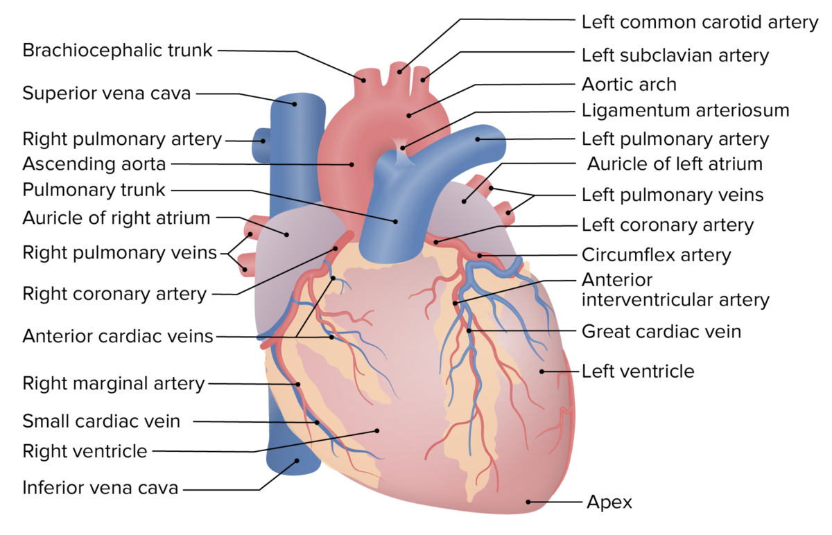
The heart is a 4-chambered muscular pump Pump ACES and RUSH: Resuscitation Ultrasound Protocols made primarily of cardiac muscle Cardiac muscle The muscle tissue of the heart. It is composed of striated, involuntary muscle cells connected to form the contractile pump to generate blood flow. Muscle Tissue: Histology tissue. The heart is divided into 4 chambers: 2 upper chambers for receiving blood from the great vessels, known as the right and left atria, and 2 stronger lower chambers, known as the right and left ventricles, which pump Pump ACES and RUSH: Resuscitation Ultrasound Protocols blood throughout the body. Blood flows through the heart in 1 direction, moving from the right side of the heart, through the lungs Lungs Lungs are the main organs of the respiratory system. Lungs are paired viscera located in the thoracic cavity and are composed of spongy tissue. The primary function of the lungs is to oxygenate blood and eliminate CO2. Lungs: Anatomy, and then returning to the left side of the heart, where it is pumped out to the rest of the body. As blood moves through the heart, 4 important valves prevent backflow. The heart muscle itself is supplied by the coronary arteries Arteries Arteries are tubular collections of cells that transport oxygenated blood and nutrients from the heart to the tissues of the body. The blood passes through the arteries in order of decreasing luminal diameter, starting in the largest artery (the aorta) and ending in the small arterioles. Arteries are classified into 3 types: large elastic arteries, medium muscular arteries, and small arteries and arterioles. Arteries: Histology. The heart also has its own conduction system, triggering its own rhythmic contractions.

Last updated: Nov 19, 2024

Editorial responsibility: Stanley Oiseth, Lindsay Jones, Evelin Maza

General Structure and Location of the Heart

Overview of the heart structure

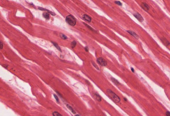
The heart is a 4-chambered muscular pump Pump ACES and RUSH: Resuscitation Ultrasound Protocols made of cardiac muscle Cardiac muscle The muscle tissue of the heart. It is composed of striated, involuntary muscle cells connected to form the contractile pump to generate blood flow. Muscle Tissue: Histology tissue.
  • 4 primary muscular chambers:
    • Right atrium (RA)
    • Right ventricle (RV)
    • Left atrium (LA)
    • Left ventricle (LV)
  • Connections to the great vessels:
    • Veins Veins Veins are tubular collections of cells, which transport deoxygenated blood and waste from the capillary beds back to the heart. Veins are classified into 3 types: small veins/venules, medium veins, and large veins. Each type contains 3 primary layers: tunica intima, tunica media, and tunica adventitia. Veins: Histology (bring blood back to the heart):
    • Arteries Arteries Arteries are tubular collections of cells that transport oxygenated blood and nutrients from the heart to the tissues of the body. The blood passes through the arteries in order of decreasing luminal diameter, starting in the largest artery (the aorta) and ending in the small arterioles. Arteries are classified into 3 types: large elastic arteries, medium muscular arteries, and small arteries and arterioles. Arteries: Histology (carry blood away):
      • Pulmonary trunk Pulmonary Trunk Truncus Arteriosus and pulmonary arteries Arteries Arteries are tubular collections of cells that transport oxygenated blood and nutrients from the heart to the tissues of the body. The blood passes through the arteries in order of decreasing luminal diameter, starting in the largest artery (the aorta) and ending in the small arterioles. Arteries are classified into 3 types: large elastic arteries, medium muscular arteries, and small arteries and arterioles. Arteries: Histology (deoxygenated) → from the RV
      • Aorta Aorta The main trunk of the systemic arteries. Mediastinum and Great Vessels: Anatomy (oxygenated) → from the LV
  • Valves:
    • Located between different vessels and chambers 
    • Blood flows in 1 direction into, through, and out of the heart; valves prevent backflow.
    • Named valves (in order through which blood passes):
      • Tricuspid valve
      • Pulmonic valve
      • Mitral valve
      • Aortic valve
  • Vasculature:
    • Heart muscle itself is supplied by the coronary arteries Arteries Arteries are tubular collections of cells that transport oxygenated blood and nutrients from the heart to the tissues of the body. The blood passes through the arteries in order of decreasing luminal diameter, starting in the largest artery (the aorta) and ending in the small arterioles. Arteries are classified into 3 types: large elastic arteries, medium muscular arteries, and small arteries and arterioles. Arteries: Histology.
    • Drained by coronary veins Veins Veins are tubular collections of cells, which transport deoxygenated blood and waste from the capillary beds back to the heart. Veins are classified into 3 types: small veins/venules, medium veins, and large veins. Each type contains 3 primary layers: tunica intima, tunica media, and tunica adventitia. Veins: Histology
  • Conduction system:
    • Electrical “wiring” that generates and allows conduction of the electrical signal throughout the heart
    • This electrical signal triggers the heart to contract.
  • Protective covering: surrounded by a tough 2-layered membrane called the pericardium 
General structure and flow of blood through the heart

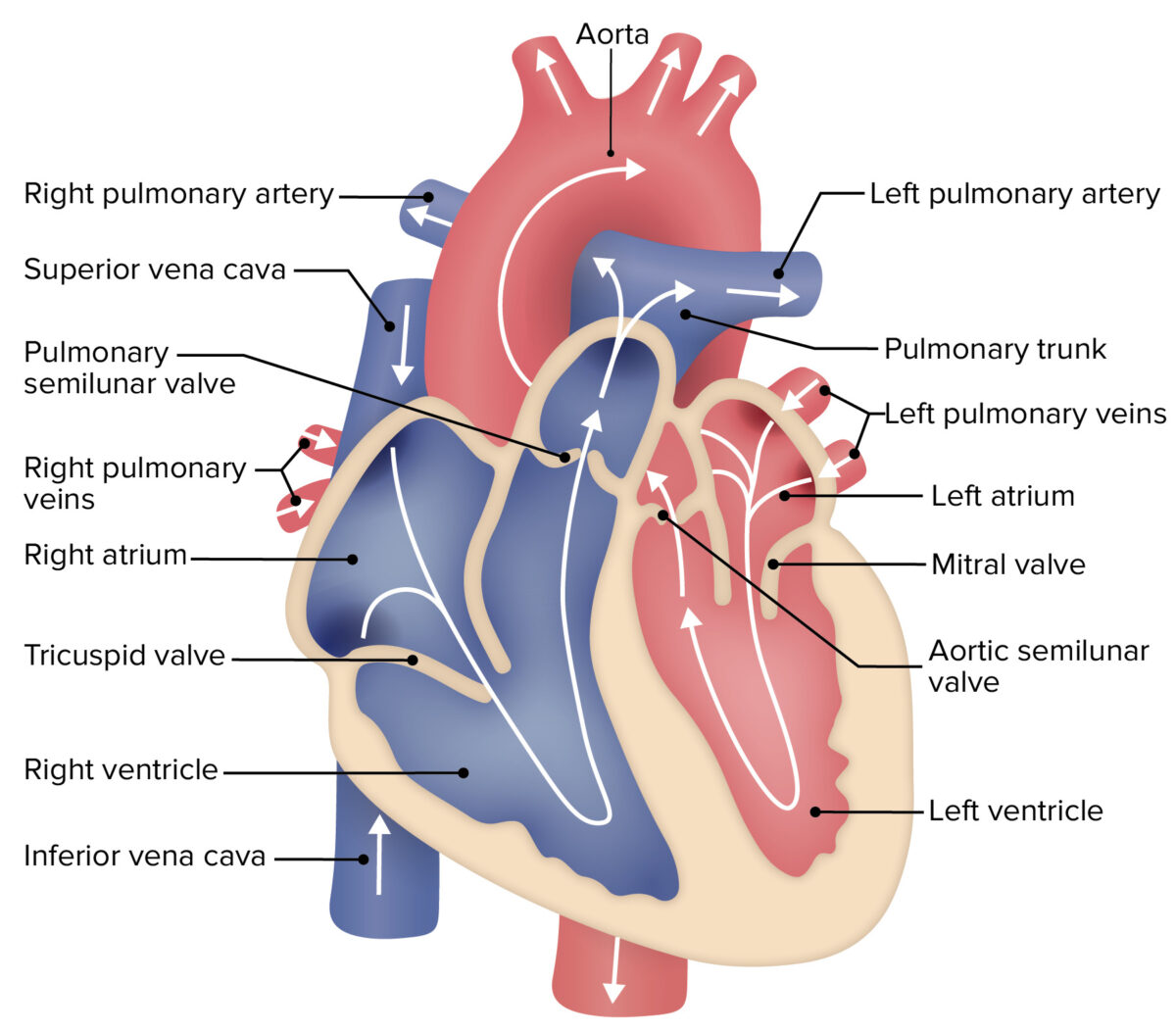
General structure and flow of blood through the heart:

Blue denotes the path of deoxygenated blood, while red indicates the path of oxygenated blood.
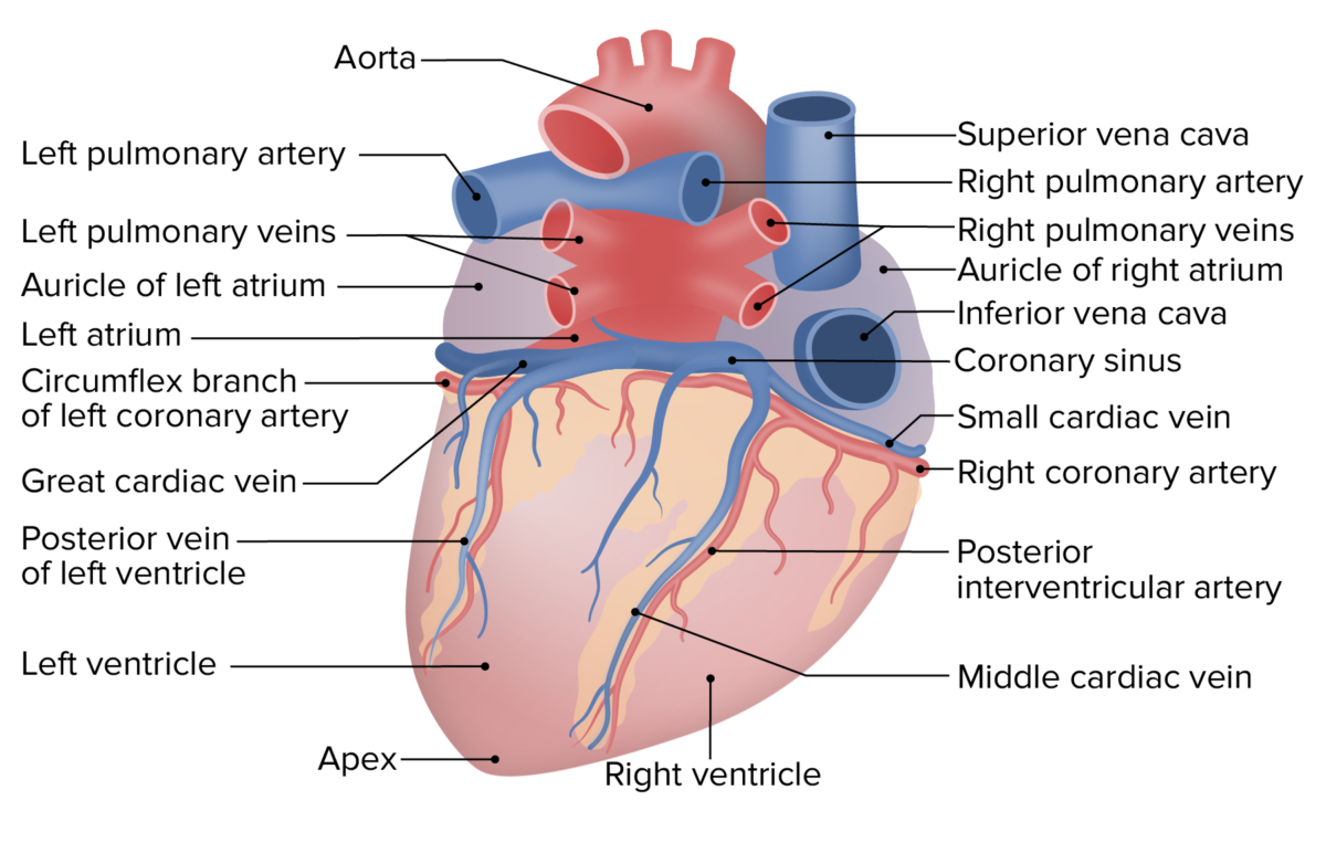
Image by Lecturio.

Size and shape

  • About the size of a fist 
  • Shape: upside-down pyramid or cone
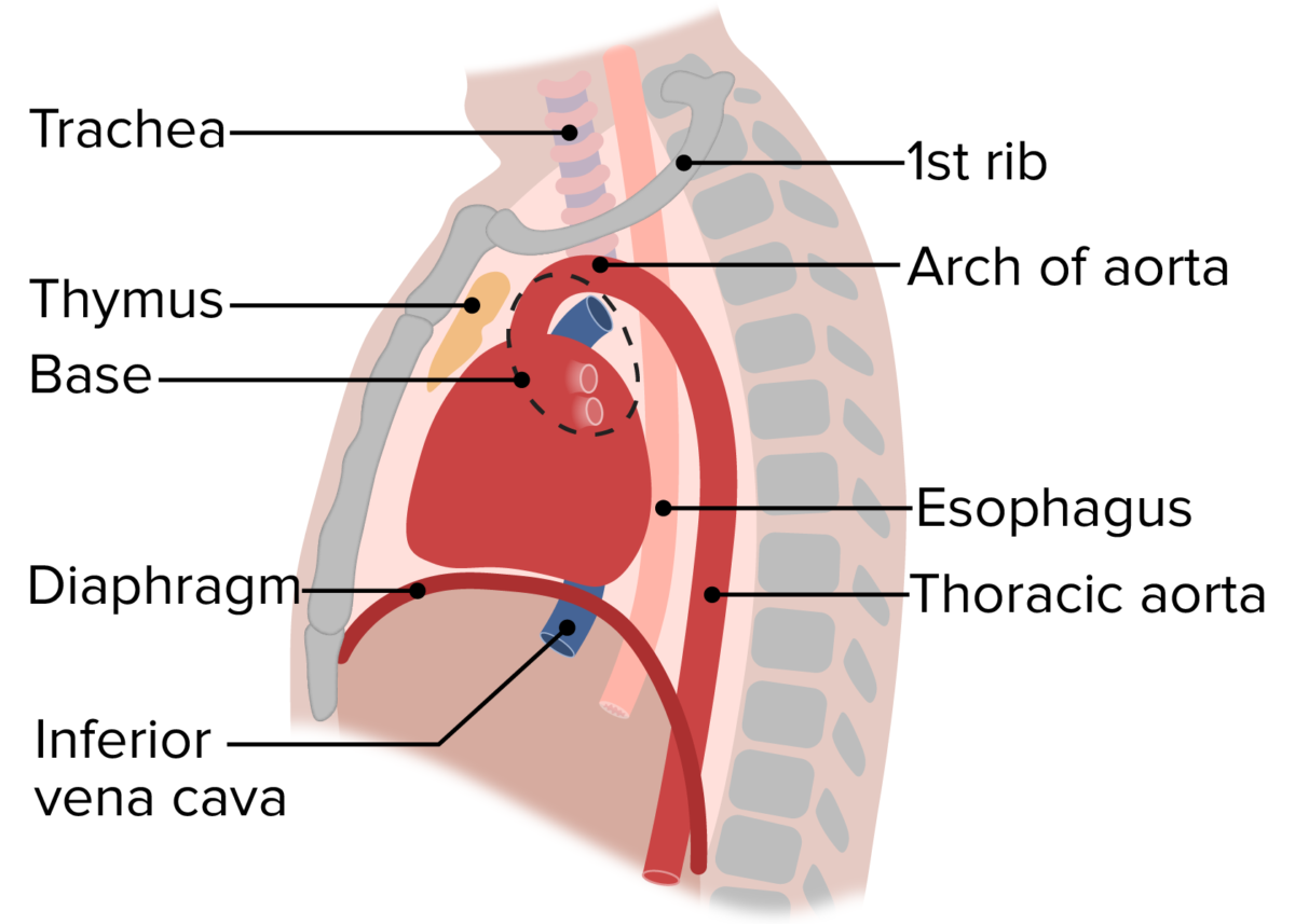
    • Base: superior portion of the heart, made up of the atria
    • Apex: inferior, rounded tip pointing to the left
  • Dimensions of the adult heart:
    • Width (at the base): approximately 9 cm 
    • Length (from base to apex): approximately 13 cm
    • Depth (thickest point): approximately 6 cm
  • Weight: approximately 300 g

Location and orientation Orientation Awareness of oneself in relation to time, place and person. Psychiatric Assessment

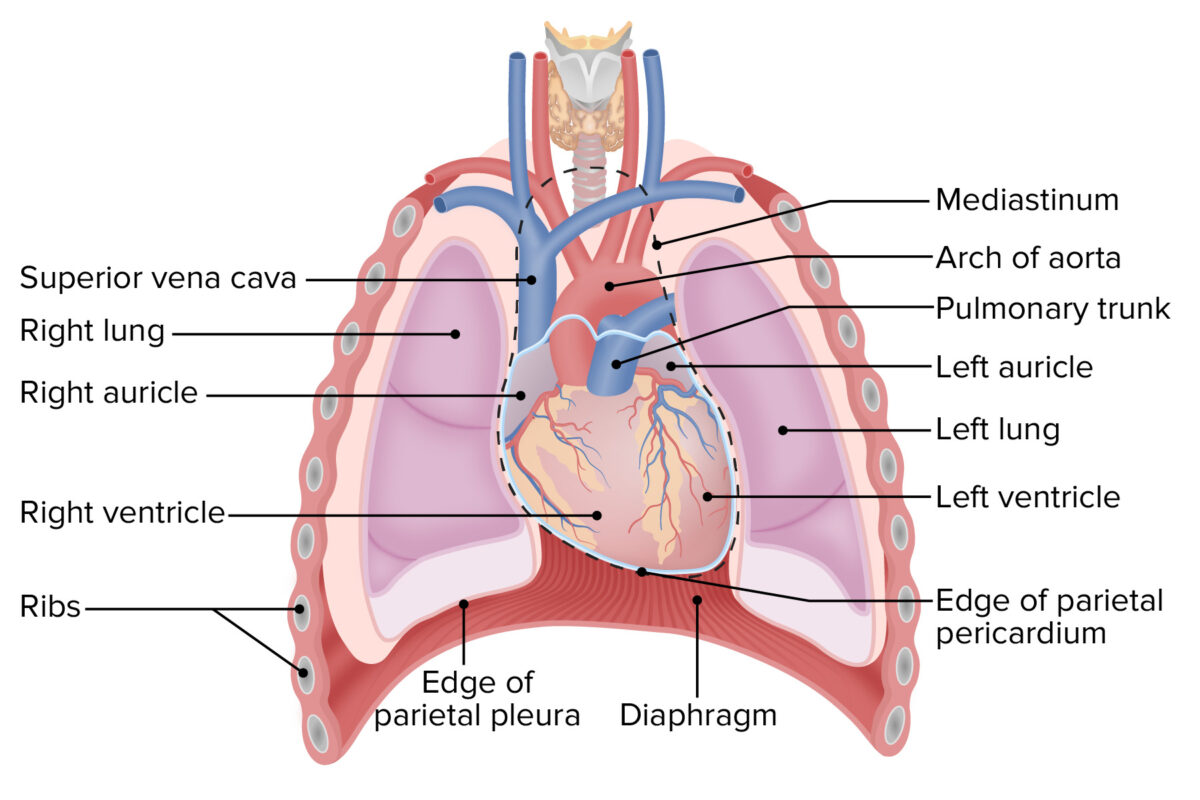
  • Located in the mediastinum Mediastinum The mediastinum is the thoracic area between the 2 pleural cavities. The mediastinum contains vital structures of the circulatory, respiratory, digestive, and nervous systems including the heart and esophagus, and major thoracic vessels. Mediastinum and Great Vessels: Anatomy in the thoracic cavity, between the lungs Lungs Lungs are the main organs of the respiratory system. Lungs are paired viscera located in the thoracic cavity and are composed of spongy tissue. The primary function of the lungs is to oxygenate blood and eliminate CO2. Lungs: Anatomy
  • Sits in its own space called the pericardial cavity
  • At the level of T5–T8
  • Approximately ⅔ is to the left of the median plane
  • The heart is slightly rotated so that:
    • Right side is more anterior
    • Left side is more posterior
  • Base of the heart: at the level of the 3rd intercostal cartilage Cartilage Cartilage is a type of connective tissue derived from embryonic mesenchyme that is responsible for structural support, resilience, and the smoothness of physical actions. Perichondrium (connective tissue membrane surrounding cartilage) compensates for the absence of vasculature in cartilage by providing nutrition and support. Cartilage: Histology
  • Apex of the heart:
    • Formed by the inferolateral part of the left ventricle
    • At the level of the left 5th intercostal space 
    • Approximately 7–9 cm to the left of the median plane
  • Primary structures forming the different surfaces of the heart:
    • Sternocostal surface (anterior): RV 
    • Diaphragmatic surface (inferior): LV and RV
    • Left pulmonary surface (lateral): LV and LA
    • Right pulmonary surface (lateral): RA

Anatomic relationships

  • Superior (to the heart):
  • Anterior (to the heart):
    • Sternum Sternum A long, narrow, and flat bone commonly known as breastbone occurring in the midsection of the anterior thoracic segment or chest region, which stabilizes the rib cage and serves as the point of origin for several muscles that move the arms, head, and neck. Chest Wall: Anatomy
    • Rib cartilage Cartilage Cartilage is a type of connective tissue derived from embryonic mesenchyme that is responsible for structural support, resilience, and the smoothness of physical actions. Perichondrium (connective tissue membrane surrounding cartilage) compensates for the absence of vasculature in cartilage by providing nutrition and support. Cartilage: Histology
  • Lateral: lungs Lungs Lungs are the main organs of the respiratory system. Lungs are paired viscera located in the thoracic cavity and are composed of spongy tissue. The primary function of the lungs is to oxygenate blood and eliminate CO2. Lungs: Anatomy 
  • Posterior:
    • Great vessels ( aorta Aorta The main trunk of the systemic arteries. Mediastinum and Great Vessels: Anatomy, vena cava, pulmonary veins Pulmonary veins The veins that return the oxygenated blood from the lungs to the left atrium of the heart. Lungs: Anatomy)
    • Primary bronchi Bronchi The larger air passages of the lungs arising from the terminal bifurcation of the trachea. They include the largest two primary bronchi which branch out into secondary bronchi, and tertiary bronchi which extend into bronchioles and pulmonary alveoli. Bronchial Tree: Anatomy
    • Esophagus Esophagus The esophagus is a muscular tube-shaped organ of around 25 centimeters in length that connects the pharynx to the stomach. The organ extends from approximately the 6th cervical vertebra to the 11th thoracic vertebra and can be divided grossly into 3 parts: the cervical part, the thoracic part, and the abdominal part. Esophagus: Anatomy
    • Vertebral column Vertebral column The human spine, or vertebral column, is the most important anatomical and functional axis of the human body. It consists of 7 cervical vertebrae, 12 thoracic vertebrae, and 5 lumbar vertebrae and is limited cranially by the skull and caudally by the sacrum. Vertebral Column: Anatomy (T5–T8)
  • Inferior: diaphragm Diaphragm The diaphragm is a large, dome-shaped muscle that separates the thoracic cavity from the abdominal cavity. The diaphragm consists of muscle fibers and a large central tendon, which is divided into right and left parts. As the primary muscle of inspiration, the diaphragm contributes 75% of the total inspiratory muscle force. Diaphragm: Anatomy

The Pericardium

Like the pleural cavity Pleural cavity Paired but separate cavity within the thoracic cavity. It consists of the space between the parietal and visceral pleura and normally contains a capillary layer of serous fluid that lubricates the pleural surfaces. Pleura: Anatomy around the lungs Lungs Lungs are the main organs of the respiratory system. Lungs are paired viscera located in the thoracic cavity and are composed of spongy tissue. The primary function of the lungs is to oxygenate blood and eliminate CO2. Lungs: Anatomy and the peritoneal cavity Peritoneal Cavity The space enclosed by the peritoneum. It is divided into two portions, the greater sac and the lesser sac or omental bursa, which lies behind the stomach. The two sacs are connected by the foramen of winslow, or epiploic foramen. Peritoneum: Anatomy inside the abdomen, the pericardial cavity around the heart is enclosed in a double layer of fibroelastic connective tissue Connective tissue Connective tissues originate from embryonic mesenchyme and are present throughout the body except inside the brain and spinal cord. The main function of connective tissues is to provide structural support to organs. Connective tissues consist of cells and an extracellular matrix. Connective Tissue: Histology known as pericardium.
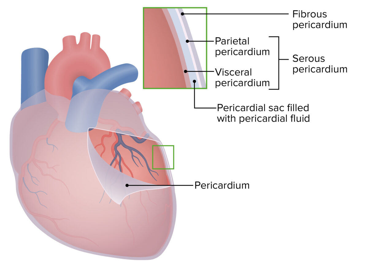
Layers of the pericardium

  • Outer layer:
    • Defines outer layer of pericardial cavity
    • Made up of:
    • Fibrous Fibrous Fibrocystic Change and serous pericardial layers are in direct contact with one another
    • Anchors the heart to:
      • Diaphragm Diaphragm The diaphragm is a large, dome-shaped muscle that separates the thoracic cavity from the abdominal cavity. The diaphragm consists of muscle fibers and a large central tendon, which is divided into right and left parts. As the primary muscle of inspiration, the diaphragm contributes 75% of the total inspiratory muscle force. Diaphragm: Anatomy below
      • Great vessels above
  • Visceral pericardium:
    • Directly covers the surface of the heart
    • Serous layer of connective tissue Connective tissue Connective tissues originate from embryonic mesenchyme and are present throughout the body except inside the brain and spinal cord. The main function of connective tissues is to provide structural support to organs. Connective tissues consist of cells and an extracellular matrix. Connective Tissue: Histology made up of:
    • Contains adipose tissue Adipose tissue Adipose tissue is a specialized type of connective tissue that has both structural and highly complex metabolic functions, including energy storage, glucose homeostasis, and a multitude of endocrine capabilities. There are three types of adipose tissue, white adipose tissue, brown adipose tissue, and beige or “brite” adipose tissue, which is a transitional form. Adipose Tissue: Histology that fills in grooves on the heart surface → protects the coronary vessels
    • Also known as the epicardium (outer layer of the heart wall)
    • Continuous with the parietal Parietal One of a pair of irregularly shaped quadrilateral bones situated between the frontal bone and occipital bone, which together form the sides of the cranium. Skull: Anatomy pericardium at the base of the heart
  • Pericardial sac:
    • The space between the parietal Parietal One of a pair of irregularly shaped quadrilateral bones situated between the frontal bone and occipital bone, which together form the sides of the cranium. Skull: Anatomy and visceral layers of pericardium
    • Contains about 15–50 mL of pericardial fluid (an ultrafiltrate of plasma Plasma The residual portion of blood that is left after removal of blood cells by centrifugation without prior blood coagulation. Transfusion Products)
    • Clinical relevance: cardiac tamponade Tamponade Pericardial effusion, usually of rapid onset, exceeding ventricular filling pressures and causing collapse of the heart with a markedly reduced cardiac output. Pericarditis
      • The fibrous Fibrous Fibrocystic Change pericardium does not expand much if the pericardial fluid accumulates rapidly.
      • Rapid increases of serous or hemorrhagic fluid (as little as 80 ml) into this space will ↑ the pressure on the heart and restrict blood flow Blood flow Blood flow refers to the movement of a certain volume of blood through the vasculature over a given unit of time (e.g., mL per minute). Vascular Resistance, Flow, and Mean Arterial Pressure through the heart
      • Slowly progressing effusions can increase to 2 L without symptoms.
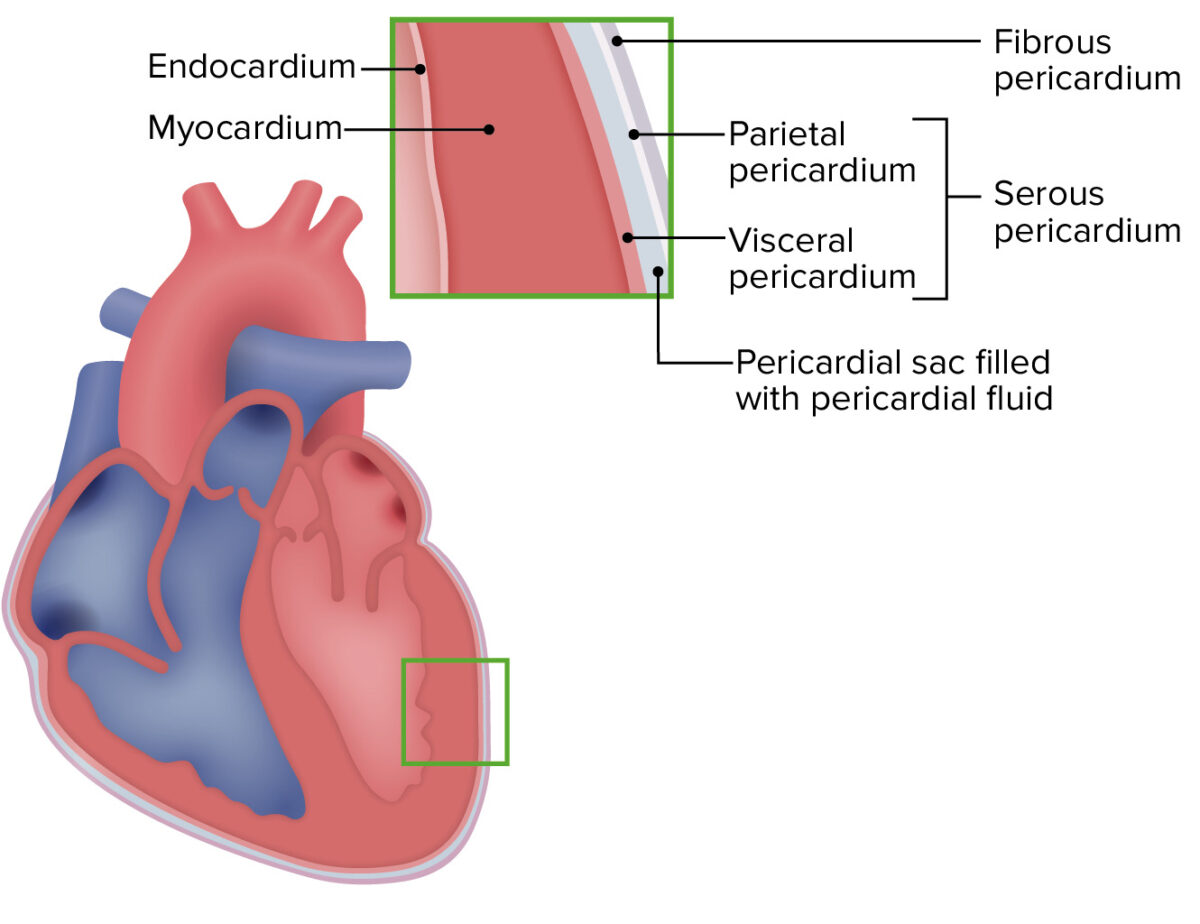
Pericardial cavity of the heart

Pericardial cavity of the heart

Image by Lecturio.

Sinuses within the pericardial cavity

There are 2 important sinuses, or spaces, within the pericardial cavity:
  • Oblique pericardial sinus:
    • A space behind the heart, between the pulmonary veins Pulmonary veins The veins that return the oxygenated blood from the lungs to the left atrium of the heart. Lungs: Anatomy
    • Created by the:
      • Fibrous Fibrous Fibrocystic Change/ parietal Parietal One of a pair of irregularly shaped quadrilateral bones situated between the frontal bone and occipital bone, which together form the sides of the cranium. Skull: Anatomy pericardium posteriorly
      • Pulmonary veins Pulmonary veins The veins that return the oxygenated blood from the lungs to the left atrium of the heart. Lungs: Anatomy laterally
      • Left atrium anteriorly
  • Transverse pericardial sinus:

Related videos

The Heart Wall

Layers of the heart wall

  • Epicardium:
    • Outermost layer of the heart
    • Formed by the visceral layer of the pericardium
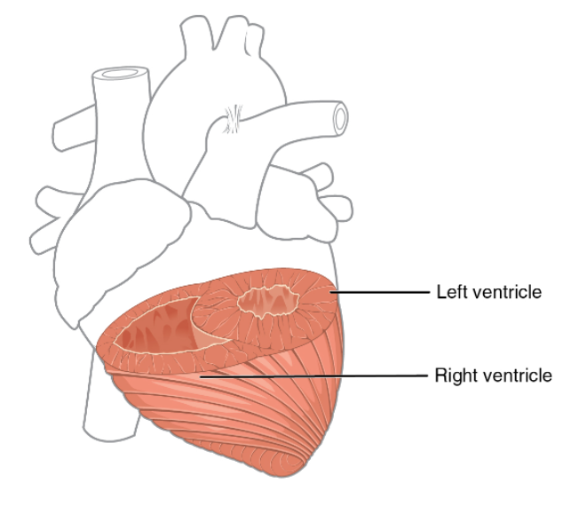
  • Myocardium:
    • Middle muscular layer
    • Thickest layer of the heart (by far)
    • Composed of:
      • Helical Helical Computed tomography where there is continuous x-ray exposure to the patient while being transported in a spiral or helical pattern through the beam of irradiation. This provides improved three-dimensional contrast and spatial resolution compared to conventional computed tomography, where data is obtained and computed from individual sequential exposures. Computed Tomography (CT) layers of cardiac muscle Cardiac muscle The muscle tissue of the heart. It is composed of striated, involuntary muscle cells connected to form the contractile pump to generate blood flow. Muscle Tissue: Histology
      • Fibrous Fibrous Fibrocystic Change skeleton made up of collagen Collagen A polypeptide substance comprising about one third of the total protein in mammalian organisms. It is the main constituent of skin; connective tissue; and the organic substance of bones (bone and bones) and teeth (tooth). Connective Tissue: Histology and elastic Elastic Connective Tissue: Histology fibers
    • Purpose of fibrous Fibrous Fibrocystic Change skeleton:
      • Structural support (especially around valves)
      • Gives cardiac muscle Cardiac muscle The muscle tissue of the heart. It is composed of striated, involuntary muscle cells connected to form the contractile pump to generate blood flow. Muscle Tissue: Histology cells something to pull against
      • Limits the routes of electrical excitation through the heart (electrically separates atria from ventricles)
  • Endocardium:
  • Subendocardial layer:
    • Loose fibrous Fibrous Fibrocystic Change connective tissue Connective tissue Connective tissues originate from embryonic mesenchyme and are present throughout the body except inside the brain and spinal cord. The main function of connective tissues is to provide structural support to organs. Connective tissues consist of cells and an extracellular matrix. Connective Tissue: Histology
    • Separates the delicate endocardium from the vigorous pumping action of the myocardium

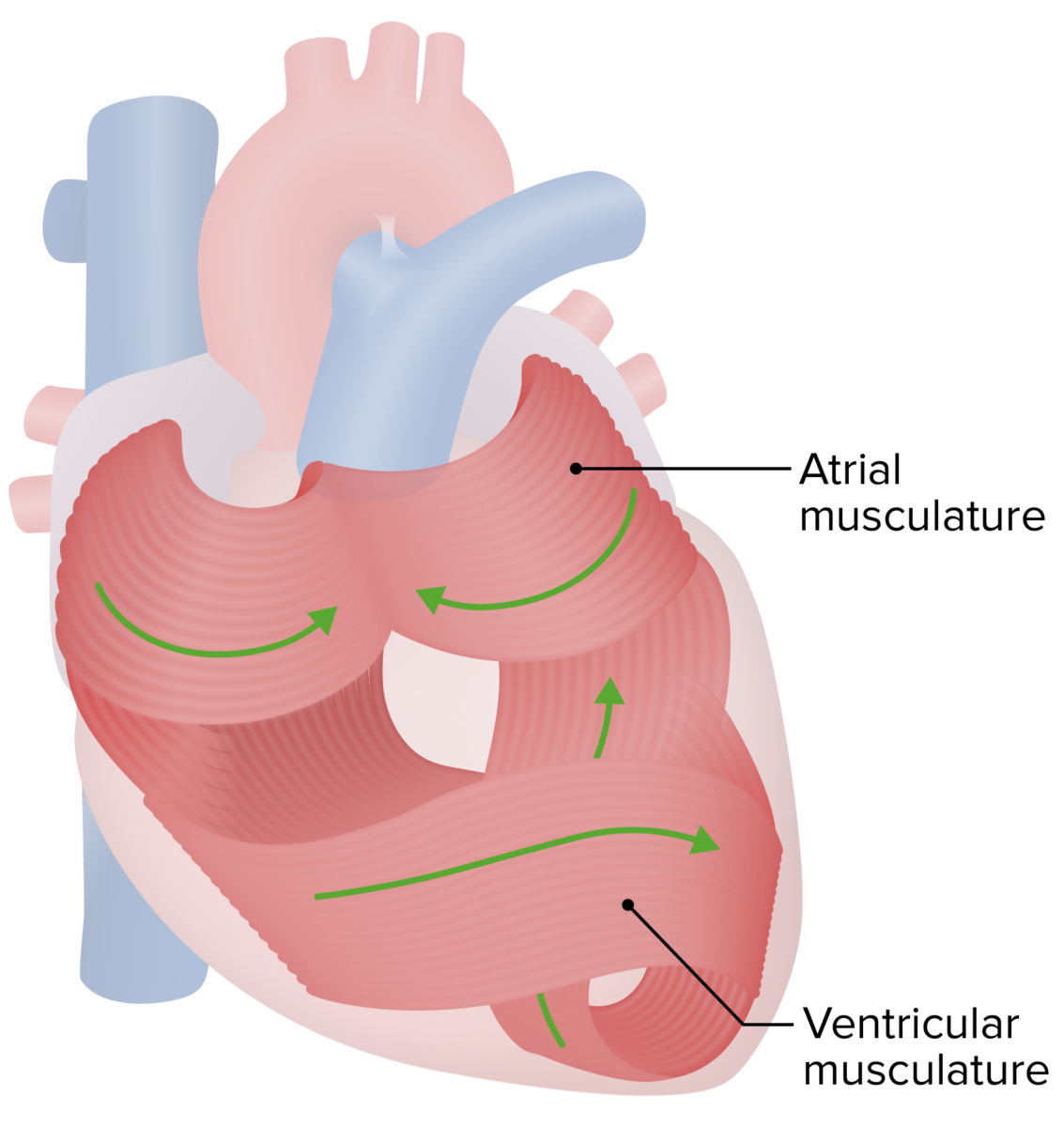
Orientation Orientation Awareness of oneself in relation to time, place and person. Psychiatric Assessment of the myocardium

  • Complex patterns
  • 1 figure eight around the atria and great vessels
  • Another figure eight around the ventricles
  • Additional superficial layers wrap around the ventricles
  • Leads to effective pumping action
Orientation of the myocardium

Orientation of the myocardium, allowing the heart to pump blood effectively

Image by Lecturio.

Heart Chambers and Valves

Cardiac chambers

The heart has 4 chambers: 2 atria (receiving chambers) and 2 ventricles (pumping chambers).

RA: 

  • Receives deoxygenated blood from the systemic circulation Circulation The movement of the blood as it is pumped through the cardiovascular system. ABCDE Assessment via:
    • Superior vena cava Superior vena cava The venous trunk which returns blood from the head, neck, upper extremities and chest. Mediastinum and Great Vessels: Anatomy (SVC): drains the upper portion of the body
    • Inferior vena cava Inferior vena cava The venous trunk which receives blood from the lower extremities and from the pelvic and abdominal organs. Mediastinum and Great Vessels: Anatomy ( IVC IVC The venous trunk which receives blood from the lower extremities and from the pelvic and abdominal organs. Mediastinum and Great Vessels: Anatomy): drains the lower portion of the body
    • Coronary sinus Coronary Sinus A short vein that collects about two thirds of the venous blood from the myocardium and drains into the right atrium. Coronary sinus, normally located between the left atrium and left ventricle on the posterior surface of the heart, can serve as an anatomical reference for cardiac procedures. Atrial Septal Defect (ASD): drains the heart muscle itself
  • After collecting the blood returning from systemic circulation Circulation The movement of the blood as it is pumped through the cardiovascular system. ABCDE Assessment, RA pumps it into the RV.
  • Right atrial appendage (auricle):
  • Sinus venarum:
    • Smooth, thin-walled area on the posterior portion of the RA
    • Entry point of the SVC and IVC IVC The venous trunk which receives blood from the lower extremities and from the pelvic and abdominal organs. Mediastinum and Great Vessels: Anatomy into the RA
  • Pectinate muscles:
    • Rough, muscular ridges of myocardium
    • Located on anterior wall and within (both right and left) auricles
  • Crista terminalis: separation between sinus venarum and pectinate muscles
  • Interatrial septum Interatrial Septum Atrial Septal Defect (ASD): muscular wall separating RA from LA
  • Fossa ovalis Fossa ovalis Prenatal and Postnatal Physiology of the Neonate (or the oval fossa):
    • Oval depression in interatrial septum Interatrial Septum Atrial Septal Defect (ASD)
    • Remnant of the foramen ovale Foramen ovale An opening in the wall between the right and the left upper chambers (heart atria) of a fetal heart. Oval foramen normally closes soon after birth; when it fails to close the condition is called patent oval foramen. Patent Foramen Ovale in the fetus (an opening in the interatrial septum Interatrial Septum Atrial Septal Defect (ASD) that allows blood to move straight from the RA to the LA, bypassing the lungs Lungs Lungs are the main organs of the respiratory system. Lungs are paired viscera located in the thoracic cavity and are composed of spongy tissue. The primary function of the lungs is to oxygenate blood and eliminate CO2. Lungs: Anatomy, which are not used for oxygenation in the fetus)

LA: 

  • Collects oxygenated blood from the:
    • Left and right superior pulmonary veins Pulmonary veins The veins that return the oxygenated blood from the lungs to the left atrium of the heart. Lungs: Anatomy
    • Left and right inferior pulmonary veins Pulmonary veins The veins that return the oxygenated blood from the lungs to the left atrium of the heart. Lungs: Anatomy 
  • After collecting blood returning from the lungs Lungs Lungs are the main organs of the respiratory system. Lungs are paired viscera located in the thoracic cavity and are composed of spongy tissue. The primary function of the lungs is to oxygenate blood and eliminate CO2. Lungs: Anatomy, LA pumps it into the LV.
  • Walls are:
    • Primarily smooth (no pectinate muscles within the main atrial chamber)
    • Slightly thicker than the walls of the RA (due to higher pressures on the left side of the heart)
  • Left atrial appendage (auricle): similar to the right auricle
  • Valve of the foramen ovale Foramen ovale An opening in the wall between the right and the left upper chambers (heart atria) of a fetal heart. Oval foramen normally closes soon after birth; when it fails to close the condition is called patent oval foramen. Patent Foramen Ovale: semilunar ridge on the interatrial septum Interatrial Septum Atrial Septal Defect (ASD) representing the remnants of the valve of the fetal foramen ovale Foramen ovale An opening in the wall between the right and the left upper chambers (heart atria) of a fetal heart. Oval foramen normally closes soon after birth; when it fails to close the condition is called patent oval foramen. Patent Foramen Ovale

RV: 

  • Receives deoxygenated blood from RA through the tricuspid valve
  • Pumps blood out through the pulmonic valve to the pulmonary trunk Pulmonary Trunk Truncus Arteriosus lungs Lungs Lungs are the main organs of the respiratory system. Lungs are paired viscera located in the thoracic cavity and are composed of spongy tissue. The primary function of the lungs is to oxygenate blood and eliminate CO2. Lungs: Anatomy
  • Walls are thicker than those in the RA, but thinner than in the LV.
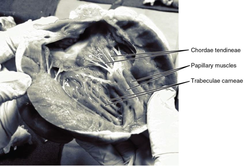
  • Trabeculae carneae: internal muscular ridges in the ventricles (similar to the pectinate muscles of the atria)
  • Papillary muscles: muscles arising from the floor of the RV that control closure of the tricuspid valve
  • Chordae tendineae: tendinous cord-like structures that connect the papillary muscles to the tricuspid valve
  • Interventricular septum Interventricular Septum Ventricular Septal Defect (VSD) (IVS):
    • Wall between RV and LV 
    • Has membranous and muscular components

LV: 

  • Receives oxygenated blood from LA through mitral valve
  • Pumps blood through aortic valve into aorta Aorta The main trunk of the systemic arteries. Mediastinum and Great Vessels: Anatomy → systemic circulation Circulation The movement of the blood as it is pumped through the cardiovascular system. ABCDE Assessment
  • Has the thickest layer of the myocardium (highest-pressure chamber)
  • Like the RV, the LV also contains:
    • Trabeculae carneae
    • Papillary muscles to control mitral valve
    • Chordae tendineae between papillary muscles and mitral valve

Sulci

  • Visible grooves on the surface of the heart
  • Mark the boundaries of the 4 chambers
  • Contain:
    • Coronary vessels
    • Adipose tissue Adipose tissue Adipose tissue is a specialized type of connective tissue that has both structural and highly complex metabolic functions, including energy storage, glucose homeostasis, and a multitude of endocrine capabilities. There are three types of adipose tissue, white adipose tissue, brown adipose tissue, and beige or “brite” adipose tissue, which is a transitional form. Adipose Tissue: Histology
  • Atrioventricular (AV) sulcus:
    • Divides the atria from the ventricles
    • Commonly called the coronary sulcus because the coronary vessels run within it
  • Anterior interventricular sulcus: marks location of IVS on anterior surface of heart
  • Posterior interventricular sulcus: marks location of IVS on posterior surface of heart

Cardiac valves

  • Valves:
    • Anchored on fibrous Fibrous Fibrocystic Change rings
    • Prevent retrograde flow Retrograde flow Veins: Histology
    • Closure produces audible heart sounds Heart sounds Heart sounds are brief, transient sounds produced by valve opening and closure and by movement of blood in the heart. They are divided into systolic and diastolic sounds. In most cases, only the first (S1) and second (S2) heart sounds are heard. These are high-frequency sounds and arise from aortic and pulmonary valve closure (S1), as well as mitral and tricuspid valve closure (S2). Heart Sounds on auscultation of the chest
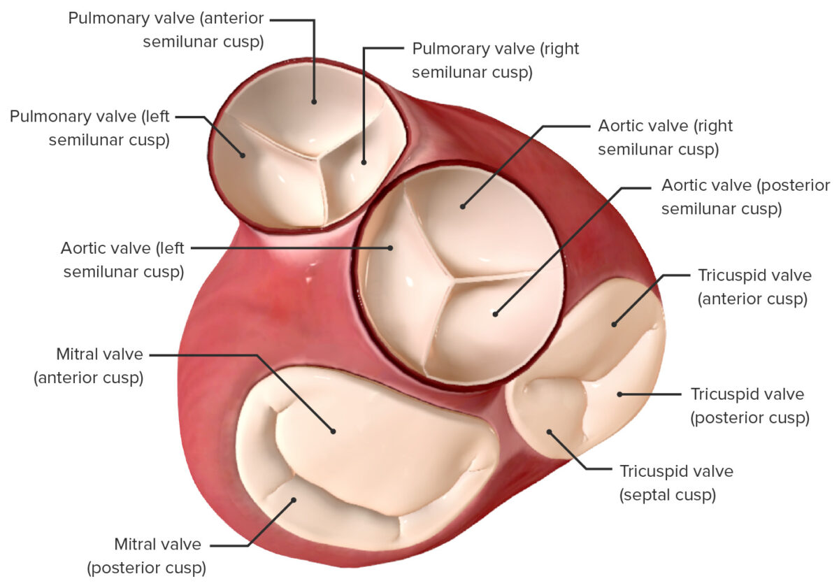
  • Tricuspid valve:
    • Between the RA and RV
    • Also called the right AV valve
    • 3 cusps: anterior, posterior, and septal
  • Mitral valve:
    • Between LA and LV
    • Also called the left AV valve
    • Bicuspid = 2 cusps: anterior and posterior
  • Pulmonary valve: 
  • Aortic valve:
View of the valves of the heart from an atrial perspective

View of the valves of the heart from an atrial perspective:
Atria removed

Image by BioDigital, edited by Lecturio

Blood Flow through the Heart

Blood flows into, through, and out of the heart by sequentially passing through the following structures (in order):
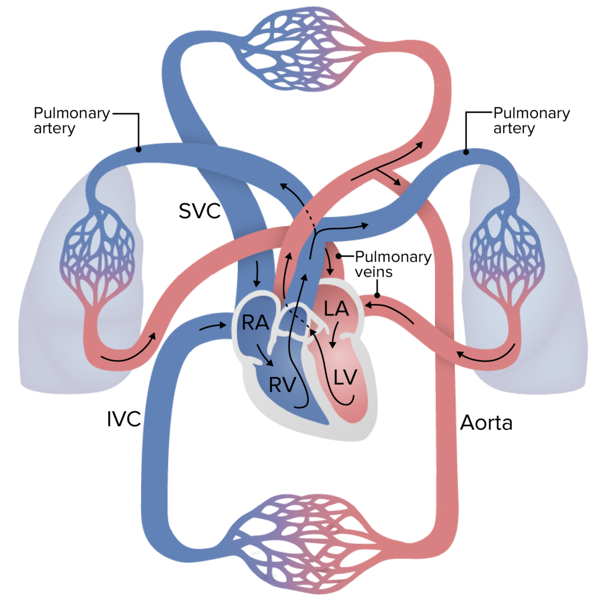
  1. Deoxygenated blood enters the heart via the SVC/ IVC IVC The venous trunk which receives blood from the lower extremities and from the pelvic and abdominal organs. Mediastinum and Great Vessels: Anatomy → 
  2. RA → 
  3. Tricuspid valve → 
  4. RV → 
  5. Pulmonary valve → 
  6. Pulmonary trunk Pulmonary Trunk Truncus Arteriosus → 
  7. Pulmonary arteries Arteries Arteries are tubular collections of cells that transport oxygenated blood and nutrients from the heart to the tissues of the body. The blood passes through the arteries in order of decreasing luminal diameter, starting in the largest artery (the aorta) and ending in the small arterioles. Arteries are classified into 3 types: large elastic arteries, medium muscular arteries, and small arteries and arterioles. Arteries: Histology → 
  8. Lungs Lungs Lungs are the main organs of the respiratory system. Lungs are paired viscera located in the thoracic cavity and are composed of spongy tissue. The primary function of the lungs is to oxygenate blood and eliminate CO2. Lungs: Anatomy (blood is oxygenated) → 
  9. Pulmonary veins Pulmonary veins The veins that return the oxygenated blood from the lungs to the left atrium of the heart. Lungs: Anatomy → 
  10. LA → 
  11. Mitral valve → 
  12. LV → 
  13. Aortic valve → 
  14. Aorta Aorta The main trunk of the systemic arteries. Mediastinum and Great Vessels: Anatomy → 
  15. Systemic arteries Arteries Arteries are tubular collections of cells that transport oxygenated blood and nutrients from the heart to the tissues of the body. The blood passes through the arteries in order of decreasing luminal diameter, starting in the largest artery (the aorta) and ending in the small arterioles. Arteries are classified into 3 types: large elastic arteries, medium muscular arteries, and small arteries and arterioles. Arteries: Histology capillaries Capillaries Capillaries are the primary structures in the circulatory system that allow the exchange of gas, nutrients, and other materials between the blood and the extracellular fluid (ECF). Capillaries are the smallest of the blood vessels. Because a capillary diameter is so small, only 1 RBC may pass through at a time. Capillaries: Histology (blood is deoxygenated) → veins Veins Veins are tubular collections of cells, which transport deoxygenated blood and waste from the capillary beds back to the heart. Veins are classified into 3 types: small veins/venules, medium veins, and large veins. Each type contains 3 primary layers: tunica intima, tunica media, and tunica adventitia. Veins: Histology → 
  16. SVC/ IVC IVC The venous trunk which receives blood from the lower extremities and from the pelvic and abdominal organs. Mediastinum and Great Vessels: Anatomy
  17. Back to the heart
Circulation of blood through the body

Circulation of blood through the body:
Deoxygenated blood enters the right side of the heart and passes through the pulmonary trunk to the lungs, where it is oxygenated. The blood then returns to the left side of the heart via the pulmonary veins, where it is pumped into the aorta and distributed throughout the body. The blood travels through systemic capillaries, where it is deoxygenated again, and travels back to the heart via the superior and inferior vena cava.
LA: left atrium
LV: left ventricle
RA: right atrium
RV: right ventricle

Image by Lecturio.

Coronary Circulation

Coronary circulation Circulation The movement of the blood as it is pumped through the cardiovascular system. ABCDE Assessment describes the flow Flow Blood flows through the heart, arteries, capillaries, and veins in a closed, continuous circuit. Flow is the movement of volume per unit of time. Flow is affected by the pressure gradient and the resistance fluid encounters between 2 points. Vascular resistance is the opposition to flow, which is caused primarily by blood friction against vessel walls. Vascular Resistance, Flow, and Mean Arterial Pressure of blood through the vessels supplying the heart muscle itself. There are 2 primary coronary arteries Arteries Arteries are tubular collections of cells that transport oxygenated blood and nutrients from the heart to the tissues of the body. The blood passes through the arteries in order of decreasing luminal diameter, starting in the largest artery (the aorta) and ending in the small arterioles. Arteries are classified into 3 types: large elastic arteries, medium muscular arteries, and small arteries and arterioles. Arteries: Histology: the left and the right, and both originate from the aorta Aorta The main trunk of the systemic arteries. Mediastinum and Great Vessels: Anatomy, just above the aortic valve.
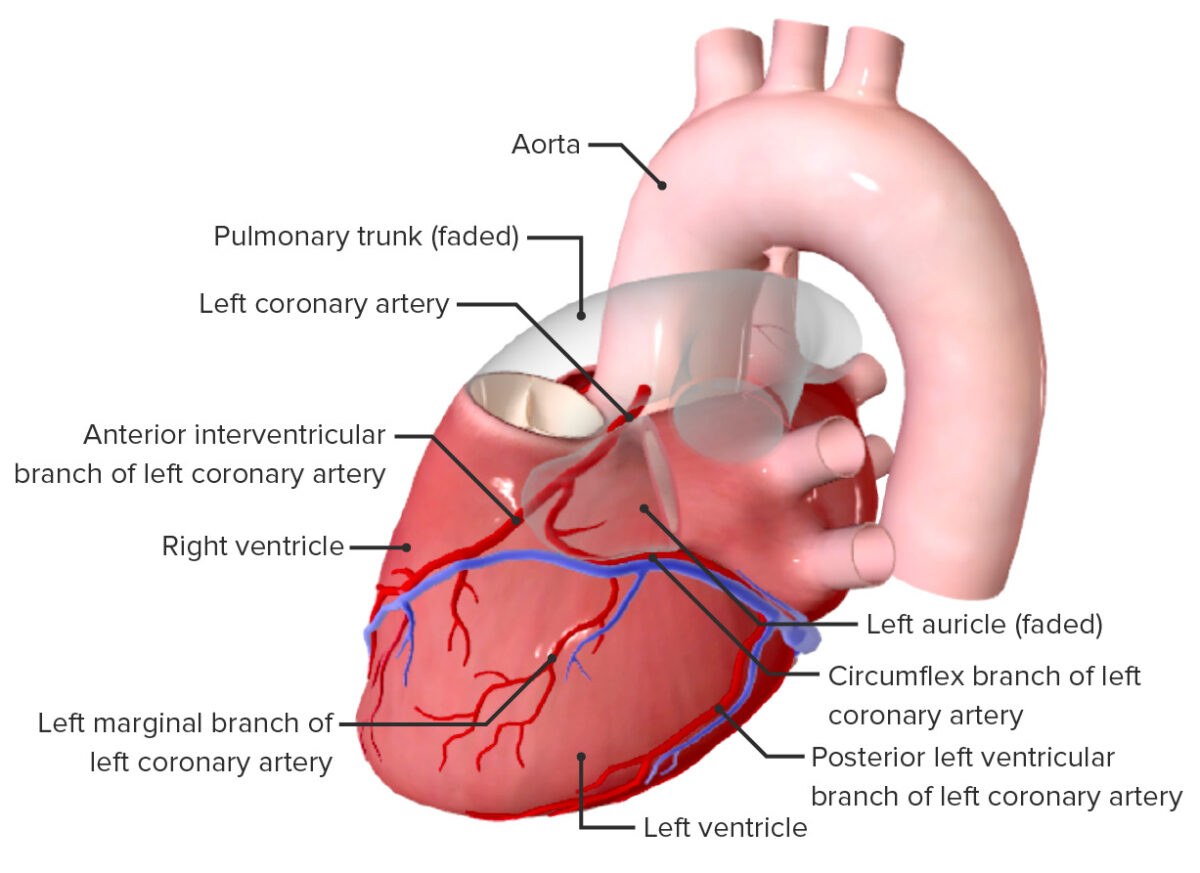
Left coronary artery Coronary Artery Truncus Arteriosus (LCA)

  • Runs posterior to the pulmonary trunk Pulmonary Trunk Truncus Arteriosus → around the left side of the heart under the left auricle in the coronary (AV) sulcus
  • Primary blood supply for:
    • Left side of the heart 
    • IVS
  • Branches into:
    • Anterior interventricular artery:
      • Commonly referred to as the left anterior descending (LAD) artery
      • Travels down the anterior interventricular sulcus toward the apex
      • Supplies the septum and anterior wall of both ventricles
      • Clinical relevance: artery most frequently occluded, causing MI MI MI is ischemia and death of an area of myocardial tissue due to insufficient blood flow and oxygenation, usually from thrombus formation on a ruptured atherosclerotic plaque in the epicardial arteries. Clinical presentation is most commonly with chest pain, but women and patients with diabetes may have atypical symptoms. Myocardial Infarction
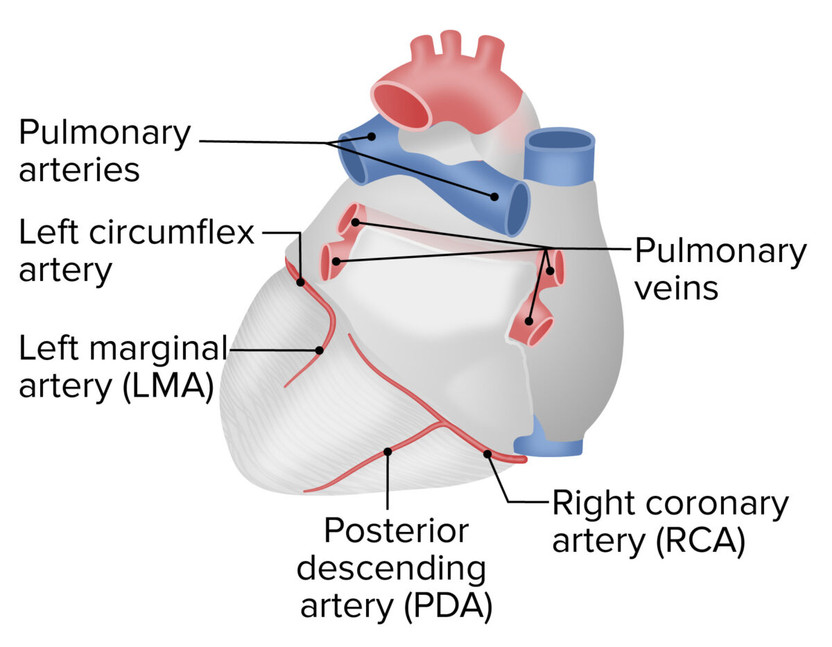
    • Circumflex artery:
      • Continuous around the left side of the heart in the coronary sulcus
      • Anastomoses with small branches of the right coronary artery Coronary Artery Truncus Arteriosus
      • Supplies the LA and posterior wall of the LV
    • Multiple other smaller branches come off of each of these
Left coronary artery and its branches

Left coronary artery and its branches

Image by BioDigital, edited by Lecturio
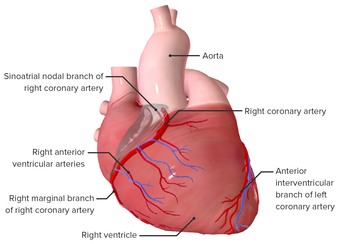
Right coronary artery Coronary Artery Truncus Arteriosus (RCA)

  • Runs around the right side of the heart, under the right auricle in the coronary sulcus
  • Primary blood supply for:
    • Right atrium
    • Parts of both ventricles
    • Conduction system (clinical relevance: occlusion of the RCA is associated with arrhythmias)
  • Branches into:
    • Posterior interventricular artery:
      • Commonly referred to as the posterior descending artery ( PDA PDA The ductus arteriosus (DA) allows blood to bypass pulmonary circulation. After birth, the DA remains open for up to 72 hours and then constricts and involutes, becoming the ligamentum arteriosum. Failure of this process to occur results in patent ductus arteriosus (PDA), a condition that causes up to 10% of congenital heart defects. Patent Ductus Arteriosus (PDA))
      • Travels down the posterior interventricular sulcus toward the apex
      • Supplies the posterior wall of both ventricles
      • Occasionally branches off the LCA rather than the RCA
    • Right marginal artery: supplies the lateral aspect of the RA and RV

Coronary dominance

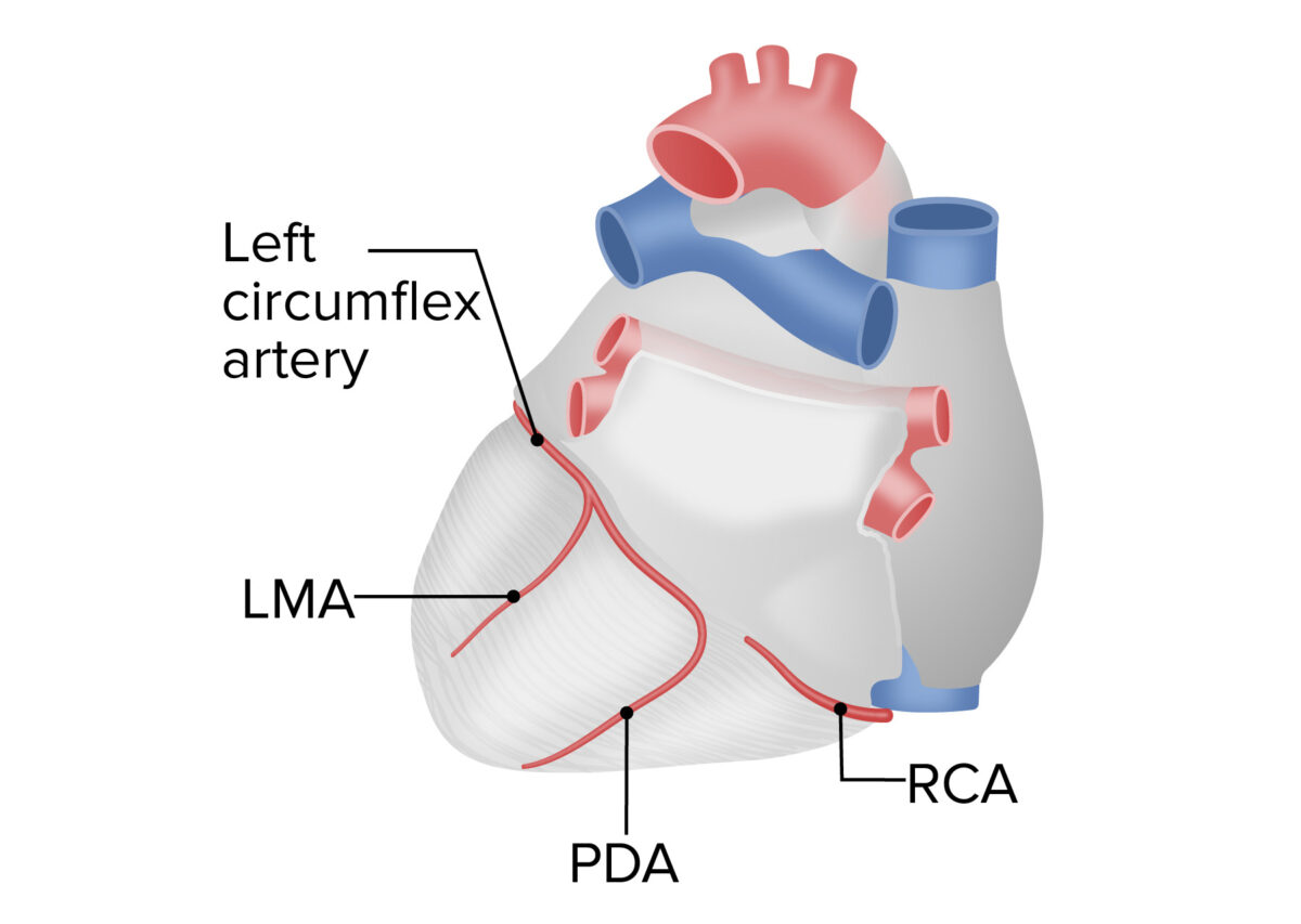
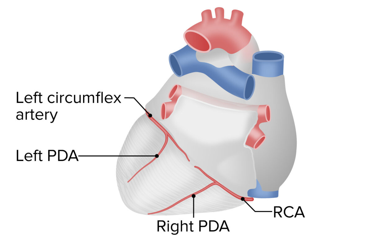
A person’s coronary dominance pattern is determined by the artery that gives off the PDA PDA The ductus arteriosus (DA) allows blood to bypass pulmonary circulation. After birth, the DA remains open for up to 72 hours and then constricts and involutes, becoming the ligamentum arteriosum. Failure of this process to occur results in patent ductus arteriosus (PDA), a condition that causes up to 10% of congenital heart defects. Patent Ductus Arteriosus (PDA).

  • Right-dominant (approximately 75% of people): PDA PDA The ductus arteriosus (DA) allows blood to bypass pulmonary circulation. After birth, the DA remains open for up to 72 hours and then constricts and involutes, becoming the ligamentum arteriosum. Failure of this process to occur results in patent ductus arteriosus (PDA), a condition that causes up to 10% of congenital heart defects. Patent Ductus Arteriosus (PDA) comes from RCA
  • Left-dominant: PDA PDA The ductus arteriosus (DA) allows blood to bypass pulmonary circulation. After birth, the DA remains open for up to 72 hours and then constricts and involutes, becoming the ligamentum arteriosum. Failure of this process to occur results in patent ductus arteriosus (PDA), a condition that causes up to 10% of congenital heart defects. Patent Ductus Arteriosus (PDA) comes from the left circumflex artery (off the LCA)
  • Codominant (least common): an individual has 2 PDAs
    • 1 comes from RCA
    • The other comes from the left circumflex artery

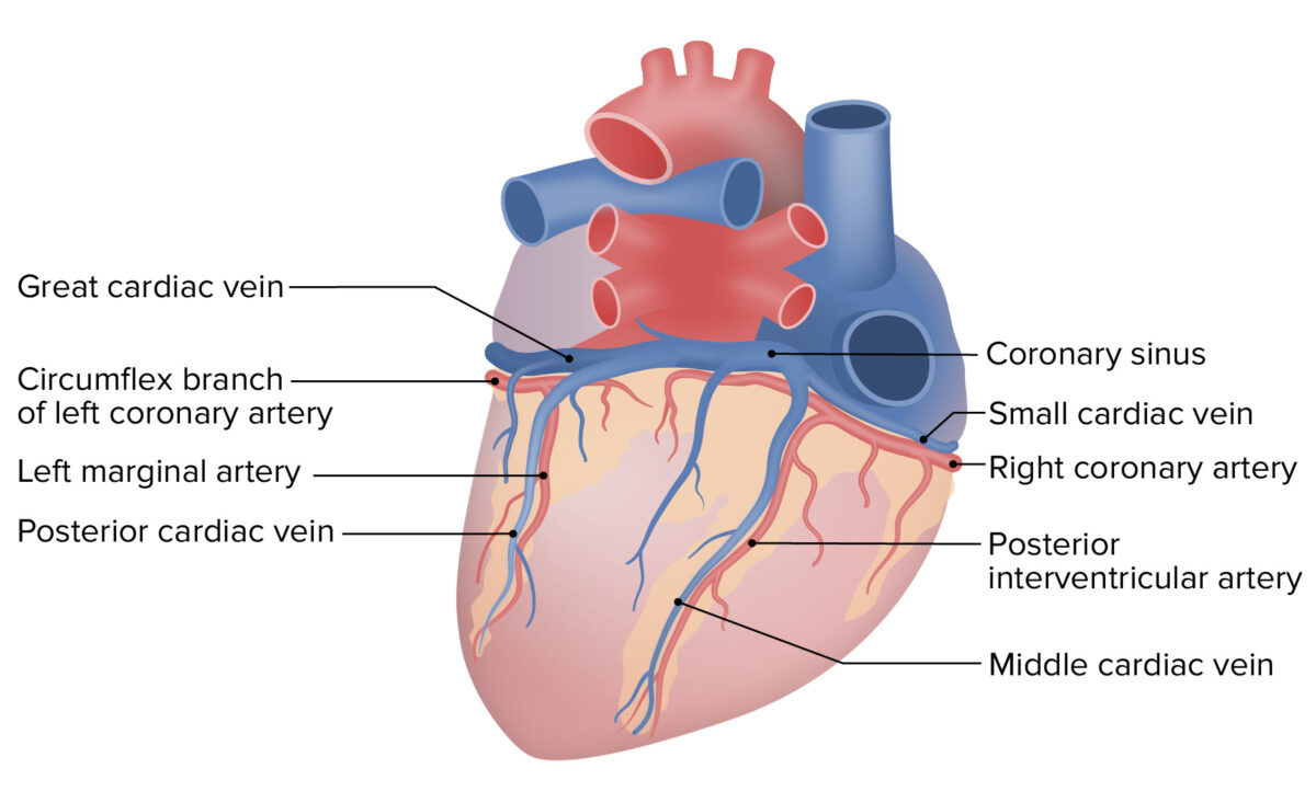
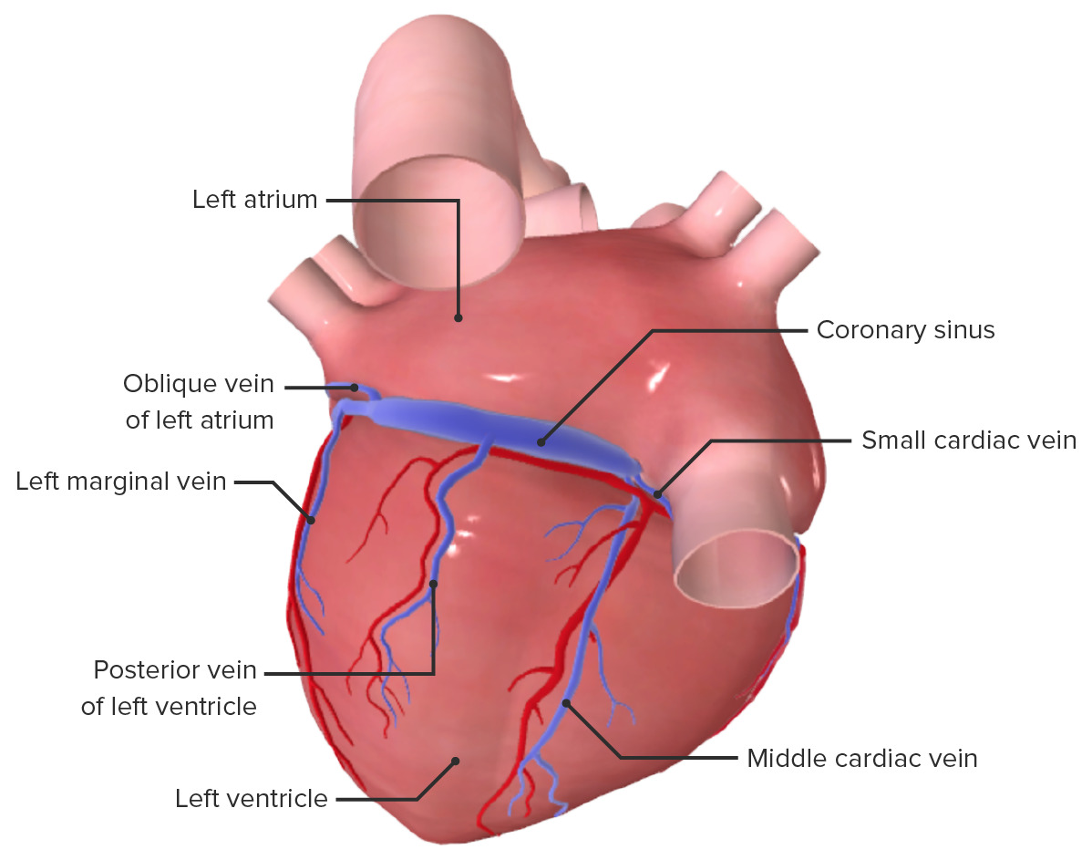
Coronary veins Veins Veins are tubular collections of cells, which transport deoxygenated blood and waste from the capillary beds back to the heart. Veins are classified into 3 types: small veins/venules, medium veins, and large veins. Each type contains 3 primary layers: tunica intima, tunica media, and tunica adventitia. Veins: Histology

  • Great cardiac vein:
    • Drains the anterior side of the heart
    • Travels with the LAD in the anterior IV sulcus
  • Middle cardiac vein:
    • Drains the posterior side of the heart
    • Travels with the PDA PDA The ductus arteriosus (DA) allows blood to bypass pulmonary circulation. After birth, the DA remains open for up to 72 hours and then constricts and involutes, becoming the ligamentum arteriosum. Failure of this process to occur results in patent ductus arteriosus (PDA), a condition that causes up to 10% of congenital heart defects. Patent Ductus Arteriosus (PDA) in the posterior IV sulcus
  • The great and middle cardiac veins Veins Veins are tubular collections of cells, which transport deoxygenated blood and waste from the capillary beds back to the heart. Veins are classified into 3 types: small veins/venules, medium veins, and large veins. Each type contains 3 primary layers: tunica intima, tunica media, and tunica adventitia. Veins: Histology drain into the coronary sinus Coronary Sinus A short vein that collects about two thirds of the venous blood from the myocardium and drains into the right atrium. Coronary sinus, normally located between the left atrium and left ventricle on the posterior surface of the heart, can serve as an anatomical reference for cardiac procedures. Atrial Septal Defect (ASD), which:
    • Lies in the left posterior AV sulcus
    • Drains directly into the RA
  • Approximately 20% of the coronary circulation Circulation The movement of the blood as it is pumped through the cardiovascular system. ABCDE Assessment drains directly into the right ventricle via small vessels.
Cardiac vein

Posterior view of the heart showing the coronary sinus and middle cardiac vein

Image by BioDigital, edited by Lecturio
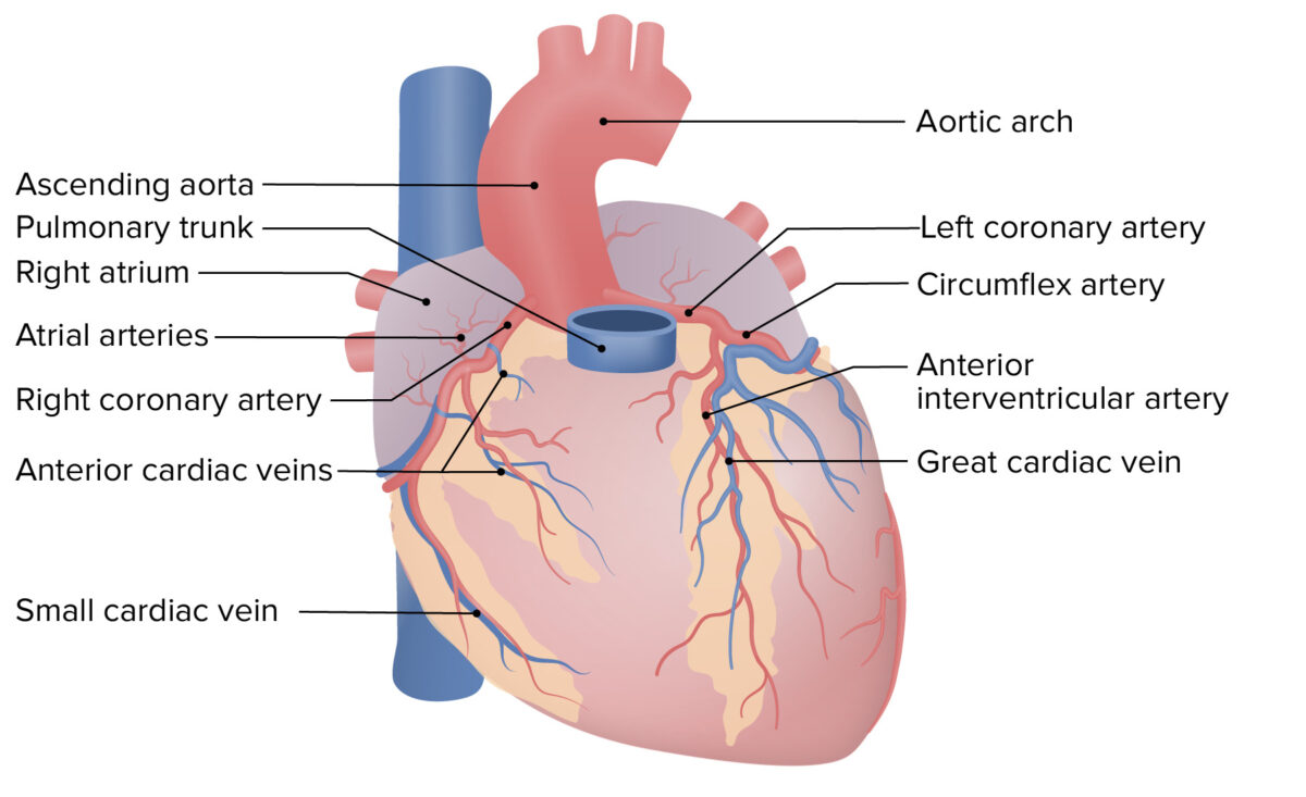
Table: Summary of coronary circulation Circulation The movement of the blood as it is pumped through the cardiovascular system. ABCDE Assessment
Vessel Trajectory Area of supply/drainage
Left coronary artery Coronary Artery Truncus Arteriosus LAD artery Runs in the anterior IV sulcus toward the apex
  • Most of the LA and LV
  • Anterior portion of the IVS
Left circumflex artery Wraps around toward the posterior aspect of the heart in the coronary (AV) sulcus Posterior aspect of LA and LV
Right coronary artery Coronary Artery Truncus Arteriosus PDA PDA The ductus arteriosus (DA) allows blood to bypass pulmonary circulation. After birth, the DA remains open for up to 72 hours and then constricts and involutes, becoming the ligamentum arteriosum. Failure of this process to occur results in patent ductus arteriosus (PDA), a condition that causes up to 10% of congenital heart defects. Patent Ductus Arteriosus (PDA) Runs in the posterior interventricular sulcus toward the apex RV and LV and posterior ⅓ of IVS
Right marginal artery Runs over the RV toward the apex RV and apex of the heart
Great cardiac vein With the LAD within the anterior IV sulcus Areas of the heart supplied by the LCA
Middle cardiac vein With the PDA PDA The ductus arteriosus (DA) allows blood to bypass pulmonary circulation. After birth, the DA remains open for up to 72 hours and then constricts and involutes, becoming the ligamentum arteriosum. Failure of this process to occur results in patent ductus arteriosus (PDA), a condition that causes up to 10% of congenital heart defects. Patent Ductus Arteriosus (PDA) within the posterior IV sulcus Posterior wall of LV and RV

Cardiac Conduction System

Overview of the cardiac conduction system

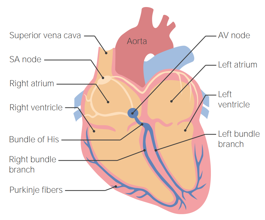
  • The heart generates its own electrical signal. 
  • This signal travels through conduction fibers throughout the heart as an electrical wave.
  • This electrical wave triggers myocardial contraction.
  • Certain anatomic features of this system ensure that the wave triggers contraction in an efficient and coordinated manner.

Anatomy of the conduction system

  • The conduction system is:
    • Myogenic = signal originates from modified myocardial cells within the heart itself (rather than from nerves)
    • Autorhythmic = depolarize spontaneously, at regular Regular Insulin time intervals
  • The sinoatrial (SA) node:
    • The primary pacemaker Pacemaker A device designed to stimulate, by electric impulses, contraction of the heart muscles. It may be temporary (external) or permanent (internal or internal-external). Bradyarrhythmias of the heart 
    • Depolarizes (creating an action potential Action Potential Abrupt changes in the membrane potential that sweep along the cell membrane of excitable cells in response to excitation stimuli. Membrane Potential) 60–80 times per minute → baseline heart rate Heart rate The number of times the heart ventricles contract per unit of time, usually per minute. Cardiac Physiology = 60–80 beats per minute 
    • A patch Patch Nonpalpable lesion > 1 cm in diameter Generalized and Localized Rashes of modified myocytes Myocytes Mature contractile cells, commonly known as myocytes, that form one of three kinds of muscle. The three types of muscle cells are skeletal, cardiac, and smooth. They are derived from embryonic (precursor) muscle cells called myoblasts. Muscle Tissue: Histology located in the RA:
      • Just deep to the epicardium 
      • At the junction of the SVC and RA
    • Supplied by the SA nodal artery, which can arise as a branch off the RCA (60%) or LCA (40%)
  • The AV node:
    • Located in the interatrial septum Interatrial Septum Atrial Septal Defect (ASD):
      • In the posteroinferior region, near the opening of the coronary sinus Coronary Sinus A short vein that collects about two thirds of the venous blood from the myocardium and drains into the right atrium. Coronary sinus, normally located between the left atrium and left ventricle on the posterior surface of the heart, can serve as an anatomical reference for cardiac procedures. Atrial Septal Defect (ASD)
      • Located in a space called Koch’s triangle
    • Natural pacemaker Pacemaker A device designed to stimulate, by electric impulses, contraction of the heart muscles. It may be temporary (external) or permanent (internal or internal-external). Bradyarrhythmias rate of 40–60 beats per minute (normally overridden by the SA nodal rate) 
    • Acts as an “electrical gateway” to the ventricles:
    • Supplied by the AV nodal artery
  • Ventricular conduction system:
    • Common AV bundle (of His): pathway within the IV septum by which the signal leaves the AV node
    • Right and left bundle branches:
      • The AV bundle divides into right and left bundles within the IV septum.
      • Both run toward the apex.
    • Purkinje fibers:
      • Arise from the bundle branches near the apex
      • Turn upward and spread throughout the ventricular walls
      • Fastest conduction fibers
      • Natural pacemaker Pacemaker A device designed to stimulate, by electric impulses, contraction of the heart muscles. It may be temporary (external) or permanent (internal or internal-external). Bradyarrhythmias rate of 25–40 beats per minute
Cardiac conduction system

Cardiac conduction system:
Starting with the sinoatrial (SA) node and ending in the Purkinje fibers
AV: atrioventricular

Image by Lecturio.

Clinical Relevance

  • Heart sounds Heart sounds Heart sounds are brief, transient sounds produced by valve opening and closure and by movement of blood in the heart. They are divided into systolic and diastolic sounds. In most cases, only the first (S1) and second (S2) heart sounds are heard. These are high-frequency sounds and arise from aortic and pulmonary valve closure (S1), as well as mitral and tricuspid valve closure (S2). Heart Sounds: on auscultation, 2 heart sounds Heart sounds Heart sounds are brief, transient sounds produced by valve opening and closure and by movement of blood in the heart. They are divided into systolic and diastolic sounds. In most cases, only the first (S1) and second (S2) heart sounds are heard. These are high-frequency sounds and arise from aortic and pulmonary valve closure (S1), as well as mitral and tricuspid valve closure (S2). Heart Sounds are heard from a normal heart—1st, closing of the AV valves (tricuspid and mitral), followed by closing of the pulmonic and aortic valves. Additional sounds may also be heard, produced by physiologic and/or pathologic conditions. For example, murmurs are generated by turbulent blood flow Blood flow Blood flow refers to the movement of a certain volume of blood through the vasculature over a given unit of time (e.g., mL per minute). Vascular Resistance, Flow, and Mean Arterial Pressure through the heart.
  • Stable and unstable angina Unstable angina Precordial pain at rest, which may precede a myocardial infarction. Stable and Unstable Angina: precordial chest pain Pain An unpleasant sensation induced by noxious stimuli which are detected by nerve endings of nociceptive neurons. Pain: Types and Pathways or pressure, due to reduced blood flow Blood flow Blood flow refers to the movement of a certain volume of blood through the vasculature over a given unit of time (e.g., mL per minute). Vascular Resistance, Flow, and Mean Arterial Pressure to the cardiac muscle Cardiac muscle The muscle tissue of the heart. It is composed of striated, involuntary muscle cells connected to form the contractile pump to generate blood flow. Muscle Tissue: Histology causing transient myocardial ischemia Myocardial ischemia A disorder of cardiac function caused by insufficient blood flow to the muscle tissue of the heart. The decreased blood flow may be due to narrowing of the coronary arteries (coronary artery disease), to obstruction by a thrombus (coronary thrombosis), or less commonly, to diffuse narrowing of arterioles and other small vessels within the heart. Coronary Heart Disease. Angina is most commonly caused by narrowing or occlusion of the coronary arteries Arteries Arteries are tubular collections of cells that transport oxygenated blood and nutrients from the heart to the tissues of the body. The blood passes through the arteries in order of decreasing luminal diameter, starting in the largest artery (the aorta) and ending in the small arterioles. Arteries are classified into 3 types: large elastic arteries, medium muscular arteries, and small arteries and arterioles. Arteries: Histology or one of their main branches. 
  • Myocardial infarction Myocardial infarction MI is ischemia and death of an area of myocardial tissue due to insufficient blood flow and oxygenation, usually from thrombus formation on a ruptured atherosclerotic plaque in the epicardial arteries. Clinical presentation is most commonly with chest pain, but women and patients with diabetes may have atypical symptoms. Myocardial Infarction: ischemia Ischemia A hypoperfusion of the blood through an organ or tissue caused by a pathologic constriction or obstruction of its blood vessels, or an absence of blood circulation. Ischemic Cell Damage and death of an area of myocardial tissue due to insufficient blood flow Blood flow Blood flow refers to the movement of a certain volume of blood through the vasculature over a given unit of time (e.g., mL per minute). Vascular Resistance, Flow, and Mean Arterial Pressure and oxygenation. MI MI MI is ischemia and death of an area of myocardial tissue due to insufficient blood flow and oxygenation, usually from thrombus formation on a ruptured atherosclerotic plaque in the epicardial arteries. Clinical presentation is most commonly with chest pain, but women and patients with diabetes may have atypical symptoms. Myocardial Infarction is usually due to thrombus formation on a ruptured atherosclerotic plaque Plaque Primary Skin Lesions in the supplying arteries Arteries Arteries are tubular collections of cells that transport oxygenated blood and nutrients from the heart to the tissues of the body. The blood passes through the arteries in order of decreasing luminal diameter, starting in the largest artery (the aorta) and ending in the small arterioles. Arteries are classified into 3 types: large elastic arteries, medium muscular arteries, and small arteries and arterioles. Arteries: Histology.
  • Pericarditis Pericarditis Pericarditis is an inflammation of the pericardium, often with fluid accumulation. It can be caused by infection (often viral), myocardial infarction, drugs, malignancies, metabolic disorders, autoimmune disorders, or trauma. Acute, subacute, and chronic forms exist. Pericarditis: inflammation Inflammation Inflammation is a complex set of responses to infection and injury involving leukocytes as the principal cellular mediators in the body’s defense against pathogenic organisms. Inflammation is also seen as a response to tissue injury in the process of wound healing. The 5 cardinal signs of inflammation are pain, heat, redness, swelling, and loss of function. Inflammation of the pericardium resulting from infection, autoimmune disease, radiation Radiation Emission or propagation of acoustic waves (sound), electromagnetic energy waves (such as light; radio waves; gamma rays; or x-rays), or a stream of subatomic particles (such as electrons; neutrons; protons; or alpha particles). Osteosarcoma, surgery, or MI MI MI is ischemia and death of an area of myocardial tissue due to insufficient blood flow and oxygenation, usually from thrombus formation on a ruptured atherosclerotic plaque in the epicardial arteries. Clinical presentation is most commonly with chest pain, but women and patients with diabetes may have atypical symptoms. Myocardial Infarction. Pericarditis Pericarditis Pericarditis is an inflammation of the pericardium, often with fluid accumulation. It can be caused by infection (often viral), myocardial infarction, drugs, malignancies, metabolic disorders, autoimmune disorders, or trauma. Acute, subacute, and chronic forms exist. Pericarditis manifests as fever Fever Fever is defined as a measured body temperature of at least 38°C (100.4°F). Fever is caused by circulating endogenous and/or exogenous pyrogens that increase levels of prostaglandin E2 in the hypothalamus. Fever is commonly associated with chills, rigors, sweating, and flushing of the skin. Fever, pleuritic chest pain Pain An unpleasant sensation induced by noxious stimuli which are detected by nerve endings of nociceptive neurons. Pain: Types and Pathways that increases when lying supine, and an audible pericardial rub on auscultation.
  • Pericardial effusion Pericardial effusion Fluid accumulation within the pericardium. Serous effusions are associated with pericardial diseases. Hemopericardium is associated with trauma. Lipid-containing effusion (chylopericardium) results from leakage of thoracic duct. Severe cases can lead to cardiac tamponade. Pericardial Effusion and Cardiac Tamponade and cardiac tamponade Tamponade Pericardial effusion, usually of rapid onset, exceeding ventricular filling pressures and causing collapse of the heart with a markedly reduced cardiac output. Pericarditis: accumulation of excess fluid in the pericardial space Pericardial Space Pericardial Effusion and Cardiac Tamponade around the heart. The pericardium does not easily expand, so rapid accumulation of fluid leads to increased pressure around the heart. This increased pressure restricts cardiac filling, resulting in decreased cardiac output Cardiac output The volume of blood passing through the heart per unit of time. It is usually expressed as liters (volume) per minute so as not to be confused with stroke volume (volume per beat). Cardiac Mechanics and cardiac tamponade Tamponade Pericardial effusion, usually of rapid onset, exceeding ventricular filling pressures and causing collapse of the heart with a markedly reduced cardiac output. Pericarditis. Signs and symptoms usually occur in the setting of cardiac tamponade Tamponade Pericardial effusion, usually of rapid onset, exceeding ventricular filling pressures and causing collapse of the heart with a markedly reduced cardiac output. Pericarditis and include dyspnea Dyspnea Dyspnea is the subjective sensation of breathing discomfort. Dyspnea is a normal manifestation of heavy physical or psychological exertion, but also may be caused by underlying conditions (both pulmonary and extrapulmonary). Dyspnea, hypotension Hypotension Hypotension is defined as low blood pressure, specifically < 90/60 mm Hg, and is most commonly a physiologic response. Hypotension may be mild, serious, or life threatening, depending on the cause. Hypotension, muffled heart sounds Muffled Heart Sounds Pericardial Effusion and Cardiac Tamponade, jugular venous distention, and pulsus paradoxus Pulsus paradoxus A drop in systolic blood pressure of > 10 mm hg during inspiration. Pericardial Effusion and Cardiac Tamponade
  • Arrhythmias: abnormal heartbeat—either too fast, too slow, or in an irregular manner. When the heart is irritated or damaged, abnormal electrical cardiac impulses can develop spontaneously in the atria or the ventricle. Some spontaneous contractions are normal, but certain contraction patterns are very dangerous and can result in permanent heart damage, stroke, or death.
  • Congenital heart defects: structural abnormalities of the heart due to abnormal development in utero. Signs and symptoms depend on the specific type of anomalies and range from asymptomatic to life-threatening. Examples include abnormalities associated with the position of the great vessels, including transposition of the great vessels Transposition of the Great Vessels Transposition of the great vessels (TGV) is a cyanotic congenital heart disease characterized by “switching” of the great arteries. There are 2 presentations: the dextro (D)- and levo (L)-looped forms. The L-looped form is rare and congenitally corrected, as the ventricles are also switched. Transposition of the Great Vessels, truncus arteriosus Truncus arteriosus Truncus arteriosus (TA) is a congenital heart defect characterized by the persistence of a common cardiac arterial trunk tract that fails to divide into the pulmonary artery and aorta during embryonic development. Truncus arteriosus is a rare congenital malformation with a high mortality rate within the 1st 5 weeks of life if not managed promptly. Truncus Arteriosus, and tetralogy of Fallot Tetralogy of Fallot Tetralogy of Fallot is the most common cyanotic congenital heart disease. The disease is the confluence of 4 pathologic cardiac features: overriding aorta, ventricular septal defect, right ventricular outflow obstruction, and right ventricular hypertrophy. Tetralogy of Fallot; holes in the heart known as atrial and ventricular septal defects or a patent foramen ovale Foramen ovale An opening in the wall between the right and the left upper chambers (heart atria) of a fetal heart. Oval foramen normally closes soon after birth; when it fails to close the condition is called patent oval foramen. Patent Foramen Ovale; and several others.
  • Dilated cardiomyopathy Cardiomyopathy Cardiomyopathy refers to a group of myocardial diseases associated with structural changes of the heart muscles (myocardium) and impaired systolic and/or diastolic function in the absence of other heart disorders (coronary artery disease, hypertension, valvular disease, and congenital heart disease). Cardiomyopathy: Overview and Types, restrictive cardiomyopathy Restrictive Cardiomyopathy Restrictive cardiomyopathy (RCM) is a fairly uncommon condition characterized by progressive stiffening of the cardiac muscle, which causes impaired relaxation and refilling of the heart during diastole, resulting in diastolic dysfunction and eventual heart failure. Restrictive Cardiomyopathy, and hypertrophic cardiomyopathy Hypertrophic Cardiomyopathy Hypertrophic cardiomyopathy (HCM) is the most commonly inherited cardiomyopathy, which is characterized by an asymmetric increase in thickness (hypertrophy) of the left ventricular wall, diastolic dysfunction, and often left ventricular outflow tract obstruction. Hypertrophic Cardiomyopathy: group of myocardial diseases associated with impaired systolic and diastolic function. These disorders may present as shortness of breath Shortness of breath Dyspnea is the subjective sensation of breathing discomfort. Dyspnea is a normal manifestation of heavy physical or psychological exertion, but also may be caused by underlying conditions (both pulmonary and extrapulmonary). Dyspnea, fatigue Fatigue The state of weariness following a period of exertion, mental or physical, characterized by a decreased capacity for work and reduced efficiency to respond to stimuli. Fibromyalgia, syncope Syncope Syncope is a short-term loss of consciousness and loss of postural stability followed by spontaneous return of consciousness to the previous neurologic baseline without the need for resuscitation. The condition is caused by transient interruption of cerebral blood flow that may be benign or related to a underlying life-threatening condition. Syncope, arrhythmias, or heart failure Heart Failure A heterogeneous condition in which the heart is unable to pump out sufficient blood to meet the metabolic need of the body. Heart failure can be caused by structural defects, functional abnormalities (ventricular dysfunction), or a sudden overload beyond its capacity. Chronic heart failure is more common than acute heart failure which results from sudden insult to cardiac function, such as myocardial infarction. Total Anomalous Pulmonary Venous Return (TAPVR) and are classified on the basis of adaptive changes experienced by the myocardium. The main types of cardiomyopathy Cardiomyopathy Cardiomyopathy refers to a group of myocardial diseases associated with structural changes of the heart muscles (myocardium) and impaired systolic and/or diastolic function in the absence of other heart disorders (coronary artery disease, hypertension, valvular disease, and congenital heart disease). Cardiomyopathy: Overview and Types include hypertrophic, dilated, and restrictive.
  • Heart failure Heart Failure A heterogeneous condition in which the heart is unable to pump out sufficient blood to meet the metabolic need of the body. Heart failure can be caused by structural defects, functional abnormalities (ventricular dysfunction), or a sudden overload beyond its capacity. Chronic heart failure is more common than acute heart failure which results from sudden insult to cardiac function, such as myocardial infarction. Total Anomalous Pulmonary Venous Return (TAPVR) ( diastolic dysfunction Diastolic dysfunction Restrictive Cardiomyopathy and systolic dysfunction Systolic dysfunction Dilated Cardiomyopathy): Cardiac insufficiency is characterized by an inability of the heart to pump Pump ACES and RUSH: Resuscitation Ultrasound Protocols the amounts of blood required to meet the body’s needs. Signs and symptoms commonly include shortness of breath Shortness of breath Dyspnea is the subjective sensation of breathing discomfort. Dyspnea is a normal manifestation of heavy physical or psychological exertion, but also may be caused by underlying conditions (both pulmonary and extrapulmonary). Dyspnea that worsens with physical exertion and lying down and lower-extremity edema Edema Edema is a condition in which excess serous fluid accumulates in the body cavity or interstitial space of connective tissues. Edema is a symptom observed in several medical conditions. It can be categorized into 2 types, namely, peripheral (in the extremities) and internal (in an organ or body cavity). Edema.

References

  1. Drake R.L., Vogl, A.W., Mitchell, A.M.W. (2020). Regional Anatomy, Mediastinum. In Drake, R-L., et al. (ed.), Gray’s Anatomy for Students (4th ed., pp. 190–215). Churchill Livingstone/Elsevier
  2. Moore, K. L., et al. (ed). (2014). Thorax. In Moore, K. L., et al. (ed), Clinically Oriented Anatomy (7th ed., pp. 135–159). Lippincott Williams & Wilkins.
  3. John Volpe BS (2021). Anatomy, Thorax, Heart and Pericardial Cavity https://www.statpearls.com/ArticleLibrary/viewarticle/36077
  4. Saladin, K.S., Miller, L. (2004). Anatomy and Physiology (3rd ed., pp. 716–727). Mc Graw-Hill.

Create your free account or log in to continue reading!

Sign up now and get free access to Lecturio with concept pages, medical videos, and questions for your medical education.

User Reviews

Details