Acute limb ischemia (ALI) is a major vascular emergency because of the rapid decrease in limb perfusion that causes a potential threat to limb viability. The majority of cases are caused by arterial thrombosis due to plaque progression or embolism, but ALI can also be caused by blockage of venous drainage. The typical signs and symptoms of ALI are often referred to as the 6 Ps: pain, pallor, poikilothermia, paralysis, paresthesia, and pulselessness. The diagnosis is made on the basis of clinical findings and Doppler studies, but additional imaging may be required. Management is focused on revascularization. IV heparin is also administered. Nonviable limbs require amputation.
Acute limb ischemiaIschemiaA hypoperfusion of the blood through an organ or tissue caused by a pathologic constriction or obstruction of its blood vessels, or an absence of blood circulation.Ischemic Cell Damage (ALI) is a vascular emergencyVascular EmergencyAcute Limb Ischemia caused by a rapid decrease in limb perfusion. Symptom duration is < 2 weeks following the acute event.
Epidemiology[5]
IncidenceIncidenceThe number of new cases of a given disease during a given period in a specified population. It also is used for the rate at which new events occur in a defined population. It is differentiated from prevalence, which refers to all cases in the population at a given time.Measures of Disease Frequency: 1.5 cases per 10,000 people per year
Frequency: men = women
More common in the elderly
The lower limb is affected in 80% of cases.
Etiology[4]
Arterial occlusion (most common):
ThrombosisThrombosisFormation and development of a thrombus or blood clot in the blood vessel.Epidemic Typhus:
Thrombosed atherosclerotic artery
Thrombosed bypass graftGraftA piece of living tissue that is surgically transplantedOrgan Transplantation
Thrombosed popliteal arteryPopliteal ArteryThe continuation of the femoral artery coursing through the popliteal fossa; it divides into the anterior and posterior tibial arteries.Popliteal Fossa: AnatomyaneurysmAneurysmAn aneurysm is a bulging, weakened area of a blood vessel that causes an abnormal widening of its diameter > 1.5 times the size of the native vessel. Aneurysms occur more often in arteries than in veins and are at risk of dissection and rupture, which can be life-threatening. Thoracic Aortic Aneurysms
HypercoagulableHypercoagulableHypercoagulable states (also referred to as thrombophilias) are a group of hematologic diseases defined by an increased risk of clot formation (i.e., thrombosis) due to either an increase in procoagulants, a decrease in anticoagulants, or a decrease in fibrinolysis. Hypercoagulable States states (e.g., antiphospholipid antibody syndrome, heparin-induced thrombocytopeniaHeparin-Induced ThrombocytopeniaThrombocytopenia)
Embolism:
ThromboembolismThromboembolismObstruction of a blood vessel (embolism) by a blood clot (thrombus) in the blood stream.Systemic Lupus Erythematosus (due to arrhythmias and/or sequelae of MIMIMI is ischemia and death of an area of myocardial tissue due to insufficient blood flow and oxygenation, usually from thrombus formation on a ruptured atherosclerotic plaque in the epicardial arteries. Clinical presentation is most commonly with chest pain, but women and patients with diabetes may have atypical symptoms.Myocardial Infarction)
AtheroembolismAtheroembolismAn embolus is an intravascular solid, liquid, or gaseous material that is carried by the blood to a site distant from its point of origin. Emboli of all types warrant immediate medical attention. The majority of emboli dislodge from a thrombus, forming a thromboembolus. Other less common nonthrombotic types of emboli are cholesterol, fat, air, amniotic fluid, and tumor emboli.Nonthrombotic Embolism (cholesterolCholesterolThe principal sterol of all higher animals, distributed in body tissues, especially the brain and spinal cord, and in animal fats and oils.Cholesterol Metabolism emboli)
IatrogenicIatrogenicAny adverse condition in a patient occurring as the result of treatment by a physician, surgeon, or other health professional, especially infections acquired by a patient during the course of treatment.Anterior Cord Syndrome injury
Injuries of the lower extremities (e.g., posterior knee dislocations)
Venous occlusion: phlegmasia cerulea dolensPhlegmasia cerulea dolensNear-total occlusion of the deep venous system resulting in venous gangrene.Acute Limb Ischemia (near-total occlusion of the deep venous system resulting in venous gangreneGangreneDeath and putrefaction of tissue usually due to a loss of blood supply.Small Bowel Obstruction)[2]
VasculitisVasculitisInflammation of any one of the blood vessels, including the arteries; veins; and rest of the vasculature system in the body.Systemic Lupus Erythematosus
Compartment syndromeCompartment SyndromeCompartment syndrome is a surgical emergency usually occurring secondary to trauma. The condition is marked by increased pressure within a compartment that compromises the circulation and function of the tissues within that space.Compartment Syndrome
Low-flow states:
Heart failureHeart FailureA heterogeneous condition in which the heart is unable to pump out sufficient blood to meet the metabolic need of the body. Heart failure can be caused by structural defects, functional abnormalities (ventricular dysfunction), or a sudden overload beyond its capacity. Chronic heart failure is more common than acute heart failure which results from sudden insult to cardiac function, such as myocardial infarction.Total Anomalous Pulmonary Venous Return (TAPVR)
HypotensionHypotensionHypotension is defined as low blood pressure, specifically < 90/60 mm Hg, and is most commonly a physiologic response. Hypotension may be mild, serious, or life threatening, depending on the cause. Hypotension
Popliteal arteryPopliteal ArteryThe continuation of the femoral artery coursing through the popliteal fossa; it divides into the anterior and posterior tibial arteries.Popliteal Fossa: Anatomy
Emboli:
Aortic bifurcation
Iliac bifurcation
Femoral bifurcation
Popliteal bifurcation
Risk factors:[5]
SmokingSmokingWillful or deliberate act of inhaling and exhaling smoke from burning substances or agents held by hand.Interstitial Lung Diseases
DiabetesDiabetesDiabetes mellitus (DM) is a metabolic disease characterized by hyperglycemia and dysfunction of the regulation of glucose metabolism by insulin. Type 1 DM is diagnosed mostly in children and young adults as the result of autoimmune destruction of β cells in the pancreas and the resulting lack of insulin. Type 2 DM has a significant association with obesity and is characterized by insulin resistance.Diabetes Mellitus mellitus
ObesityObesityObesity is a condition associated with excess body weight, specifically with the deposition of excessive adipose tissue. Obesity is considered a global epidemic. Major influences come from the western diet and sedentary lifestyles, but the exact mechanisms likely include a mixture of genetic and environmental factors. Obesity
Arterial hypertensionHypertensionHypertension, or high blood pressure, is a common disease that manifests as elevated systemic arterial pressures. Hypertension is most often asymptomatic and is found incidentally as part of a routine physical examination or during triage for an unrelated medical encounter. Hypertension
High cholesterolCholesterolThe principal sterol of all higher animals, distributed in body tissues, especially the brain and spinal cord, and in animal fats and oils.Cholesterol Metabolism
The initiating event of reduced perfusion leads to a switch to anaerobic metabolism.
Lactate production and acidosisAcidosisA pathologic condition of acid accumulation or depletion of base in the body. The two main types are respiratory acidosis and metabolic acidosis, due to metabolic acid build up.Respiratory Acidosis
Depletion of ATP stores
Dysfunction of Na+/K+-ATPase pumpPumpACES and RUSH: Resuscitation Ultrasound Protocols and sodiumSodiumA member of the alkali group of metals. It has the atomic symbol na, atomic number 11, and atomic weight 23.Hyponatremia/calciumCalciumA basic element found in nearly all tissues. It is a member of the alkaline earth family of metals with the atomic symbol ca, atomic number 20, and atomic weight 40. Calcium is the most abundant mineral in the body and combines with phosphorus to form calcium phosphate in the bones and teeth. It is essential for the normal functioning of nerves and muscles and plays a role in blood coagulation (as factor IV) and in many enzymatic processes.ElectrolytespumpPumpACES and RUSH: Resuscitation Ultrasound Protocols
Leakage of calciumCalciumA basic element found in nearly all tissues. It is a member of the alkaline earth family of metals with the atomic symbol ca, atomic number 20, and atomic weight 40. Calcium is the most abundant mineral in the body and combines with phosphorus to form calcium phosphate in the bones and teeth. It is essential for the normal functioning of nerves and muscles and plays a role in blood coagulation (as factor IV) and in many enzymatic processes.Electrolytes into myocytesMyocytesMature contractile cells, commonly known as myocytes, that form one of three kinds of muscle. The three types of muscle cells are skeletal, cardiac, and smooth. They are derived from embryonic (precursor) muscle cells called myoblasts.Muscle Tissue: Histology
Dysfunction of actinActinFilamentous proteins that are the main constituent of the thin filaments of muscle fibers. The filaments (known also as filamentous or f-actin) can be dissociated into their globular subunits; each subunit is composed of a single polypeptide 375 amino acids long. This is known as globular or g-actin. In conjunction with myosins, actin is responsible for the contraction and relaxation of muscle.Skeletal Muscle Contraction, myosinMyosinA diverse superfamily of proteins that function as translocating proteins. They share the common characteristics of being able to bind actins and hydrolyze mgATP. Myosins generally consist of heavy chains which are involved in locomotion, and light chains which are involved in regulation. Within the structure of myosin heavy chain are three domains: the head, the neck and the tail. The head region of the heavy chain contains the actin binding domain and mgATPase domain which provides energy for locomotion. The neck region is involved in binding the light-chains. The tail region provides the anchoring point that maintains the position of the heavy chain. The superfamily of myosins is organized into structural classes based upon the type and arrangement of the subunits they contain.Skeletal Muscle Contraction, proteasesProteasesProteins and Peptides
Occlusive arterial embolism presents with sudden onset of severe painPainAn unpleasant sensation induced by noxious stimuli which are detected by nerve endings of nociceptive neurons.Pain: Types and Pathways.
Later progresses to cyanosisCyanosisA bluish or purplish discoloration of the skin and mucous membranes due to an increase in the amount of deoxygenated hemoglobin in the blood or a structural defect in the hemoglobin molecule.Pulmonary Examination
Poikilothermia (cold to the touch)
Paralysis
Paresthesia
The longer ischemiaIschemiaA hypoperfusion of the blood through an organ or tissue caused by a pathologic constriction or obstruction of its blood vessels, or an absence of blood circulation.Ischemic Cell Damage is present, the greater the chance that painPainAn unpleasant sensation induced by noxious stimuli which are detected by nerve endings of nociceptive neurons.Pain: Types and Pathways will turn into paresthesia/sensorySensoryNeurons which conduct nerve impulses to the central nervous system.Nervous System: Histology loss.
Pulselessness
All 6 signs are rarely seen at the same time.
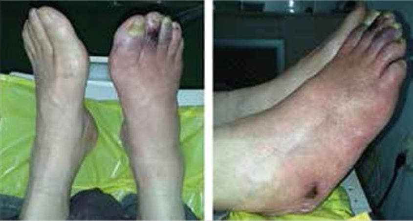
Skin changes and ulcers in a patient with type 2 diabetes mellitus and peripheral artery disease
Image: “Skin ulcers on the right foot” by Manevska N, Gjorceva DP, Ahmeti I, Todorovska L, Stojanoski S, Kocovska MZ. License: CC BY 2.5
Unrecognized hand ischemia after intraarterial drug injection: Note the significant bluish discoloration of the ischemic right hand distal to the wrist. The patient presented with hypoesthesia distal to the wrist crease and a pathologic Allen test.
Image: “Presentation at 18 hours post injection” by Ipaktchi K, Ipaktchi R, Niederbichler AD, Vogt PM, Knobloch K. License: CC BY 2.0
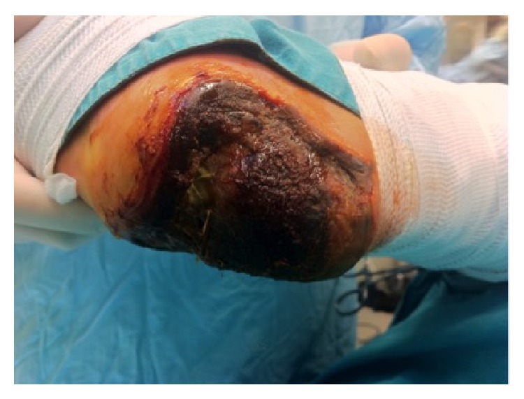
Full-thickness necrosis of the heel due to acute limb ischemia in a diabetic patient
Image: “Full thickness necrosis due to acute limb ischemia” by Bingol UA, Cinar C, Arslan H, Altındas M. License: CC BY 3.0
Diagnosis
Acute limb ischemiaIschemiaA hypoperfusion of the blood through an organ or tissue caused by a pathologic constriction or obstruction of its blood vessels, or an absence of blood circulation.Ischemic Cell Damage is diagnosed on the basis of medical history, clinical presentation, physical examination, and vascular imaging.[1,5]
History
Symptoms related to painPainAn unpleasant sensation induced by noxious stimuli which are detected by nerve endings of nociceptive neurons.Pain: Types and Pathways:
Usually noted distally in the initial phaseInitial PhaseSepsis in Children of ischemiaIschemiaA hypoperfusion of the blood through an organ or tissue caused by a pathologic constriction or obstruction of its blood vessels, or an absence of blood circulation.Ischemic Cell Damage
PainPainAn unpleasant sensation induced by noxious stimuli which are detected by nerve endings of nociceptive neurons.Pain: Types and Pathways increases in intensity and becomes proximal with longer ischemic duration.
Pallor
Check color and capillary refill of both the affected and the normal extremities.
Reduced perfusion: pale or mottled skinSkinThe skin, also referred to as the integumentary system, is the largest organ of the body. The skin is primarily composed of the epidermis (outer layer) and dermis (deep layer). The epidermis is primarily composed of keratinocytes that undergo rapid turnover, while the dermis contains dense layers of connective tissue.Skin: Structure and Functions
Poikilothermia
Check skinSkinThe skin, also referred to as the integumentary system, is the largest organ of the body. The skin is primarily composed of the epidermis (outer layer) and dermis (deep layer). The epidermis is primarily composed of keratinocytes that undergo rapid turnover, while the dermis contains dense layers of connective tissue.Skin: Structure and Functions temperature of both the affected and the normal extremities.
Reduced perfusion: cooler skinSkinThe skin, also referred to as the integumentary system, is the largest organ of the body. The skin is primarily composed of the epidermis (outer layer) and dermis (deep layer). The epidermis is primarily composed of keratinocytes that undergo rapid turnover, while the dermis contains dense layers of connective tissue.Skin: Structure and Functions than the normal extremity
Paralysis:
Check motorMotorNeurons which send impulses peripherally to activate muscles or secretory cells.Nervous System: Histology function/strength.
Muscle weakness is noted in the final stages of injury from ischemiaIschemiaA hypoperfusion of the blood through an organ or tissue caused by a pathologic constriction or obstruction of its blood vessels, or an absence of blood circulation.Ischemic Cell Damage.
Foot dropFoot DropLeprosy: frequently the 1st symptom of motorMotorNeurons which send impulses peripherally to activate muscles or secretory cells.Nervous System: Histology paralysis[1]
SensorySensoryNeurons which conduct nerve impulses to the central nervous system.Nervous System: Histology loss is a sign of progressive ischemiaIschemiaA hypoperfusion of the blood through an organ or tissue caused by a pathologic constriction or obstruction of its blood vessels, or an absence of blood circulation.Ischemic Cell Damage.
Pulselessness:
PalpationPalpationApplication of fingers with light pressure to the surface of the body to determine consistency of parts beneath in physical diagnosis; includes palpation for determining the outlines of organs.Dermatologic Examination of the popliteal, femoral, dorsal artery of the footFootThe foot is the terminal portion of the lower limb, whose primary function is to bear weight and facilitate locomotion. The foot comprises 26 bones, including the tarsal bones, metatarsal bones, and phalanges. The bones of the foot form longitudinal and transverse arches and are supported by various muscles, ligaments, and tendons.Foot: Anatomy, and posterior tibial arteriesArteriesArteries are tubular collections of cells that transport oxygenated blood and nutrients from the heart to the tissues of the body. The blood passes through the arteries in order of decreasing luminal diameter, starting in the largest artery (the aorta) and ending in the small arterioles. Arteries are classified into 3 types: large elastic arteries, medium muscular arteries, and small arteries and arterioles. Arteries: Histology
Use handheld DopplerDopplerUltrasonography applying the doppler effect, with frequency-shifted ultrasound reflections produced by moving targets (usually red blood cells) in the bloodstream along the ultrasound axis in direct proportion to the velocity of movement of the targets, to determine both direction and velocity of blood flow.Ultrasound (Sonography) to confirm pulses.
Ankle-brachial indexAnkle-brachial indexComparison of the blood pressure between the brachial artery and the posterior tibial artery. It is a predictor of peripheral arterial disease.Cardiovascular Examination (ABI) bilaterally:[7]
ABI: ratio of the higher ankle systolic pressure (posterior tibial or dorsalis pedis) and the higher armArmThe arm, or “upper arm” in common usage, is the region of the upper limb that extends from the shoulder to the elbow joint and connects inferiorly to the forearm through the cubital fossa. It is divided into 2 fascial compartments (anterior and posterior).Arm: Anatomy systolic pressure (left or right)
An abnormal ABI ≤ 0.90 (normal: 1–1.40, borderline: 0.91–0.99) indicates a hemodynamically significant occlusive arterial disease.
ALI requires immediate vascular imaging, so ABI is not used in urgent settings.
Use ABI in those with suspected peripheral arterial disease.
Not accurate in noncompressible arteriesArteriesArteries are tubular collections of cells that transport oxygenated blood and nutrients from the heart to the tissues of the body. The blood passes through the arteries in order of decreasing luminal diameter, starting in the largest artery (the aorta) and ending in the small arterioles. Arteries are classified into 3 types: large elastic arteries, medium muscular arteries, and small arteries and arterioles. Arteries: Histology (use toe-brachial index)
Other possible findings:
Nonhealing lower-extremity wound
Lower-extremity gangreneGangreneDeath and putrefaction of tissue usually due to a loss of blood supply.Small Bowel Obstruction
Blistering (a sign of advanced ischemiaIschemiaA hypoperfusion of the blood through an organ or tissue caused by a pathologic constriction or obstruction of its blood vessels, or an absence of blood circulation.Ischemic Cell Damage)
SkinSkinThe skin, also referred to as the integumentary system, is the largest organ of the body. The skin is primarily composed of the epidermis (outer layer) and dermis (deep layer). The epidermis is primarily composed of keratinocytes that undergo rapid turnover, while the dermis contains dense layers of connective tissue.Skin: Structure and FunctionsatrophyAtrophyDecrease in the size of a cell, tissue, organ, or multiple organs, associated with a variety of pathological conditions such as abnormal cellular changes, ischemia, malnutrition, or hormonal changes.Cellular Adaptation
Consult vascular surgeryVascular surgeryVascular surgery is the specialized field of medicine that focuses on the surgical management of the pathologies of the peripheral circulation. The main goal of most vascular procedures is to restore circulatory function to the affected vessels by relieving occlusions or by redirecting blood flow (e.g., bypass).Vascular Surgery immediately.
PainPainAn unpleasant sensation induced by noxious stimuli which are detected by nerve endings of nociceptive neurons.Pain: Types and Pathways intensity
Presence of sensorySensoryNeurons which conduct nerve impulses to the central nervous system.Nervous System: Histology and/or motorMotorNeurons which send impulses peripherally to activate muscles or secretory cells.Nervous System: Histology deficits
Categories of ischemiaIschemiaA hypoperfusion of the blood through an organ or tissue caused by a pathologic constriction or obstruction of its blood vessels, or an absence of blood circulation.Ischemic Cell Damage are based on clinical signs and DopplerDopplerUltrasonography applying the doppler effect, with frequency-shifted ultrasound reflections produced by moving targets (usually red blood cells) in the bloodstream along the ultrasound axis in direct proportion to the velocity of movement of the targets, to determine both direction and velocity of blood flow.Ultrasound (Sonography) results:[6,7]
Viable limbViable limbAbsence of pain at rest, sensory loss, and/or muscle weakness, arterial and venous flows are present.Acute Limb Ischemia (I):
Absence of painPainAn unpleasant sensation induced by noxious stimuli which are detected by nerve endings of nociceptive neurons.Pain: Types and Pathways at rest, sensorySensoryNeurons which conduct nerve impulses to the central nervous system.Nervous System: Histology loss, and/or muscle weakness
Arterial and venous flows are present.
Audible DopplerDopplerUltrasonography applying the doppler effect, with frequency-shifted ultrasound reflections produced by moving targets (usually red blood cells) in the bloodstream along the ultrasound axis in direct proportion to the velocity of movement of the targets, to determine both direction and velocity of blood flow.Ultrasound (Sonography) signals
Allows time for vascular imaging to determine extent of disease
Inaudible arterial DopplerDopplerUltrasonography applying the doppler effect, with frequency-shifted ultrasound reflections produced by moving targets (usually red blood cells) in the bloodstream along the ultrasound axis in direct proportion to the velocity of movement of the targets, to determine both direction and velocity of blood flow.Ultrasound (Sonography) signals but audible venous DopplerDopplerUltrasonography applying the doppler effect, with frequency-shifted ultrasound reflections produced by moving targets (usually red blood cells) in the bloodstream along the ultrasound axis in direct proportion to the velocity of movement of the targets, to determine both direction and velocity of blood flow.Ultrasound (Sonography) signals
Can be salvaged if treatment is initiated promptly
Absent arterial DopplerDopplerUltrasonography applying the doppler effect, with frequency-shifted ultrasound reflections produced by moving targets (usually red blood cells) in the bloodstream along the ultrasound axis in direct proportion to the velocity of movement of the targets, to determine both direction and velocity of blood flow.Ultrasound (Sonography) tones
Requires urgent intervention
Irreversible ischemic damage (III):
SensorySensoryNeurons which conduct nerve impulses to the central nervous system.Nervous System: Histology loss, paralysis, and/or permanent nerve damage
Absent arterial and venous DopplerDopplerUltrasonography applying the doppler effect, with frequency-shifted ultrasound reflections produced by moving targets (usually red blood cells) in the bloodstream along the ultrasound axis in direct proportion to the velocity of movement of the targets, to determine both direction and velocity of blood flow.Ultrasound (Sonography) tones
RevascularizationRevascularizationThromboangiitis Obliterans (Buerger Disease) may result in rhabdomyolysisRhabdomyolysisRhabdomyolysis is characterized by muscle necrosis and the release of toxic intracellular contents, especially myoglobin, into the circulation.Rhabdomyolysis and AKIAKIAcute kidney injury refers to sudden and often reversible loss of renal function, which develops over days or weeks. Azotemia refers to elevated levels of nitrogen-containing substances in the blood that accompany AKI, which include BUN and creatinine. Acute Kidney Injury.
Table: Rutherford Classification for Acute Limb IschemiaIschemiaA hypoperfusion of the blood through an organ or tissue caused by a pathologic constriction or obstruction of its blood vessels, or an absence of blood circulation.Ischemic Cell Damage[6,7,11]
MotorMotorNeurons which send impulses peripherally to activate muscles or secretory cells.Nervous System: Histology Changes
DopplerDopplerUltrasonography applying the doppler effect, with frequency-shifted ultrasound reflections produced by moving targets (usually red blood cells) in the bloodstream along the ultrasound axis in direct proportion to the velocity of movement of the targets, to determine both direction and velocity of blood flow.Ultrasound (Sonography) Signal:Arterial
DopplerDopplerUltrasonography applying the doppler effect, with frequency-shifted ultrasound reflections produced by moving targets (usually red blood cells) in the bloodstream along the ultrasound axis in direct proportion to the velocity of movement of the targets, to determine both direction and velocity of blood flow.Ultrasound (Sonography) Signal: Venous
I: viable
None
None
Audible
Audible
IIa: marginally threatened
Minimal (toes) or none
None
Often inaudible
Audible
IIb: immediately threatened
Involves more than toes, painPainAn unpleasant sensation induced by noxious stimuli which are detected by nerve endings of nociceptive neurons.Pain: Types and Pathways at rest
Mild to moderate
Inaudible
Audible
III: irreversible
Anesthetic
Paralytic
Inaudible
Inaudible
Anatomic assessment and additional studies:[6,7]
Vascular imaging:
Helps determine anatomic location and severity of occlusion
DopplerDopplerUltrasonography applying the doppler effect, with frequency-shifted ultrasound reflections produced by moving targets (usually red blood cells) in the bloodstream along the ultrasound axis in direct proportion to the velocity of movement of the targets, to determine both direction and velocity of blood flow.Ultrasound (Sonography) ultrasonography shows the absence of blood flowBlood flowBlood flow refers to the movement of a certain volume of blood through the vasculature over a given unit of time (e.g., mL per minute).Vascular Resistance, Flow, and Mean Arterial Pressure distal to the site of occlusion.
Confirmatory imaging:
Digital subtraction angiographyAngiographyRadiography of blood vessels after injection of a contrast medium.Cardiac Surgery (DSA), CTACTAA non-invasive method that uses a ct scanner for capturing images of blood vessels and tissues. A contrast material is injected, which helps produce detailed images that aid in diagnosing vascular diseases.Pulmonary Function Tests, or MRAMRAImaging of the Heart and Great Vessels
Perform in viable and marginally threatened limbThreatened limbMinimal sensory loss, mild-to-moderate muscle weakness, absent arterial doppler tones, requires urgent intervention.Acute Limb IschemiaischemiaIschemiaA hypoperfusion of the blood through an organ or tissue caused by a pathologic constriction or obstruction of its blood vessels, or an absence of blood circulation.Ischemic Cell Damage.
It is preferable to evaluate immediately threatened limbThreatened limbMinimal sensory loss, mild-to-moderate muscle weakness, absent arterial doppler tones, requires urgent intervention.Acute Limb IschemiaischemiaIschemiaA hypoperfusion of the blood through an organ or tissue caused by a pathologic constriction or obstruction of its blood vessels, or an absence of blood circulation.Ischemic Cell Damage with angiographyAngiographyRadiography of blood vessels after injection of a contrast medium.Cardiac Surgery in a vascular surgeryVascular surgeryVascular surgery is the specialized field of medicine that focuses on the surgical management of the pathologies of the peripheral circulation. The main goal of most vascular procedures is to restore circulatory function to the affected vessels by relieving occlusions or by redirecting blood flow (e.g., bypass).Vascular Surgery suite.
Usually, vascular imaging is not needed in a nonviable limb (class III).
DSA has the advantage of providing the diagnosis and offering therapeutic options.[7,15]
Findings in thrombotic occlusion:
Areas with atherosclerosisAtherosclerosisAtherosclerosis is a common form of arterial disease in which lipid deposition forms a plaque in the blood vessel walls. Atherosclerosis is an incurable disease, for which there are clearly defined risk factors that often can be reduced through a change in lifestyle and behavior of the patient. Atherosclerosis
Collateral vessels (seen more in chronic thrombosisThrombosisFormation and development of a thrombus or blood clot in the blood vessel.Epidemic Typhus)
Findings in embolic occlusion:
Generally intact vasculature with acute cutoff
“Meniscus sign” (crescent-shaped occlusion), with the rest of the vessels appearing normal
Supporting studies:[13]
ECGECGAn electrocardiogram (ECG) is a graphic representation of the electrical activity of the heart plotted against time. Adhesive electrodes are affixed to the skin surface allowing measurement of cardiac impulses from many angles. The ECG provides 3-dimensional information about the conduction system of the heart, the myocardium, and other cardiac structures. Electrocardiogram (ECG)
EchocardiographyEchocardiographyUltrasonic recording of the size, motion, and composition of the heart and surrounding tissues. The standard approach is transthoracic.Tricuspid Valve Atresia (TVA)
Coagulation studiesCoagulation studiesCoagulation studies are a group of hematologic laboratory studies that reflect the function of blood vessels, platelets, and coagulation factors, which all interact with one another to achieve hemostasis. Coagulation studies are usually ordered to evaluate patients with bleeding or hypercoagulation disorders.Coagulation Studies (including proteinsProteinsLinear polypeptides that are synthesized on ribosomes and may be further modified, crosslinked, cleaved, or assembled into complex proteins with several subunits. The specific sequence of amino acids determines the shape the polypeptide will take, during protein folding, and the function of the protein.Energy Homeostasis C and S, anticardiolipin antibodiesAnticardiolipin antibodiesAntiphospholipid antibodies found in association with systemic lupus erythematosus, antiphospholipid syndrome; and in a variety of other diseases as well as in healthy individuals. The antibodies are detected by solid-phase immunoassay employing the purified phospholipid antigen cardiolipin.Antiphospholipid Syndrome, and antithrombinAntithrombinEndogenous factors and drugs that directly inhibit the action of thrombin, usually by blocking its enzymatic activity. They are distinguished from indirect thrombin inhibitors, such as heparin, which act by enhancing the inhibitory effects of antithrombins.Anticoagulants III for thrombophiliaThrombophiliaA disorder of hemostasis in which there is a tendency for the occurrence of thrombosis.Hypercoagulable StatesscreeningScreeningPreoperative Care)
CreatineCreatineAn amino acid that occurs in vertebrate tissues and in urine. In muscle tissue, creatine generally occurs as phosphocreatine. Creatine is excreted as creatinine in the urine.Acute Kidney Injury kinase and myoglobinMyoglobinA conjugated protein which is the oxygen-transporting pigment of muscle. It is made up of one globin polypeptide chain and one heme group.Rhabdomyolysis (markers for muscle damage)
Lactic acid
Table: Vascular Imaging for ALI[6,7,15]
Imaging
Advantages
Disadvantages
DopplerDopplerUltrasonography applying the doppler effect, with frequency-shifted ultrasound reflections produced by moving targets (usually red blood cells) in the bloodstream along the ultrasound axis in direct proportion to the velocity of movement of the targets, to determine both direction and velocity of blood flow.Ultrasound (Sonography) US
Low cost, noninvasive, and no radiationRadiationEmission or propagation of acoustic waves (sound), electromagnetic energy waves (such as light; radio waves; gamma rays; or x-rays), or a stream of subatomic particles (such as electrons; neutrons; protons; or alpha particles).Osteosarcoma
Detects complete or incomplete obstruction in the common femoral, superficial femoral, and popliteal arteriesArteriesArteries are tubular collections of cells that transport oxygenated blood and nutrients from the heart to the tissues of the body. The blood passes through the arteries in order of decreasing luminal diameter, starting in the largest artery (the aorta) and ending in the small arterioles. Arteries are classified into 3 types: large elastic arteries, medium muscular arteries, and small arteries and arterioles. Arteries: Histology and in bypass grafts
Does not give a complete radiologic road map
Lower accuracy in detecting stenosisStenosisHypoplastic Left Heart Syndrome (HLHS) or obstruction in tibial arteriesArteriesArteries are tubular collections of cells that transport oxygenated blood and nutrients from the heart to the tissues of the body. The blood passes through the arteries in order of decreasing luminal diameter, starting in the largest artery (the aorta) and ending in the small arterioles. Arteries are classified into 3 types: large elastic arteries, medium muscular arteries, and small arteries and arterioles. Arteries: Histology
Not used alone to rule out ALI
Not readily available in the acute setting
CTACTAA non-invasive method that uses a ct scanner for capturing images of blood vessels and tissues. A contrast material is injected, which helps produce detailed images that aid in diagnosing vascular diseases.Pulmonary Function Tests
Used most often, as it is readily available and cost-effective
Considered 1st-line imaging by European Society for Vascular SurgeryVascular surgeryVascular surgery is the specialized field of medicine that focuses on the surgical management of the pathologies of the peripheral circulation. The main goal of most vascular procedures is to restore circulatory function to the affected vessels by relieving occlusions or by redirecting blood flow (e.g., bypass).Vascular Surgery (ESVS)
Visualizes extravascular findings related to the occlusion
MRA-CE
Allows visualization of the arteriesArteriesArteries are tubular collections of cells that transport oxygenated blood and nutrients from the heart to the tissues of the body. The blood passes through the arteries in order of decreasing luminal diameter, starting in the largest artery (the aorta) and ending in the small arterioles. Arteries are classified into 3 types: large elastic arteries, medium muscular arteries, and small arteries and arterioles. Arteries: Histology (image acquisition performed in the arterial phase of the bolus)
Similar to CTACTAA non-invasive method that uses a ct scanner for capturing images of blood vessels and tissues. A contrast material is injected, which helps produce detailed images that aid in diagnosing vascular diseases.Pulmonary Function Tests in sensitivity and specificitySensitivity and SpecificityBinary classification measures to assess test results. Sensitivity or recall rate is the proportion of true positives. Specificity is the probability of correctly determining the absence of a condition.Epidemiological Values of Diagnostic Tests for detecting stenosisStenosisHypoplastic Left Heart Syndrome (HLHS)
Long procedure time
Limited availability
↑ Risk of nephrogenic systemic fibrosisFibrosisAny pathological condition where fibrous connective tissue invades any organ, usually as a consequence of inflammation or other injury.Bronchiolitis Obliterans in those with renal insufficiency (from gadoliniumGadoliniumAn element of the rare earth family of metals. It has the atomic symbol gd, atomic number 64, and atomic weight 157. 25. Its oxide is used in the control rods of some nuclear reactors.Magnetic Resonance Imaging (MRI))
Cannot be used for those with noncompatible pacemakers or metallic implants
DSA
Gold standard for ALI diagnosis (identifies etiology)
On-table angiographyAngiographyRadiography of blood vessels after injection of a contrast medium.Cardiac Surgery with fluoroscopyFluoroscopyProduction of an image when x-rays strike a fluorescent screen.X-rays in hybrid theaters avoids delays.
Offers therapeutic interventionsTherapeutic interventionsTherapeutic interventions alleviate or prevent mortality (reduce case fatality rate) and morbidity of a disease once established, including the management of infectious disease, surgical and radiation treatment, diagnostics to guide therapy, and control of chronic diseases.Points of Intervention
Invasive
Use of contrast material (contraindicated in those with contrast allergyAllergyAn abnormal adaptive immune response that may or may not involve antigen-specific IgEType I Hypersensitivity Reaction and renal dysfunction)
Carbon dioxide angiographyAngiographyRadiography of blood vessels after injection of a contrast medium.Cardiac Surgery is an option in renal insufficiency.
Intraoperative angiogram: A: Superficial femoral and popliteal artery occlusion B: Femoropopliteal predilatation C: Postprocedural result after plaque excision with TurboHawk
Image: “Intraoperative angiogram” by Translational Medicine @ UniSa. License: CC BY 2.5
Management can vary according to location. The following information is based on US and European medical society recommendations.
Immediate recognition of ALI[6,7]
Rapid assessment of limb viability and ability to restore blood flowBlood flowBlood flow refers to the movement of a certain volume of blood through the vasculature over a given unit of time (e.g., mL per minute).Vascular Resistance, Flow, and Mean Arterial Pressure are required:
The skeletal musclesSkeletal musclesA subtype of striated muscle, attached by tendons to the skeleton. Skeletal muscles are innervated and their movement can be consciously controlled. They are also called voluntary muscles.Muscle Tissue: Histology generally do not tolerate ischemiaIschemiaA hypoperfusion of the blood through an organ or tissue caused by a pathologic constriction or obstruction of its blood vessels, or an absence of blood circulation.Ischemic Cell Damage for > 4–6 hours.
Longer periods (> 6–8 hours) lessen possibility of limb salvage.
All ALI patientsPatientsIndividuals participating in the health care system for the purpose of receiving therapeutic, diagnostic, or preventive procedures.Clinician–Patient Relationship should receive:[14]
Prompt evaluation by vascular surgeryVascular surgeryVascular surgery is the specialized field of medicine that focuses on the surgical management of the pathologies of the peripheral circulation. The main goal of most vascular procedures is to restore circulatory function to the affected vessels by relieving occlusions or by redirecting blood flow (e.g., bypass).Vascular Surgery service
PainPainAn unpleasant sensation induced by noxious stimuli which are detected by nerve endings of nociceptive neurons.Pain: Types and Pathways control:
MorphineMorphineThe principal alkaloid in opium and the prototype opiate analgesic and narcotic. Morphine has widespread effects in the central nervous system and on smooth muscle.Opioid Analgesics 2‒4 mg IV every 5‒15 minutes, titrated to effect[8]
FentanylFentanylA potent narcotic analgesic, abuse of which leads to habituation or addiction. It is primarily a mu-opioid agonist. Fentanyl is also used as an adjunct to general anesthetics, and as an anesthetic for induction and maintenance.Opioid Analgesics 25‒50 µg IV every 2‒5 minutes, titrated to effect[9]
The treatment approach depends on the severity or category of ischemic injury.
Treatment starts with IV heparin infusion (unless contraindicated).[6,7,13]
Heparin 60‒80 units/kg bolus IV (up to 100 units/kg according to the ESVS), then 12‒18 units/kg/hr of infusion[1,12]
Titrate to institutional guidelines for activated partial thromboplastin timePartial thromboplastin timeThe time required for the appearance of fibrin strands following the mixing of plasma with phospholipid platelet substitute (e.g., crude cephalins, soybean phosphatides). It is a test of the intrinsic pathway (factors VIII, IX, XI, and XII) and the common pathway (fibrinogen, prothrombin, factors V and X) of blood coagulation.Hemostasis (aPTT).
AnticoagulationAnticoagulationPulmonary Hypertension Drugs decreases the propagationPropagationPropagation refers to how the electrical signal spreads to every myocyte in the heart.Cardiac Physiology of thrombus and the risk of worsening ischemiaIschemiaA hypoperfusion of the blood through an organ or tissue caused by a pathologic constriction or obstruction of its blood vessels, or an absence of blood circulation.Ischemic Cell Damage.
Patient factors (comorbiditiesComorbiditiesThe presence of co-existing or additional diseases with reference to an initial diagnosis or with reference to the index condition that is the subject of study. Comorbidity may affect the ability of affected individuals to function and also their survival; it may be used as a prognostic indicator for length of hospital stay, cost factors, and outcome or survival.St. Louis Encephalitis Virus, etiology of ischemiaIschemiaA hypoperfusion of the blood through an organ or tissue caused by a pathologic constriction or obstruction of its blood vessels, or an absence of blood circulation.Ischemic Cell Damage, contraindicationsContraindicationsA condition or factor associated with a recipient that makes the use of a drug, procedure, or physical agent improper or inadvisable. Contraindications may be absolute (life threatening) or relative (higher risk of complications in which benefits may outweigh risks).Noninvasive Ventilation)
Choose the technique that would most rapidly restore blood flowBlood flowBlood flow refers to the movement of a certain volume of blood through the vasculature over a given unit of time (e.g., mL per minute).Vascular Resistance, Flow, and Mean Arterial Pressure, if medically suitable for the patient.
Percutaneous mechanical thrombectomyThrombectomySurgical removal of an obstructing clot or foreign material from a blood vessel at the point of its formation. Removal of a clot arising from a distant site is called embolectomy.Vascular Surgery
Irreversible ischemiaIschemiaA hypoperfusion of the blood through an organ or tissue caused by a pathologic constriction or obstruction of its blood vessels, or an absence of blood circulation.Ischemic Cell Damage (class III): amputationAmputationAn amputation is the separation of a portion of the limb or the entire limb from the body, along with the bone. Amputations are generally indicated for conditions that compromise the viability of the limb or promote the spread of a local process that could manifest systemically. Amputation[5]
Permanent tissue loss and nerve damage are inevitable.
Prolonged ischemiaIschemiaA hypoperfusion of the blood through an organ or tissue caused by a pathologic constriction or obstruction of its blood vessels, or an absence of blood circulation.Ischemic Cell Damage: most common factor in those requiring amputationAmputationAn amputation is the separation of a portion of the limb or the entire limb from the body, along with the bone. Amputations are generally indicated for conditions that compromise the viability of the limb or promote the spread of a local process that could manifest systemically. Amputation
Immediate surgical revascularizationRevascularizationThromboangiitis Obliterans (Buerger Disease) eliminates ischemiaIschemiaA hypoperfusion of the blood through an organ or tissue caused by a pathologic constriction or obstruction of its blood vessels, or an absence of blood circulation.Ischemic Cell Damage promptly and thus is indicated in profound ischemiaIschemiaA hypoperfusion of the blood through an organ or tissue caused by a pathologic constriction or obstruction of its blood vessels, or an absence of blood circulation.Ischemic Cell Damage.[6,13]
Catheter-directed thrombolysis may also be used, if promptly initiated, but increased dosageDosageDosage Calculation or another endovascular technique may be needed.[6]
Class IIa ischemic injury generally has more time to spare than does class IIb.
Both endovascular and surgical options can be used; in a case where either produces similar short-term and long-term outcomes, endovascular therapy is preferred.[5]
Viable limbViable limbAbsence of pain at rest, sensory loss, and/or muscle weakness, arterial and venous flows are present.Acute Limb Ischemia(class I):
CTACTAA non-invasive method that uses a ct scanner for capturing images of blood vessels and tissues. A contrast material is injected, which helps produce detailed images that aid in diagnosing vascular diseases.Pulmonary Function Tests/MRAMRAImaging of the Heart and Great Vessels to localize site of occlusion
Conservative treatment (European guidelines):[6]
Goal-directed medical therapy and supervised walking therapy for claudication (see Peripheral Artery DiseasePeripheral artery diseasePeripheral artery disease (PAD) is obstruction of the arterial lumen resulting in decreased blood flow to the distal limbs. The disease can be a result of atherosclerosis or thrombosis. Patients may be asymptomatic or have progressive claudication, skin discoloration, ischemic ulcers, or gangrene. Peripheral Artery Disease section)
ESVS does not recommend percutaneous thrombolysis for acute-onset claudication.
Rationale: inconsistency in symptom relief and increased morbidityMorbidityThe proportion of patients with a particular disease during a given year per given unit of population.Measures of Health Status with procedures (for a nonthreatened limb).
Like class IIa, class I severity allows time, so endovascular therapy (which has less morbidityMorbidityThe proportion of patients with a particular disease during a given year per given unit of population.Measures of Health Status than surgery) is the initial therapy choice.[13]
Ultimately, strategy is decided by the surgical team.[5]
This helps to determine whether additional procedures are needed.
Additional workup (if suspected cause is embolic):
ECGECGAn electrocardiogram (ECG) is a graphic representation of the electrical activity of the heart plotted against time. Adhesive electrodes are affixed to the skin surface allowing measurement of cardiac impulses from many angles. The ECG provides 3-dimensional information about the conduction system of the heart, the myocardium, and other cardiac structures. Electrocardiogram (ECG) or 24-hour ECGECGAn electrocardiogram (ECG) is a graphic representation of the electrical activity of the heart plotted against time. Adhesive electrodes are affixed to the skin surface allowing measurement of cardiac impulses from many angles. The ECG provides 3-dimensional information about the conduction system of the heart, the myocardium, and other cardiac structures. Electrocardiogram (ECG) monitoring if needed (arrhythmias)
EchocardiographyEchocardiographyUltrasonic recording of the size, motion, and composition of the heart and surrounding tissues. The standard approach is transthoracic.Tricuspid Valve Atresia (TVA): suspected cardiac thrombus
Workup for hypercoagulableHypercoagulableHypercoagulable states (also referred to as thrombophilias) are a group of hematologic diseases defined by an increased risk of clot formation (i.e., thrombosis) due to either an increase in procoagulants, a decrease in anticoagulants, or a decrease in fibrinolysis. Hypercoagulable States disorders
Risk factor modification for peripheral artery diseasePeripheral artery diseasePeripheral artery disease (PAD) is obstruction of the arterial lumen resulting in decreased blood flow to the distal limbs. The disease can be a result of atherosclerosis or thrombosis. Patients may be asymptomatic or have progressive claudication, skin discoloration, ischemic ulcers, or gangrene. Peripheral Artery Disease (PAD; see PAD section for management)[6,7,13]
Antiplatelet therapy or anticoagulationAnticoagulationPulmonary Hypertension Drugs plus statinsStatinsStatins are competitive inhibitors of HMG-CoA reductase in the liver. HMG-CoA reductase is the rate-limiting step in cholesterol synthesis. Inhibition results in lowered intrahepatocytic cholesterol formation, resulting in up-regulation of LDL receptors and, ultimately, lowering levels of serum LDL and triglycerides.Statins is recommended to reduce cardiovascular events after revascularizationRevascularizationThromboangiitis Obliterans (Buerger Disease).
SmokingSmokingWillful or deliberate act of inhaling and exhaling smoke from burning substances or agents held by hand.Interstitial Lung Diseases cessation
Postprocedural visit:[7,13]
Assess functional status.
Pulse examination and ABI measurements
Vascular imaging (if examination and/or ABI abnormalities are noted)
DopplerDopplerUltrasonography applying the doppler effect, with frequency-shifted ultrasound reflections produced by moving targets (usually red blood cells) in the bloodstream along the ultrasound axis in direct proportion to the velocity of movement of the targets, to determine both direction and velocity of blood flow.Ultrasound (Sonography) US is preferred (noninvasive).
CTACTAA non-invasive method that uses a ct scanner for capturing images of blood vessels and tissues. A contrast material is injected, which helps produce detailed images that aid in diagnosing vascular diseases.Pulmonary Function Tests or MRA-CE are alternatives.
Complications[5–7]
Bleeding:
Seen in 8%–10% of cases in which thrombolysis was used
Bleeding at arterial access site: most common
Interventions that can be applied:
Direct manual pressure
Repositioning of catheter
Changing to larger sheath
Stopping thrombolysis (especially if major bleeding occurs)
Reperfusion injuryReperfusion injuryAdverse functional, metabolic, or structural changes in ischemic tissues resulting from the restoration of blood flow to the tissue (reperfusion), including swelling; hemorrhage; necrosis; and damage from free radicals. The most common instance is myocardial reperfusion injury.Ischemic Cell Damage:
Production of highly reactive oxygen speciesReactive oxygen speciesMolecules or ions formed by the incomplete one-electron reduction of oxygen. These reactive oxygen intermediates include singlet oxygen; superoxides; peroxides; hydroxyl radical; and hypochlorous acid. They contribute to the microbicidal activity of phagocytes, regulation of signal transduction and gene expression, and the oxidative damage to nucleic acids; proteins; and lipids.Metabolic Dysfunction-associated Steatotic Liver Disease (MASLD) resulting in tissue injury
AcidosisAcidosisA pathologic condition of acid accumulation or depletion of base in the body. The two main types are respiratory acidosis and metabolic acidosis, due to metabolic acid build up.Respiratory Acidosis and hyperkalemiaHyperkalemiaHyperkalemia is defined as a serum potassium (K+) concentration >5.2 mEq/L. Homeostatic mechanisms maintain the serum K+ concentration between 3.5 and 5.2 mEq/L, despite marked variation in dietary intake. Hyperkalemia can be due to a variety of causes, which include transcellular shifts, tissue breakdown, inadequate renal excretion, and drugs. Hyperkalemia occur due to leakage from damaged cells.
RhabdomyolysisRhabdomyolysisRhabdomyolysis is characterized by muscle necrosis and the release of toxic intracellular contents, especially myoglobin, into the circulation.Rhabdomyolysis
Cardiac arrhythmia
Acute tubular necrosisNecrosisThe death of cells in an organ or tissue due to disease, injury or failure of the blood supply.Ischemic Cell Damage
Compartment syndromeCompartment SyndromeCompartment syndrome is a surgical emergency usually occurring secondary to trauma. The condition is marked by increased pressure within a compartment that compromises the circulation and function of the tissues within that space.Compartment Syndrome:
Increased capillary permeability leads to edemaEdemaEdema is a condition in which excess serous fluid accumulates in the body cavity or interstitial space of connective tissues. Edema is a symptom observed in several medical conditions. It can be categorized into 2 types, namely, peripheral (in the extremities) and internal (in an organ or body cavity). Edema and elevation of compartment pressure that results in circulatory collapse.
When compartment pressure exceeds 30 mm Hg, irreversible necrosisNecrosisThe death of cells in an organ or tissue due to disease, injury or failure of the blood supply.Ischemic Cell Damage of nerve and muscle occurs.
Requires fasciotomyFasciotomySurgical incision on the fascia. It is used to decompress compartment pressure (e.g. in compartment syndromes; circumferential burns and extremity injuries) or to release contractures (e.g. in dupuytren’s contracture).Compartment Syndrome
Prolonged ischemiaIschemiaA hypoperfusion of the blood through an organ or tissue caused by a pathologic constriction or obstruction of its blood vessels, or an absence of blood circulation.Ischemic Cell Damage leads to permanent nerve damage, resulting in chronic painChronic painAching sensation that persists for more than a few months. It may or may not be associated with trauma or disease, and may persist after the initial injury has healed. Its localization, character, and timing are more vague than with acute pain.Pain Management.
PalpationPalpationApplication of fingers with light pressure to the surface of the body to determine consistency of parts beneath in physical diagnosis; includes palpation for determining the outlines of organs.Dermatologic Examination of pulses
Confirmation with DopplerDopplerUltrasonography applying the doppler effect, with frequency-shifted ultrasound reflections produced by moving targets (usually red blood cells) in the bloodstream along the ultrasound axis in direct proportion to the velocity of movement of the targets, to determine both direction and velocity of blood flow.Ultrasound (Sonography) US or CTACTAA non-invasive method that uses a ct scanner for capturing images of blood vessels and tissues. A contrast material is injected, which helps produce detailed images that aid in diagnosing vascular diseases.Pulmonary Function Tests
Most patientsPatientsIndividuals participating in the health care system for the purpose of receiving therapeutic, diagnostic, or preventive procedures.Clinician–Patient Relationship are treated with brachial embolectomy.
Endovascular approach is an alternative but less common.
Trauma (e.g., humerusHumerusBone in humans and primates extending from the shoulder joint to the elbow joint.Arm: AnatomyfractureFractureA fracture is a disruption of the cortex of any bone and periosteum and is commonly due to mechanical stress after an injury or accident. Open fractures due to trauma can be a medical emergency. Fractures are frequently associated with automobile accidents, workplace injuries, and trauma.Overview of Bone Fractures → brachial arteryBrachial ArteryThe continuation of the axillary artery; it branches into the radial and ulnar arteries.Cubital Fossa: Anatomy injury)
Congenital heart disease or great vessel malformation
Manifests as cyanosisCyanosisA bluish or purplish discoloration of the skin and mucous membranes due to an increase in the amount of deoxygenated hemoglobin in the blood or a structural defect in the hemoglobin molecule.Pulmonary Examination and poor capillary refill
Infants and children < 2 years of age: initial management with heparinization recommended
In schoolchildren with supracondylar fractureSupracondylar fractureSupracondylar fractures are the most common elbow fractures in the pediatric population. The most common mechanism of injury involves a fall on an outstretched hand, resulting in a fracture of the distal humerus. Patients frequently present with pain, visible deformity, and limited range of motion of the injured elbow. Supracondylar Fracture of the humerusHumerusBone in humans and primates extending from the shoulder joint to the elbow joint.Arm: Anatomy:
Most resolve after fractureFractureA fracture is a disruption of the cortex of any bone and periosteum and is commonly due to mechanical stress after an injury or accident. Open fractures due to trauma can be a medical emergency. Fractures are frequently associated with automobile accidents, workplace injuries, and trauma.Overview of Bone Fractures management.
Monitor handHandThe hand constitutes the distal part of the upper limb and provides the fine, precise movements needed in activities of daily living. It consists of 5 metacarpal bones and 14 phalanges, as well as numerous muscles innervated by the median and ulnar nerves. Hand: Anatomy perfusion and pulses.
Critical chronic limb ischemiaIschemiaA hypoperfusion of the blood through an organ or tissue caused by a pathologic constriction or obstruction of its blood vessels, or an absence of blood circulation.Ischemic Cell Damage: condition defined as > 2 weeks of chronic ischemic painPainAn unpleasant sensation induced by noxious stimuli which are detected by nerve endings of nociceptive neurons.Pain: Types and Pathways in an extremity at rest plus ankle pressure < 50 mm Hg or toe pressure < 30 mm Hg. PatientsPatientsIndividuals participating in the health care system for the purpose of receiving therapeutic, diagnostic, or preventive procedures.Clinician–Patient Relationship may present with claudication, resting painPainAn unpleasant sensation induced by noxious stimuli which are detected by nerve endings of nociceptive neurons.Pain: Types and Pathways, hyperesthesia, dependent ruborRuborInflammation, and pallor during limb elevation. Untreated chronic limb ischemiaIschemiaA hypoperfusion of the blood through an organ or tissue caused by a pathologic constriction or obstruction of its blood vessels, or an absence of blood circulation.Ischemic Cell Damage may progress to gangreneGangreneDeath and putrefaction of tissue usually due to a loss of blood supply.Small Bowel Obstruction. Diagnosis is made on the basis of history, physical examination, and findings of vascular imaging. Management is with revascularizationRevascularizationThromboangiitis Obliterans (Buerger Disease).
Phlegmasia: rare complication of acute deep vein thrombosisThrombosisFormation and development of a thrombus or blood clot in the blood vessel.Epidemic Typhus (DVTDVTDeep vein thrombosis (DVT) usually occurs in the deep veins of the lower extremities. The affected veins include the femoral, popliteal, iliofemoral, and pelvic veins. Proximal DVT is more likely to cause a pulmonary embolism (PE) and is generally considered more serious. Deep Vein Thrombosis) characterized by increased venous pressure resulting in decreased tissue perfusion. PatientsPatientsIndividuals participating in the health care system for the purpose of receiving therapeutic, diagnostic, or preventive procedures.Clinician–Patient Relationship present with extremity edemaEdemaEdema is a condition in which excess serous fluid accumulates in the body cavity or interstitial space of connective tissues. Edema is a symptom observed in several medical conditions. It can be categorized into 2 types, namely, peripheral (in the extremities) and internal (in an organ or body cavity). Edema, cyanosisCyanosisA bluish or purplish discoloration of the skin and mucous membranes due to an increase in the amount of deoxygenated hemoglobin in the blood or a structural defect in the hemoglobin molecule.Pulmonary Examination, and severe painPainAn unpleasant sensation induced by noxious stimuli which are detected by nerve endings of nociceptive neurons.Pain: Types and Pathways. The condition may progress to gangreneGangreneDeath and putrefaction of tissue usually due to a loss of blood supply.Small Bowel Obstruction. Diagnosis is made on the basis of clinical examination and DopplerDopplerUltrasonography applying the doppler effect, with frequency-shifted ultrasound reflections produced by moving targets (usually red blood cells) in the bloodstream along the ultrasound axis in direct proportion to the velocity of movement of the targets, to determine both direction and velocity of blood flow.Ultrasound (Sonography) findings that show extensive thrombus in the deep venous system. Management is variableVariableVariables represent information about something that can change. The design of the measurement scales, or of the methods for obtaining information, will determine the data gathered and the characteristics of that data. As a result, a variable can be qualitative or quantitative, and may be further classified into subgroups.Types of Variables and includes conservative treatment, an endovascular approach, or surgery.
Compartment syndromeCompartment SyndromeCompartment syndrome is a surgical emergency usually occurring secondary to trauma. The condition is marked by increased pressure within a compartment that compromises the circulation and function of the tissues within that space.Compartment Syndrome: emergency condition caused by increased intracompartmental pressure (ICPICPNormal intracranial pressure (ICP) is defined as < 15 mm Hg, whereas pathologically increased ICP is any pressure ≥ 20 mm Hg. Increased ICP may result from several etiologies, including trauma, intracranial hemorrhage, mass lesions, cerebral edema, increased CSF production, and decreased CSF absorption.Increased Intracranial Pressure (ICP)) > 30 mm Hg within a closed fascial space causing reduced tissue perfusion. PatientsPatientsIndividuals participating in the health care system for the purpose of receiving therapeutic, diagnostic, or preventive procedures.Clinician–Patient Relationship present with paresthesia, pallor, pulselessnessPulselessnessCardiac Arrest, and severe painPainAn unpleasant sensation induced by noxious stimuli which are detected by nerve endings of nociceptive neurons.Pain: Types and Pathways that worsens with passive stretching. Diagnosis is made on the basis of clinical findings. Measurement of ICPICPNormal intracranial pressure (ICP) is defined as < 15 mm Hg, whereas pathologically increased ICP is any pressure ≥ 20 mm Hg. Increased ICP may result from several etiologies, including trauma, intracranial hemorrhage, mass lesions, cerebral edema, increased CSF production, and decreased CSF absorption.Increased Intracranial Pressure (ICP) is not necessary. Radiographs should be obtained if a fractureFractureA fracture is a disruption of the cortex of any bone and periosteum and is commonly due to mechanical stress after an injury or accident. Open fractures due to trauma can be a medical emergency. Fractures are frequently associated with automobile accidents, workplace injuries, and trauma.Overview of Bone Fractures is suspected. Management involves immediate surgical fasciotomyFasciotomySurgical incision on the fascia. It is used to decompress compartment pressure (e.g. in compartment syndromes; circumferential burns and extremity injuries) or to release contractures (e.g. in dupuytren’s contracture).Compartment Syndrome.
Billing and Coding
Diagnosis Codes:
These codes are used to diagnose acute limb ischemiaIschemiaA hypoperfusion of the blood through an organ or tissue caused by a pathologic constriction or obstruction of its blood vessels, or an absence of blood circulation.Ischemic Cell Damage, a vascular emergencyVascular EmergencyAcute Limb Ischemia caused by a sudden decrease in limb perfusion, typically due to an arterial embolism or thrombosisThrombosisFormation and development of a thrombus or blood clot in the blood vessel.Epidemic Typhus. The codes specify the affected vessel.
Coding System
Code
Description
ICD-10-CM
I74.3
Embolism and thrombosisThrombosisFormation and development of a thrombus or blood clot in the blood vessel.Epidemic Typhus of arteriesArteriesArteries are tubular collections of cells that transport oxygenated blood and nutrients from the heart to the tissues of the body. The blood passes through the arteries in order of decreasing luminal diameter, starting in the largest artery (the aorta) and ending in the small arterioles. Arteries are classified into 3 types: large elastic arteries, medium muscular arteries, and small arteries and arterioles. Arteries: Histology of the lower extremities
ICD-10-CM
I74.2
Embolism and thrombosisThrombosisFormation and development of a thrombus or blood clot in the blood vessel.Epidemic Typhus of arteriesArteriesArteries are tubular collections of cells that transport oxygenated blood and nutrients from the heart to the tissues of the body. The blood passes through the arteries in order of decreasing luminal diameter, starting in the largest artery (the aorta) and ending in the small arterioles. Arteries are classified into 3 types: large elastic arteries, medium muscular arteries, and small arteries and arterioles. Arteries: Histology of the upper extremities
Computed tomography angiographyAngiographyRadiography of blood vessels after injection of a contrast medium.Cardiac Surgery, lower extremity, with contrast material(s)
Procedures/Interventions:
These codes represent the emergent procedures to restore blood flowBlood flowBlood flow refers to the movement of a certain volume of blood through the vasculature over a given unit of time (e.g., mL per minute).Vascular Resistance, Flow, and Mean Arterial Pressure, including a surgical thrombectomyThrombectomySurgical removal of an obstructing clot or foreign material from a blood vessel at the point of its formation. Removal of a clot arising from a distant site is called embolectomy.Vascular Surgery or embolectomy to remove the clot, or catheter-directed thrombolysis to dissolve it.
Coding System
Code
Description
CPT
34203
Embolectomy or thrombectomyThrombectomySurgical removal of an obstructing clot or foreign material from a blood vessel at the point of its formation. Removal of a clot arising from a distant site is called embolectomy.Vascular Surgery, with or without catheter; popliteal-tibio-peroneal artery, by legLegThe lower leg, or just “leg” in anatomical terms, is the part of the lower limb between the knee and the ankle joint. The bony structure is composed of the tibia and fibula bones, and the muscles of the leg are grouped into the anterior, lateral, and posterior compartments by extensions of fascia.Leg: Anatomy incision
CPT
37211
Transcatheter therapy, arterial infusion for thrombolysis other than coronary
Complications:
These codes document severe consequences of acute limb ischemiaIschemiaA hypoperfusion of the blood through an organ or tissue caused by a pathologic constriction or obstruction of its blood vessels, or an absence of blood circulation.Ischemic Cell Damage, including reperfusion injuryReperfusion injuryAdverse functional, metabolic, or structural changes in ischemic tissues resulting from the restoration of blood flow to the tissue (reperfusion), including swelling; hemorrhage; necrosis; and damage from free radicals. The most common instance is myocardial reperfusion injury.Ischemic Cell Damage leading to compartment syndromeCompartment SyndromeCompartment syndrome is a surgical emergency usually occurring secondary to trauma. The condition is marked by increased pressure within a compartment that compromises the circulation and function of the tissues within that space.Compartment Syndrome, and amputationAmputationAn amputation is the separation of a portion of the limb or the entire limb from the body, along with the bone. Amputations are generally indicated for conditions that compromise the viability of the limb or promote the spread of a local process that could manifest systemically. Amputation if the limb cannot be salvaged.
Coding System
Code
Description
ICD-10-CM
M62.261
Ischemic infarction of muscle, right lower legLegThe lower leg, or just “leg” in anatomical terms, is the part of the lower limb between the knee and the ankle joint. The bony structure is composed of the tibia and fibula bones, and the muscles of the leg are grouped into the anterior, lateral, and posterior compartments by extensions of fascia.Leg: Anatomy (Nontraumatic compartment syndromeCompartment SyndromeCompartment syndrome is a surgical emergency usually occurring secondary to trauma. The condition is marked by increased pressure within a compartment that compromises the circulation and function of the tissues within that space.Compartment Syndrome)
ICD-10-CM
Z89.511
Acquired absence of right legLegThe lower leg, or just “leg” in anatomical terms, is the part of the lower limb between the knee and the ankle joint. The bony structure is composed of the tibia and fibula bones, and the muscles of the leg are grouped into the anterior, lateral, and posterior compartments by extensions of fascia.Leg: Anatomy below knee
Norgren, L., et al. (2007). Inter-society consensus for the management of peripheral arterial disease (TASC II). Journal of Vascular Surgery, 45(Suppl S), S5–S67. https://doi.org/10.1016/j.jvs.2006.12.037
Björck, M., Earnshaw, J. J., Acosta, S., Bastos Gonçalves, F., Cochennec, F., Debus, E. S., Hinchliffe, R., Jongkind, V., Koelemay, M. J. W., Menyhei, G., Svetlikov, A. V., Tshomba, Y., Van Den Berg, J. C., ESVS Guidelines Committee, de Borst, G. J., Chakfé, N., Kakkos, S. K., Koncar, I., Lindholt, J. S., Tulamo, R., … Rai, K. (2020). Editor’s choice—European Society for Vascular Surgery (ESVS) 2020 clinical practice guidelines on the management of acute limb ischaemia. European Journal of Vascular and Endovascular Surgery, 59(2), 173–218. https://doi.org/10.1016/j.ejvs.2019.09.006
Gerhard-Herman, M. D., et al. (2016) 2016 AHA/ACC guideline on the management of patients with lower extremity peripheral artery disease: executive summary: a report of the American College of Cardiology/American Heart Association task force on clinical practice guidelines. Circulation, 135(12), e686–e725. https://doi.org/10.1161/CIR.0000000000000470
Garcia, D. A., Baglin, T. P., Weitz, J. I., Samama, M. M. (2012). Parenteral anticoagulants: antithrombotic therapy and prevention of thrombosis, 9th ed: American College of Chest Physicians evidence-based clinical practice guidelines. Chest, 141(2 Suppl), e24S–e43S. https://doi.org/10.1378/chest.11-2291
Olinic, D. M., Stanek, A., Tătaru, D. A., Homorodean, C., Olinic, M. (2019). Acute limb ischemia: an update on diagnosis and management. Journal of Clinical Medicine, 8(8), 1215. https://doi.org/10.3390/jcm8081215
American College of Radiology. (2016). ACS Appropriateness Criteria®: sudden onset of cold, painful leg. Retrieved December 17, 2022, from https://acsearch.acr.org/docs/69338/Narrative/
Create your free account or log in to continue reading!