An aortic aneurysm is the abnormal dilation of a segment of the aorta. Abdominal aortic aneurysm is the most common aortic aneurysm, occurring frequently in the infrarenal area. Degenerative aortic disorders are the prevalent etiology, affecting patients > 60 years of age. Most aneurysms are asymptomatic, but can cause compression of surrounding structures or rupture, which is a life-threatening emergency. Diagnosis is often made by ultrasound. As aneurysmal rupture carries a high mortality rate, surveillance is recommended for asymptomatic cases to monitor aortic diameter. Elective surgery (the majority via endovascular aortic repair) is an effective way to reduce complications and aneurysm-related death. This surgery is performed based on aortic size, underlying condition, and associated symptoms.
Abdominal aortic aneurysmAortic aneurysmAn abnormal balloon- or sac-like dilatation in the wall of aorta.Thoracic Aortic Aneurysms (AAAAAAAn aortic aneurysm is the abnormal dilation of a segment of the aorta. Abdominal aortic aneurysm is the most common aortic aneurysm, occurring frequently in the infrarenal area. Most aneurysms are asymptomatic, but can cause compression of surrounding structures or rupture, which is a life-threatening emergency. Abdominal Aortic Aneurysms): infradiaphragmatic dilation of the aortaAortaThe main trunk of the systemic arteries.Mediastinum and Great Vessels: Anatomy (to an aortic diameter of ≥ 3 cm)
PseudoaneurysmPseudoaneurysmNot an aneurysm but a well-defined collection of blood and connective tissue outside the wall of a blood vessel or the heart. It is the containment of a ruptured blood vessel or heart, such as sealing a rupture of the left ventricle. False aneurysm is formed by organized thrombus and hematoma in surrounding tissue.Thoracic Aortic Aneurysms:
Dilation caused by a disruption of the aortic wall
Extravasated blood contained by periarterial connective tissueConnective tissueConnective tissues originate from embryonic mesenchyme and are present throughout the body except inside the brain and spinal cord. The main function of connective tissues is to provide structural support to organs. Connective tissues consist of cells and an extracellular matrix.Connective Tissue: Histology, not by all wall layers
Extravascular hematomaHematomaA collection of blood outside the blood vessels. Hematoma can be localized in an organ, space, or tissue.Intussusception communicates with the intravascular space.
Location:
Suprarenal: involves visceral arteriesArteriesArteries are tubular collections of cells that transport oxygenated blood and nutrients from the heart to the tissues of the body. The blood passes through the arteries in order of decreasing luminal diameter, starting in the largest artery (the aorta) and ending in the small arterioles. Arteries are classified into 3 types: large elastic arteries, medium muscular arteries, and small arteries and arterioles. Arteries: Histology; below the chest
Pararenal: involves origin of the renal arteriesArteriesArteries are tubular collections of cells that transport oxygenated blood and nutrients from the heart to the tissues of the body. The blood passes through the arteries in order of decreasing luminal diameter, starting in the largest artery (the aorta) and ending in the small arterioles. Arteries are classified into 3 types: large elastic arteries, medium muscular arteries, and small arteries and arterioles. Arteries: Histology
Juxtarenal:
No aneurysmAneurysmAn aneurysm is a bulging, weakened area of a blood vessel that causes an abnormal widening of its diameter > 1.5 times the size of the native vessel. Aneurysms occur more often in arteries than in veins and are at risk of dissection and rupture, which can be life-threatening. Thoracic Aortic Aneurysms in origin of renal arteriesArteriesArteries are tubular collections of cells that transport oxygenated blood and nutrients from the heart to the tissues of the body. The blood passes through the arteries in order of decreasing luminal diameter, starting in the largest artery (the aorta) and ending in the small arterioles. Arteries are classified into 3 types: large elastic arteries, medium muscular arteries, and small arteries and arterioles. Arteries: Histology but aneurysmAneurysmAn aneurysm is a bulging, weakened area of a blood vessel that causes an abnormal widening of its diameter > 1.5 times the size of the native vessel. Aneurysms occur more often in arteries than in veins and are at risk of dissection and rupture, which can be life-threatening. Thoracic Aortic Aneurysms starts just beyond renal arteriesArteriesArteries are tubular collections of cells that transport oxygenated blood and nutrients from the heart to the tissues of the body. The blood passes through the arteries in order of decreasing luminal diameter, starting in the largest artery (the aorta) and ending in the small arterioles. Arteries are classified into 3 types: large elastic arteries, medium muscular arteries, and small arteries and arterioles. Arteries: Histology
No normal aortic segment between renal arteriesArteriesArteries are tubular collections of cells that transport oxygenated blood and nutrients from the heart to the tissues of the body. The blood passes through the arteries in order of decreasing luminal diameter, starting in the largest artery (the aorta) and ending in the small arterioles. Arteries are classified into 3 types: large elastic arteries, medium muscular arteries, and small arteries and arterioles. Arteries: Histology and aneurysmAneurysmAn aneurysm is a bulging, weakened area of a blood vessel that causes an abnormal widening of its diameter > 1.5 times the size of the native vessel. Aneurysms occur more often in arteries than in veins and are at risk of dissection and rupture, which can be life-threatening. Thoracic Aortic Aneurysms
Infrarenal (most common):
Below renal arteriesArteriesArteries are tubular collections of cells that transport oxygenated blood and nutrients from the heart to the tissues of the body. The blood passes through the arteries in order of decreasing luminal diameter, starting in the largest artery (the aorta) and ending in the small arterioles. Arteries are classified into 3 types: large elastic arteries, medium muscular arteries, and small arteries and arterioles. Arteries: Histology
There is a normal aortic segment between renal arteriesArteriesArteries are tubular collections of cells that transport oxygenated blood and nutrients from the heart to the tissues of the body. The blood passes through the arteries in order of decreasing luminal diameter, starting in the largest artery (the aorta) and ending in the small arterioles. Arteries are classified into 3 types: large elastic arteries, medium muscular arteries, and small arteries and arterioles. Arteries: Histology and aneurysmAneurysmAn aneurysm is a bulging, weakened area of a blood vessel that causes an abnormal widening of its diameter > 1.5 times the size of the native vessel. Aneurysms occur more often in arteries than in veins and are at risk of dissection and rupture, which can be life-threatening. Thoracic Aortic Aneurysms.
Epidemiology[1,3,5,8]
AAAs: more common than thoracic aortic aneurysms (TAAs)
In the United States:
PrevalencePrevalenceThe total number of cases of a given disease in a specified population at a designated time. It is differentiated from incidence, which refers to the number of new cases in the population at a given time.Measures of Disease Frequency 3 to 8%, mostly males
More than 50% of patientsPatientsIndividuals participating in the health care system for the purpose of receiving therapeutic, diagnostic, or preventive procedures.Clinician–Patient Relationship with ruptured AAAAAAAn aortic aneurysm is the abnormal dilation of a segment of the aorta. Abdominal aortic aneurysm is the most common aortic aneurysm, occurring frequently in the infrarenal area. Most aneurysms are asymptomatic, but can cause compression of surrounding structures or rupture, which is a life-threatening emergency. Abdominal Aortic Aneurysms die before reaching the hospital.
Over the past 30 years, AAA-related mortalityMortalityAll deaths reported in a given population.Measures of Health Status has decreased, possibly due to:
Decline in smokingSmokingWillful or deliberate act of inhaling and exhaling smoke from burning substances or agents held by hand.Interstitial Lung Diseases
Use of endovascular aortic repair
Etiology[3,5,8]
Degenerative disorders:
Most common cause of AAAAAAAn aortic aneurysm is the abnormal dilation of a segment of the aorta. Abdominal aortic aneurysm is the most common aortic aneurysm, occurring frequently in the infrarenal area. Most aneurysms are asymptomatic, but can cause compression of surrounding structures or rupture, which is a life-threatening emergency. Abdominal Aortic Aneurysms
Risk factors:
Age (> 60) and male sexSexThe totality of characteristics of reproductive structure, functions, phenotype, and genotype, differentiating the male from the female organism.Gender Dysphoria
SmokingSmokingWillful or deliberate act of inhaling and exhaling smoke from burning substances or agents held by hand.Interstitial Lung Diseases
AtherosclerosisAtherosclerosisAtherosclerosis is a common form of arterial disease in which lipid deposition forms a plaque in the blood vessel walls. Atherosclerosis is an incurable disease, for which there are clearly defined risk factors that often can be reduced through a change in lifestyle and behavior of the patient. Atherosclerosis (more common in AAAAAAAn aortic aneurysm is the abnormal dilation of a segment of the aorta. Abdominal aortic aneurysm is the most common aortic aneurysm, occurring frequently in the infrarenal area. Most aneurysms are asymptomatic, but can cause compression of surrounding structures or rupture, which is a life-threatening emergency. Abdominal Aortic Aneurysms)
HypertensionHypertensionHypertension, or high blood pressure, is a common disease that manifests as elevated systemic arterial pressures. Hypertension is most often asymptomatic and is found incidentally as part of a routine physical examination or during triage for an unrelated medical encounter. Hypertension
Decreased risk noted in: females, non-White population and patientsPatientsIndividuals participating in the health care system for the purpose of receiving therapeutic, diagnostic, or preventive procedures.Clinician–Patient Relationship with diabetesDiabetesDiabetes mellitus (DM) is a metabolic disease characterized by hyperglycemia and dysfunction of the regulation of glucose metabolism by insulin. Type 1 DM is diagnosed mostly in children and young adults as the result of autoimmune destruction of β cells in the pancreas and the resulting lack of insulin. Type 2 DM has a significant association with obesity and is characterized by insulin resistance.Diabetes Mellitus
Genetic or developmental disorders:
Marfan syndromeMarfan syndromeMarfan syndrome is a genetic condition with autosomal dominant inheritance. Marfan syndrome affects the elasticity of connective tissues throughout the body, most notably in the cardiovascular, ocular, and musculoskeletal systems. Marfan Syndrome
Turner syndromeTurner syndromeTurner syndrome is a genetic condition affecting women, in which 1 X chromosome is partly or completely missing. The classic result is the karyotype 45,XO with a female phenotype. Turner syndrome is associated with decreased sex hormone levels and is the most common cause of primary amenorrhea.Turner Syndrome
Ehlers-Danlos syndromeEhlers-Danlos syndromeEhlers-Danlos syndrome (EDS) is a heterogeneous group of inherited connective tissue disorders that are characterized by hyperextensible skin, hypermobile joints, and fragility of the skin and connective tissue. Ehlers-Danlos Syndrome
Loeys-Dietz syndrome
Autosomal dominant Polycystic kidney diseaseAutosomal dominant polycystic kidney diseasePolycystic kidney disease (PKD) is an inherited genetic disorder leading to the development of numerous fluid-filled cysts in the kidneys. The 2 main types of PKD are autosomal dominant polycystic kidney disease (ADPKD), which is often diagnosed in adulthood, and autosomal recessive polycystic kidney disease (ARPKD), which is often diagnosed antenatally or shortly after birth. Autosomal dominant polycystic kidney disease (ADPKD)
Family historyFamily HistoryAdult Health Maintenance of AAAAAAAn aortic aneurysm is the abnormal dilation of a segment of the aorta. Abdominal aortic aneurysm is the most common aortic aneurysm, occurring frequently in the infrarenal area. Most aneurysms are asymptomatic, but can cause compression of surrounding structures or rupture, which is a life-threatening emergency. Abdominal Aortic Aneurysms and presence of other arterial aneurysms (popliteal, femoral, intracranial)
VasculitisVasculitisInflammation of any one of the blood vessels, including the arteries; veins; and rest of the vasculature system in the body.Systemic Lupus Erythematosus:
Giant cell arteritisGiant Cell ArteritisGiant cell arteritis (GCA), also known as temporal arteritis, is a type of large-vessel vasculitis that predominantly affects the aorta and its major branches, with a predilection for the branches of the carotid (including the temporal artery). Giant cell arteritis is defined by inflammatory leukocytes in the vessel walls leading to reactive damage, ischemia, and necrosis. Giant Cell Arteritis
Behcet’s disease
Takayasu’s arteritisTakayasu’s arteritisA chronic inflammatory process that affects the aorta and its primary branches, such as the brachiocephalic artery (brachiocephalic trunk) and carotid arteries. It results in progressive arterial stenosis, occlusion, and aneurysm formation. The pulse in the arm is hard to detect. Patients with aortitis syndrome often exhibit retinopathy.Vasculitides
SyphilisSyphilisSyphilis is a bacterial infection caused by the spirochete Treponema pallidum pallidum (T. p. pallidum), which is usually spread through sexual contact. Syphilis has 4 clinical stages: primary, secondary, latent, and tertiary. Syphilis (tertiary)
TuberculosisTuberculosisTuberculosis (TB) is an infectious disease caused by Mycobacterium tuberculosis complex bacteria. The bacteria usually attack the lungs but can also damage other parts of the body. Approximately 30% of people around the world are infected with this pathogen, with the majority harboring a latent infection. Tuberculosis spreads through the air when a person with active pulmonary infection coughs or sneezes. Tuberculosis
Mycotic (SalmonellaSalmonellaSalmonellae are gram-negative bacilli of the family Enterobacteriaceae. Salmonellae are flagellated, non-lactose-fermenting, and hydrogen sulfide-producing microbes. Salmonella enterica, the most common disease-causing species in humans, is further classified based on serotype as typhoidal (S. typhi and paratyphi) and nontyphoidal (S. enteritidis and typhimurium). Salmonella, staphylococcal, streptococcal, fungal infection)
Embryologic origin can affect the response of aortaAortaThe main trunk of the systemic arteries.Mediastinum and Great Vessels: Anatomy to cytokinesCytokinesNon-antibody proteins secreted by inflammatory leukocytes and some non-leukocytic cells, that act as intercellular mediators. They differ from classical hormones in that they are produced by a number of tissue or cell types rather than by specialized glands. They generally act locally in a paracrine or autocrine rather than endocrine manner.Adaptive Immune Response and growth factors.
Lower elastin, collagenCollagenA polypeptide substance comprising about one third of the total protein in mammalian organisms. It is the main constituent of skin; connective tissue; and the organic substance of bones (bone and bones) and teeth (tooth).Connective Tissue: Histology content
AtherosclerosisAtherosclerosisAtherosclerosis is a common form of arterial disease in which lipid deposition forms a plaque in the blood vessel walls. Atherosclerosis is an incurable disease, for which there are clearly defined risk factors that often can be reduced through a change in lifestyle and behavior of the patient. Atherosclerosis:
AAAAAAAn aortic aneurysm is the abnormal dilation of a segment of the aorta. Abdominal aortic aneurysm is the most common aortic aneurysm, occurring frequently in the infrarenal area. Most aneurysms are asymptomatic, but can cause compression of surrounding structures or rupture, which is a life-threatening emergency. Abdominal Aortic Aneurysms is associated with severe atherosclerosisAtherosclerosisAtherosclerosis is a common form of arterial disease in which lipid deposition forms a plaque in the blood vessel walls. Atherosclerosis is an incurable disease, for which there are clearly defined risk factors that often can be reduced through a change in lifestyle and behavior of the patient. Atherosclerosis.
High likelihood of progression of fatty streak to atheroma (compared with TAATAAThoracic aortic aneurysm (TAA) is the abnormal dilation of a segment of the thoracic aorta, usually the ascending aorta. Most TAAs are due to degenerative aortic disorders, commonly in patients > 65 years of age. Most TAAs are asymptomatic (incidentally found in imaging) but could present with symptoms from its effects on surrounding structures.Thoracic Aortic Aneurysms)
InflammationInflammationInflammation is a complex set of responses to infection and injury involving leukocytes as the principal cellular mediators in the body’s defense against pathogenic organisms. Inflammation is also seen as a response to tissue injury in the process of wound healing. The 5 cardinal signs of inflammation are pain, heat, redness, swelling, and loss of function. Inflammation affects both TAATAAThoracic aortic aneurysm (TAA) is the abnormal dilation of a segment of the thoracic aorta, usually the ascending aorta. Most TAAs are due to degenerative aortic disorders, commonly in patients > 65 years of age. Most TAAs are asymptomatic (incidentally found in imaging) but could present with symptoms from its effects on surrounding structures.Thoracic Aortic Aneurysms and AAAAAAAn aortic aneurysm is the abnormal dilation of a segment of the aorta. Abdominal aortic aneurysm is the most common aortic aneurysm, occurring frequently in the infrarenal area. Most aneurysms are asymptomatic, but can cause compression of surrounding structures or rupture, which is a life-threatening emergency. Abdominal Aortic Aneurysms:
Predominantly T cellsT cellsLymphocytes responsible for cell-mediated immunity. Two types have been identified – cytotoxic (t-lymphocytes, cytotoxic) and helper T-lymphocytes (t-lymphocytes, helper-inducer). They are formed when lymphocytes circulate through the thymus gland and differentiate to thymocytes. When exposed to an antigen, they divide rapidly and produce large numbers of new T cells sensitized to that antigen.T cells: Types and Functions and macrophagesMacrophagesThe relatively long-lived phagocytic cell of mammalian tissues that are derived from blood monocytes. Main types are peritoneal macrophages; alveolar macrophages; histiocytes; kupffer cells of the liver; and osteoclasts. They may further differentiate within chronic inflammatory lesions to epithelioid cells or may fuse to form foreign body giant cells or langhans giant cells.Innate Immunity: Phagocytes and Antigen Presentation
In AAAAAAAn aortic aneurysm is the abnormal dilation of a segment of the aorta. Abdominal aortic aneurysm is the most common aortic aneurysm, occurring frequently in the infrarenal area. Most aneurysms are asymptomatic, but can cause compression of surrounding structures or rupture, which is a life-threatening emergency. Abdominal Aortic Aneurysms, both pro- and anti-inflammatory cytokinesCytokinesNon-antibody proteins secreted by inflammatory leukocytes and some non-leukocytic cells, that act as intercellular mediators. They differ from classical hormones in that they are produced by a number of tissue or cell types rather than by specialized glands. They generally act locally in a paracrine or autocrine rather than endocrine manner.Adaptive Immune Response noted
Proteolysis by matrix metalloproteinasesMatrix metalloproteinasesA family of zinc-dependent metalloendopeptidases that is involved in the degradation of extracellular matrix components.Hypertrophic and Keloid Scars (MMPs):
↑ Breakdown of extracellular matrixExtracellular matrixA meshwork-like substance found within the extracellular space and in association with the basement membrane of the cell surface. It promotes cellular proliferation and provides a supporting structure to which cells or cell lysates in culture dishes adhere.Hypertrophic and Keloid Scars
MMP-9 proportional to aneurysmAneurysmAn aneurysm is a bulging, weakened area of a blood vessel that causes an abnormal widening of its diameter > 1.5 times the size of the native vessel. Aneurysms occur more often in arteries than in veins and are at risk of dissection and rupture, which can be life-threatening. Thoracic Aortic Aneurysms diameter
MMP-2: ↑ growth of aneurysmAneurysmAn aneurysm is a bulging, weakened area of a blood vessel that causes an abnormal widening of its diameter > 1.5 times the size of the native vessel. Aneurysms occur more often in arteries than in veins and are at risk of dissection and rupture, which can be life-threatening. Thoracic Aortic Aneurysms
Pathogenesis[3,6–8]
RegularRegularInsulinvascular remodelingVascular remodelingThe active alterations of vascular wall structures, often leading to elevated vascular resistance. It is associated with aging; atherosclerosis; diabetes mellitus; hypertension; pregnancy; pulmonary hypertension; and stroke, but is also a normal part of embryogenesis.Cor Pulmonale (synthesisSynthesisPolymerase Chain Reaction (PCR), degradation, and repair) of extracellular matrixExtracellular matrixA meshwork-like substance found within the extracellular space and in association with the basement membrane of the cell surface. It promotes cellular proliferation and provides a supporting structure to which cells or cell lysates in culture dishes adhere.Hypertrophic and Keloid Scars (ECM) components maintain the functional and structural integrity of the artery.
Above factors, in combination with age and environment (i.e. smokingSmokingWillful or deliberate act of inhaling and exhaling smoke from burning substances or agents held by hand.Interstitial Lung Diseases, trauma) → result in breakdown of ECM → arterial medial degeneration → weakened vascular wall → dilation
The dilation + rapid expansion of aortaAortaThe main trunk of the systemic arteries.Mediastinum and Great Vessels: Anatomy: ↑ risk of rupture or aortic dissectionAortic dissectionAortic dissection occurs due to shearing stress from pulsatile pressure causing a tear in the tunica intima of the aortic wall. This tear allows blood to flow into the media, creating a “false lumen.” Aortic dissection is most commonly caused by uncontrolled hypertension.Aortic Dissection
Widening of the vessel disrupts laminar blood flowBlood flowBlood flow refers to the movement of a certain volume of blood through the vasculature over a given unit of time (e.g., mL per minute).Vascular Resistance, Flow, and Mean Arterial Pressure → turbulence + inflammationInflammationInflammation is a complex set of responses to infection and injury involving leukocytes as the principal cellular mediators in the body’s defense against pathogenic organisms. Inflammation is also seen as a response to tissue injury in the process of wound healing. The 5 cardinal signs of inflammation are pain, heat, redness, swelling, and loss of function. Inflammation → possible thrombus formation within the vessel (with risk of embolism)
Rupture typically into retroperitoneum: sudden severe flank or back painPainAn unpleasant sensation induced by noxious stimuli which are detected by nerve endings of nociceptive neurons.Pain: Types and Pathways
Distal abdominal aneurysmAneurysmAn aneurysm is a bulging, weakened area of a blood vessel that causes an abnormal widening of its diameter > 1.5 times the size of the native vessel. Aneurysms occur more often in arteries than in veins and are at risk of dissection and rupture, which can be life-threatening. Thoracic Aortic AneurysmscompressionCompressionBlunt Chest Trauma and/or rupture: pelvic, groinGroinThe external junctural region between the lower part of the abdomen and the thigh.Male Genitourinary Examination or thighThighThe thigh is the region of the lower limb found between the hip and the knee joint. There is a single bone in the thigh called the femur, which is surrounded by large muscles grouped into 3 fascial compartments. Thigh: Anatomy, lower extremity painPainAn unpleasant sensation induced by noxious stimuli which are detected by nerve endings of nociceptive neurons.Pain: Types and Pathways
Thromboembolic events: claudication (limb ischemiaIschemiaA hypoperfusion of the blood through an organ or tissue caused by a pathologic constriction or obstruction of its blood vessels, or an absence of blood circulation.Ischemic Cell Damage), painful pulseless extremity (embolism)
Aortoduodenal fistulaAortoduodenal fistulaAbdominal Aortic Aneurysms: upper gastrointestinal bleedingGastrointestinal bleedingGastrointestinal bleeding (GIB) is a symptom of multiple diseases within the gastrointestinal (GI) tract. Gastrointestinal bleeding is designated as upper or lower based on the etiology’s location to the ligament of Treitz. Depending on the location of the bleeding, the patient may present with hematemesis (vomiting blood), melena (black, tarry stool), or hematochezia (fresh blood in stools). Gastrointestinal Bleeding
Aortic infectionAortic infectionAbdominal Aortic Aneurysms: feverFeverFever is defined as a measured body temperature of at least 38°C (100.4°F). Fever is caused by circulating endogenous and/or exogenous pyrogens that increase levels of prostaglandin E2 in the hypothalamus. Fever is commonly associated with chills, rigors, sweating, and flushing of the skin. Fever, weight lossWeight lossDecrease in existing body weight.Bariatric Surgery, vague abdominal painAbdominal PainAcute Abdomen
AAAAAAAn aortic aneurysm is the abnormal dilation of a segment of the aorta. Abdominal aortic aneurysm is the most common aortic aneurysm, occurring frequently in the infrarenal area. Most aneurysms are asymptomatic, but can cause compression of surrounding structures or rupture, which is a life-threatening emergency. Abdominal Aortic Aneurysms often found incidentally in imaging studies
AAAAAAAn aortic aneurysm is the abnormal dilation of a segment of the aorta. Abdominal aortic aneurysm is the most common aortic aneurysm, occurring frequently in the infrarenal area. Most aneurysms are asymptomatic, but can cause compression of surrounding structures or rupture, which is a life-threatening emergency. Abdominal Aortic Aneurysms rupture:
Life-threatening emergency!
HypotensionHypotensionHypotension is defined as low blood pressure, specifically < 90/60 mm Hg, and is most commonly a physiologic response. Hypotension may be mild, serious, or life threatening, depending on the cause. Hypotension, tachycardiaTachycardiaAbnormally rapid heartbeat, usually with a heart rate above 100 beats per minute for adults. Tachycardia accompanied by disturbance in the cardiac depolarization (cardiac arrhythmia) is called tachyarrhythmia.Sepsis in Children
Pulsatile abdominal massMassThree-dimensional lesion that occupies a space within the breastImaging of the Breast (in 62% of ruptured AAAs) on examination
Examination can detect a large AAAAAAAn aortic aneurysm is the abnormal dilation of a segment of the aorta. Abdominal aortic aneurysm is the most common aortic aneurysm, occurring frequently in the infrarenal area. Most aneurysms are asymptomatic, but can cause compression of surrounding structures or rupture, which is a life-threatening emergency. Abdominal Aortic Aneurysms (> 5.5 cm).
EcchymosisEcchymosisExtravasation of blood into the skin, resulting in a nonelevated, rounded or irregular, blue or purplish patch, larger than a petechia.Orbital Fractures (sign of retroperitonealRetroperitonealPeritoneum: AnatomyhematomaHematomaA collection of blood outside the blood vessels. Hematoma can be localized in an organ, space, or tissue.Intussusception and blood extravasation into subcutaneous tissues)
Flank (Grey-Turner sign)
Proximal thighThighThe thigh is the region of the lower limb found between the hip and the knee joint. There is a single bone in the thigh called the femur, which is surrounded by large muscles grouped into 3 fascial compartments. Thigh: Anatomy (Fox’s sign)
Periumbilical (Cullen’s signCullen’s signBluish discoloration around the umbilicus due to blood in the peritoneum.Acute Pancreatitis)
ScrotumScrotumA cutaneous pouch of skin containing the testicles and spermatic cords.Testicles: Anatomy (Bryant’s sign)
Reduced femoral and pedal pulses in thromboembolismThromboembolismObstruction of a blood vessel (embolism) by a blood clot (thrombus) in the blood stream.Systemic Lupus Erythematosus
Risk factors (hypertensionHypertensionHypertension, or high blood pressure, is a common disease that manifests as elevated systemic arterial pressures. Hypertension is most often asymptomatic and is found incidentally as part of a routine physical examination or during triage for an unrelated medical encounter. Hypertension, smokingSmokingWillful or deliberate act of inhaling and exhaling smoke from burning substances or agents held by hand.Interstitial Lung Diseases, hypercholesterolemiaHypercholesterolemiaA condition with abnormally high levels of cholesterol in the blood. It is defined as a cholesterol value exceeding the 95th percentile for the population.Lipid Disorders)
Known history of aneurysmAneurysmAn aneurysm is a bulging, weakened area of a blood vessel that causes an abnormal widening of its diameter > 1.5 times the size of the native vessel. Aneurysms occur more often in arteries than in veins and are at risk of dissection and rupture, which can be life-threatening. Thoracic Aortic Aneurysms in other areas (intracranial, iliac, femoral, popliteal aneurysms)
Prior aortic dissectionAortic dissectionAortic dissection occurs due to shearing stress from pulsatile pressure causing a tear in the tunica intima of the aortic wall. This tear allows blood to flow into the media, creating a “false lumen.” Aortic dissection is most commonly caused by uncontrolled hypertension.Aortic Dissection
Other conditions: Marfan’s syndrome, Loeys-Dietz syndrome, Ehlers-Danlos syndromeEhlers-Danlos syndromeEhlers-Danlos syndrome (EDS) is a heterogeneous group of inherited connective tissue disorders that are characterized by hyperextensible skin, hypermobile joints, and fragility of the skin and connective tissue. Ehlers-Danlos Syndrome, and other connective tissueConnective tissueConnective tissues originate from embryonic mesenchyme and are present throughout the body except inside the brain and spinal cord. The main function of connective tissues is to provide structural support to organs. Connective tissues consist of cells and an extracellular matrix.Connective Tissue: Histology diseases
Prior aortic procedure
Diagnostic testsDiagnostic testsDiagnostic tests are important aspects in making a diagnosis. Some of the most important epidemiological values of diagnostic tests include sensitivity and specificity, false positives and false negatives, positive and negative predictive values, likelihood ratios, and pre-test and post-test probabilities. Epidemiological Values of Diagnostic Tests[2,5,8,10–14]
Determines the location and size of aneurysmAneurysmAn aneurysm is a bulging, weakened area of a blood vessel that causes an abnormal widening of its diameter > 1.5 times the size of the native vessel. Aneurysms occur more often in arteries than in veins and are at risk of dissection and rupture, which can be life-threatening. Thoracic Aortic Aneurysms
Best initial step for asymptomatic patientsPatientsIndividuals participating in the health care system for the purpose of receiving therapeutic, diagnostic, or preventive procedures.Clinician–Patient Relationship
One-timeultrasound is:
US:[2,12]
Recommended in men aged 65–75 years who have smoked
Selective screeningScreeningPreoperative Care for men aged 65–75 years with family historyFamily HistoryAdult Health Maintenance of AAAAAAAn aortic aneurysm is the abnormal dilation of a segment of the aorta. Abdominal aortic aneurysm is the most common aortic aneurysm, occurring frequently in the infrarenal area. Most aneurysms are asymptomatic, but can cause compression of surrounding structures or rupture, which is a life-threatening emergency. Abdominal Aortic Aneurysms or AAAAAAAn aortic aneurysm is the abnormal dilation of a segment of the aorta. Abdominal aortic aneurysm is the most common aortic aneurysm, occurring frequently in the infrarenal area. Most aneurysms are asymptomatic, but can cause compression of surrounding structures or rupture, which is a life-threatening emergency. Abdominal Aortic Aneurysms rupture
US Preventive Services Task Force (USPSTF) states that there is insufficient evidence to make the same recommendation for women.
UK:[13]
Recommended in men > 65 years of age with risk factors (COPDCOPDChronic obstructive pulmonary disease (COPD) is a lung disease characterized by progressive, largely irreversible airflow obstruction. The condition usually presents in middle-aged or elderly persons with a history of cigarette smoking. Signs and symptoms include prolonged expiration, wheezing, diminished breath sounds, progressive dyspnea, and chronic cough. Chronic Obstructive Pulmonary Disease (COPD), vascular disease, family historyFamily HistoryAdult Health Maintenance of AAAAAAAn aortic aneurysm is the abnormal dilation of a segment of the aorta. Abdominal aortic aneurysm is the most common aortic aneurysm, occurring frequently in the infrarenal area. Most aneurysms are asymptomatic, but can cause compression of surrounding structures or rupture, which is a life-threatening emergency. Abdominal Aortic Aneurysms, hyperlipidemia, HTN, smokingSmokingWillful or deliberate act of inhaling and exhaling smoke from burning substances or agents held by hand.Interstitial Lung Diseases history)
Considered in women ≥ 70 years of age with risk factors
Diagnosis of aneurysmAneurysmAn aneurysm is a bulging, weakened area of a blood vessel that causes an abnormal widening of its diameter > 1.5 times the size of the native vessel. Aneurysms occur more often in arteries than in veins and are at risk of dissection and rupture, which can be life-threatening. Thoracic Aortic Aneurysms: > 3 cm outer aortic diameter
Limited in detecting rupture, leakage, and other vessel involvement
For stable symptomatic patientsPatientsIndividuals participating in the health care system for the purpose of receiving therapeutic, diagnostic, or preventive procedures.Clinician–Patient Relationship and planning operative repair
Defines extent of aneurysmAneurysmAn aneurysm is a bulging, weakened area of a blood vessel that causes an abnormal widening of its diameter > 1.5 times the size of the native vessel. Aneurysms occur more often in arteries than in veins and are at risk of dissection and rupture, which can be life-threatening. Thoracic Aortic Aneurysms, leakage, rupture, vessel involvement
No radiationRadiationEmission or propagation of acoustic waves (sound), electromagnetic energy waves (such as light; radio waves; gamma rays; or x-rays), or a stream of subatomic particles (such as electrons; neutrons; protons; or alpha particles).Osteosarcoma or dye (may be used for patientsPatientsIndividuals participating in the health care system for the purpose of receiving therapeutic, diagnostic, or preventive procedures.Clinician–Patient Relationship with contrast allergyAllergyAn abnormal adaptive immune response that may or may not involve antigen-specific IgEType I Hypersensitivity Reaction)
Limited availability and higher cost
Arteriography:
Cannot accurately measure aortic diameter
Used intraoperatively (endovascular repair)
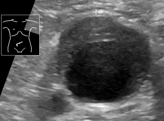
Ultrasound showing an abdominal aortic aneurysm with a mural thrombus
Image: “Ultrasonography of abdominal aortic aneurysm with mural thrombus” by Mikael Häggström, M.D.. License: Public Domain
Management
Management may vary based on practice location. The following information is based on US and European guidelines.
Goals[4,5,8,12–14]
Assess the acuity of the AAAAAAAn aortic aneurysm is the abnormal dilation of a segment of the aorta. Abdominal aortic aneurysm is the most common aortic aneurysm, occurring frequently in the infrarenal area. Most aneurysms are asymptomatic, but can cause compression of surrounding structures or rupture, which is a life-threatening emergency. Abdominal Aortic Aneurysms:
Does not fit criteria for surgery: medical management
Urgent/emergent surgery
Risk factor modification
Slow the progression of the aneurysmAneurysmAn aneurysm is a bulging, weakened area of a blood vessel that causes an abnormal widening of its diameter > 1.5 times the size of the native vessel. Aneurysms occur more often in arteries than in veins and are at risk of dissection and rupture, which can be life-threatening. Thoracic Aortic Aneurysms
Administration of beta blockers, statinsStatinsStatins are competitive inhibitors of HMG-CoA reductase in the liver. HMG-CoA reductase is the rate-limiting step in cholesterol synthesis. Inhibition results in lowered intrahepatocytic cholesterol formation, resulting in up-regulation of LDL receptors and, ultimately, lowering levels of serum LDL and triglycerides.Statins, ACE inhibitorsACE inhibitorsTruncus Arteriosus, or angiotensin receptorReceptorReceptors are proteins located either on the surface of or within a cell that can bind to signaling molecules known as ligands (e.g., hormones) and cause some type of response within the cell.Receptors blockers solely for reducing the risk of AAAAAAAn aortic aneurysm is the abnormal dilation of a segment of the aorta. Abdominal aortic aneurysm is the most common aortic aneurysm, occurring frequently in the infrarenal area. Most aneurysms are asymptomatic, but can cause compression of surrounding structures or rupture, which is a life-threatening emergency. Abdominal Aortic Aneurysms expansion and rupture is not recommended.[12]
Administer these medication only for primary cardiovascular risk reduction
Reduce cardiovascular risk:
SmokingSmokingWillful or deliberate act of inhaling and exhaling smoke from burning substances or agents held by hand.Interstitial Lung Diseases cessation (most effective nonsurgical intervention to reduce aneurysm-related complications and death)
Exercise:
Avoid heavy lifting.
Avoid activities that lead to Valsalva (↑ blood pressure).
Moderate amounts of physical activity do not increase risk of rupture.[12]
HypertensionHypertensionHypertension, or high blood pressure, is a common disease that manifests as elevated systemic arterial pressures. Hypertension is most often asymptomatic and is found incidentally as part of a routine physical examination or during triage for an unrelated medical encounter. Hypertension control:
AntihypertensivesAntihypertensivesThe 1st-line medication classes for hypertension include thiazide-like diuretics, angiotensin-converting enzyme inhibitors (ACEis), angiotensin II receptor blockers (ARBs), and calcium channel blockers (CCBS). Contraindications, adverse effects, and drug-to-drug interactions are agent specific.Hypertension Drugs given to reach recommended blood pressure goals
Unlike TAATAAThoracic aortic aneurysm (TAA) is the abnormal dilation of a segment of the thoracic aorta, usually the ascending aorta. Most TAAs are due to degenerative aortic disorders, commonly in patients > 65 years of age. Most TAAs are asymptomatic (incidentally found in imaging) but could present with symptoms from its effects on surrounding structures.Thoracic Aortic Aneurysms, no specific medication recommended to limitLimitA value (e.g., pressure or time) that should not be exceeded and which is specified by the operator to protect the lungInvasive Mechanical VentilationAAAAAAAn aortic aneurysm is the abnormal dilation of a segment of the aorta. Abdominal aortic aneurysm is the most common aortic aneurysm, occurring frequently in the infrarenal area. Most aneurysms are asymptomatic, but can cause compression of surrounding structures or rupture, which is a life-threatening emergency. Abdominal Aortic Aneurysms expansion
Lipid control with statinsStatinsStatins are competitive inhibitors of HMG-CoA reductase in the liver. HMG-CoA reductase is the rate-limiting step in cholesterol synthesis. Inhibition results in lowered intrahepatocytic cholesterol formation, resulting in up-regulation of LDL receptors and, ultimately, lowering levels of serum LDL and triglycerides.Statins (target LDL < 70 mg/dL)
Avoid fluoroquinolonesFluoroquinolonesFluoroquinolones are a group of broad-spectrum, bactericidal antibiotics inhibiting bacterial DNA replication. Fluoroquinolones cover gram-negative, anaerobic, and atypical organisms, as well as some gram-positive and multidrug-resistant (MDR) organisms. Fluoroquinolones (may ↑ risk of dissection or rupture)
SurveillanceSurveillanceDevelopmental Milestones and Normal Growth: Asymptomatic AAAAAAAn aortic aneurysm is the abnormal dilation of a segment of the aorta. Abdominal aortic aneurysm is the most common aortic aneurysm, occurring frequently in the infrarenal area. Most aneurysms are asymptomatic, but can cause compression of surrounding structures or rupture, which is a life-threatening emergency. Abdominal Aortic Aneurysms < 5.5 cm, periodic evaluation and aneurysmAneurysmAn aneurysm is a bulging, weakened area of a blood vessel that causes an abnormal widening of its diameter > 1.5 times the size of the native vessel. Aneurysms occur more often in arteries than in veins and are at risk of dissection and rupture, which can be life-threatening. Thoracic Aortic Aneurysms diameter surveillanceSurveillanceDevelopmental Milestones and Normal Growth
Table: Management of asymptomatic patientsPatientsIndividuals participating in the health care system for the purpose of receiving therapeutic, diagnostic, or preventive procedures.Clinician–Patient Relationship by AAAAAAAn aortic aneurysm is the abnormal dilation of a segment of the aorta. Abdominal aortic aneurysm is the most common aortic aneurysm, occurring frequently in the infrarenal area. Most aneurysms are asymptomatic, but can cause compression of surrounding structures or rupture, which is a life-threatening emergency. Abdominal Aortic Aneurysms size
AAAAAAAn aortic aneurysm is the abnormal dilation of a segment of the aorta. Abdominal aortic aneurysm is the most common aortic aneurysm, occurring frequently in the infrarenal area. Most aneurysms are asymptomatic, but can cause compression of surrounding structures or rupture, which is a life-threatening emergency. Abdominal Aortic Aneurysms management of asymptomatic patientsPatientsIndividuals participating in the health care system for the purpose of receiving therapeutic, diagnostic, or preventive procedures.Clinician–Patient Relationship (US)*[12]
AAAAAAAn aortic aneurysm is the abnormal dilation of a segment of the aorta. Abdominal aortic aneurysm is the most common aortic aneurysm, occurring frequently in the infrarenal area. Most aneurysms are asymptomatic, but can cause compression of surrounding structures or rupture, which is a life-threatening emergency. Abdominal Aortic Aneurysms size
Indicated for younger patientsPatientsIndividuals participating in the health care system for the purpose of receiving therapeutic, diagnostic, or preventive procedures.Clinician–Patient Relationship, low perioperative risk, or those who have contraindicationsContraindicationsA condition or factor associated with a recipient that makes the use of a drug, procedure, or physical agent improper or inadvisable. Contraindications may be absolute (life threatening) or relative (higher risk of complications in which benefits may outweigh risks).Noninvasive Ventilation for endovascular aortic repair (EVAR)
SurveillanceSurveillanceDevelopmental Milestones and Normal Growth: CT angiographyAngiographyRadiography of blood vessels after injection of a contrast medium.Cardiac Surgery 5 years later, look for aortic dilation or pseudoaneurysmPseudoaneurysmNot an aneurysm but a well-defined collection of blood and connective tissue outside the wall of a blood vessel or the heart. It is the containment of a ruptured blood vessel or heart, such as sealing a rupture of the left ventricle. False aneurysm is formed by organized thrombus and hematoma in surrounding tissue.Thoracic Aortic Aneurysms
EVAR:
In the United States: 80% of AAAAAAAn aortic aneurysm is the abnormal dilation of a segment of the aorta. Abdominal aortic aneurysm is the most common aortic aneurysm, occurring frequently in the infrarenal area. Most aneurysms are asymptomatic, but can cause compression of surrounding structures or rupture, which is a life-threatening emergency. Abdominal Aortic Aneurysms surgical repair
Access through iliac or femoral arteriesArteriesArteries are tubular collections of cells that transport oxygenated blood and nutrients from the heart to the tissues of the body. The blood passes through the arteries in order of decreasing luminal diameter, starting in the largest artery (the aorta) and ending in the small arterioles. Arteries are classified into 3 types: large elastic arteries, medium muscular arteries, and small arteries and arterioles. Arteries: Histology and endograft placed within AAAAAAAn aortic aneurysm is the abnormal dilation of a segment of the aorta. Abdominal aortic aneurysm is the most common aortic aneurysm, occurring frequently in the infrarenal area. Most aneurysms are asymptomatic, but can cause compression of surrounding structures or rupture, which is a life-threatening emergency. Abdominal Aortic Aneurysms lumen
Requires anatomic suitability (site and structure of aneurysmAneurysmAn aneurysm is a bulging, weakened area of a blood vessel that causes an abnormal widening of its diameter > 1.5 times the size of the native vessel. Aneurysms occur more often in arteries than in veins and are at risk of dissection and rupture, which can be life-threatening. Thoracic Aortic Aneurysms and access vessels)
Decreased operative mortalityMortalityAll deaths reported in a given population.Measures of Health Status but higher rate of re-intervention
CT angiographyAngiographyRadiography of blood vessels after injection of a contrast medium.Cardiac Surgery 1 month and 1 year post-operatively, then duplex ultrasonographyDuplex ultrasonographyUltrasonography applying the doppler effect combined with real-time imaging. The real-time image is created by rapid movement of the ultrasound beam. A powerful advantage of this technique is the ability to estimate the velocity of flow from the doppler shift frequency.Hypercoagulable States/CT annually if with uncomplicated surgery
Look for endoleak, sac enlargement, migration of stents, and device integrity
Indications for operative repair:[5,12‒14]
Elective repair: most effective way of preventing rupture and aneurysm-related death
Asymptomatic AAAAAAAn aortic aneurysm is the abnormal dilation of a segment of the aorta. Abdominal aortic aneurysm is the most common aortic aneurysm, occurring frequently in the infrarenal area. Most aneurysms are asymptomatic, but can cause compression of surrounding structures or rupture, which is a life-threatening emergency. Abdominal Aortic Aneurysms: when diameter ≥ 5.5 cm (5 cm in women: higher rate of rupture)
AAAAAAAn aortic aneurysm is the abnormal dilation of a segment of the aorta. Abdominal aortic aneurysm is the most common aortic aneurysm, occurring frequently in the infrarenal area. Most aneurysms are asymptomatic, but can cause compression of surrounding structures or rupture, which is a life-threatening emergency. Abdominal Aortic Aneurysms with coexisting iliac, femoral, or popliteal arteryPopliteal ArteryThe continuation of the femoral artery coursing through the popliteal fossa; it divides into the anterior and posterior tibial arteries.Popliteal Fossa: AnatomyaneurysmAneurysmAn aneurysm is a bulging, weakened area of a blood vessel that causes an abnormal widening of its diameter > 1.5 times the size of the native vessel. Aneurysms occur more often in arteries than in veins and are at risk of dissection and rupture, which can be life-threatening. Thoracic Aortic Aneurysms
AAAAAAAn aortic aneurysm is the abnormal dilation of a segment of the aorta. Abdominal aortic aneurysm is the most common aortic aneurysm, occurring frequently in the infrarenal area. Most aneurysms are asymptomatic, but can cause compression of surrounding structures or rupture, which is a life-threatening emergency. Abdominal Aortic Aneurysms associated with symptomatic peripheral arterial disease (PAD) undergoing revascularizationRevascularizationThromboangiitis Obliterans (Buerger Disease)
Emergency repair: Symptomatic AAAAAAAn aortic aneurysm is the abnormal dilation of a segment of the aorta. Abdominal aortic aneurysm is the most common aortic aneurysm, occurring frequently in the infrarenal area. Most aneurysms are asymptomatic, but can cause compression of surrounding structures or rupture, which is a life-threatening emergency. Abdominal Aortic Aneurysms (impending rupture) or AAAAAAAn aortic aneurysm is the abnormal dilation of a segment of the aorta. Abdominal aortic aneurysm is the most common aortic aneurysm, occurring frequently in the infrarenal area. Most aneurysms are asymptomatic, but can cause compression of surrounding structures or rupture, which is a life-threatening emergency. Abdominal Aortic Aneurysms rupture
Preparation for emergency surgery[12–14]
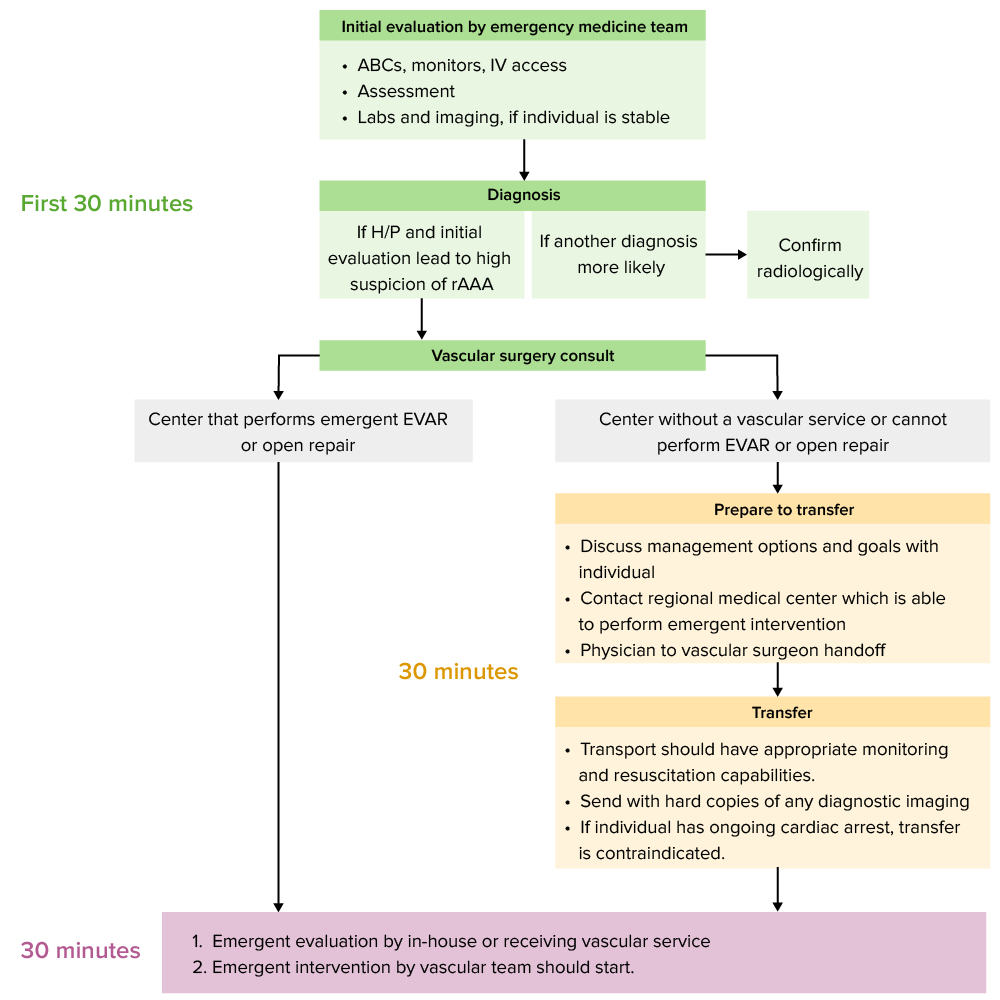
Management of symptomatic/ruptured abdominal aortic aneurysm: 30-30-30 rule[12] ABCs: airway, breathing, and circulation EVAR: endovascular aortic repair H/P: history and physical exam rAAA: ruptured abdominal aortic aneurysm
Image by Lecturio.
30-30-30 rule:
Time from entering emergency department until intervention should be 90 minutes:
30 minutes for initial evaluation and management
30 minutes for a transfer to a medical center that performs EVAR or open surgery
30 minutes for vascular surgeryVascular surgeryVascular surgery is the specialized field of medicine that focuses on the surgical management of the pathologies of the peripheral circulation. The main goal of most vascular procedures is to restore circulatory function to the affected vessels by relieving occlusions or by redirecting blood flow (e.g., bypass).Vascular Surgery evaluation and start of intervention
Access cardiopulmonary status:
Place:
Pulse oximetry monitor
Non-invasive blood pressure cuff
Cardiac telemetryTelemetryTransmission of the readings of instruments to a remote location by means of wires, radio waves, or other means.Crush Syndrome monitoring
Place 2 large-bore IVs (preferably 16 gauge or larger)
Obtain ECGECGAn electrocardiogram (ECG) is a graphic representation of the electrical activity of the heart plotted against time. Adhesive electrodes are affixed to the skin surface allowing measurement of cardiac impulses from many angles. The ECG provides 3-dimensional information about the conduction system of the heart, the myocardium, and other cardiac structures. Electrocardiogram (ECG)
Labs:
CBC
Type and cross-match
Metabolic panel
Cardiac enzymesEnzymesEnzymes are complex protein biocatalysts that accelerate chemical reactions without being consumed by them. Due to the body’s constant metabolic needs, the absence of enzymes would make life unsustainable, as reactions would occur too slowly without these molecules. Basics of Enzymes
Coagulation studiesCoagulation studiesCoagulation studies are a group of hematologic laboratory studies that reflect the function of blood vessels, platelets, and coagulation factors, which all interact with one another to achieve hemostasis. Coagulation studies are usually ordered to evaluate patients with bleeding or hypercoagulation disorders.Coagulation Studies
Alert the blood bank that a massive transfusion may be needed.
Imaging: If the individual is hemodynamically stable, CT is useful to determine if EVAR is feasible.
Medical treatment (US dosing):[14]
If hypertensive:
Decrease aortic wall stress and impending rupture (beta-blockade and blood pressure control)
Goal SBPSBPAscites: 100‒120 (reduce SBPSBPAscites to < 120 mm Hg in the first hour)
Goal HR: approximately 60/min
Beta blockers:
EsmololEsmololAntiadrenergic Drugs: 500‒1000 µg/kg/min loading doseLoading DoseDosage Calculation over 1 minute, followed by 50 µg/kg/min infusion. If additional BP control is needed, repeat the bolus dose and increase the infusion by 50 µg/kg/min. Repeat bolus and incremental increase up to an infusion of 200 µg/kg/min.
LabetalolLabetalolA salicylamide derivative that is a non-cardioselective blocker of beta-adrenergic receptors and alpha-1 adrenergic receptors.Subarachnoid Hemorrhage: 20-mg loading doseLoading DoseDosage Calculation, followed by 0.5‒2 mg/min infusion
CalciumCalciumA basic element found in nearly all tissues. It is a member of the alkaline earth family of metals with the atomic symbol ca, atomic number 20, and atomic weight 40. Calcium is the most abundant mineral in the body and combines with phosphorus to form calcium phosphate in the bones and teeth. It is essential for the normal functioning of nerves and muscles and plays a role in blood coagulation (as factor IV) and in many enzymatic processes.Electrolytes channel blocker and nitroglycerinNitroglycerinA volatile vasodilator which relieves angina pectoris by stimulating guanylate cyclase and lowering cytosolic calcium. It is also sometimes used for tocolysis and explosives.Nitrates are also acceptable choices.
Ensure adequate painPainAn unpleasant sensation induced by noxious stimuli which are detected by nerve endings of nociceptive neurons.Pain: Types and Pathways control[18,19]:
MorphineMorphineThe principal alkaloid in opium and the prototype opiate analgesic and narcotic. Morphine has widespread effects in the central nervous system and on smooth muscle.Opioid Analgesics 2‒4 mg IV every 5‒15 minutes, titrated to effect
FentanylFentanylA potent narcotic analgesic, abuse of which leads to habituation or addiction. It is primarily a mu-opioid agonist. Fentanyl is also used as an adjunct to general anesthetics, and as an anesthetic for induction and maintenance.Opioid Analgesics 25‒50 µg IV every 2‒5 minutes, titrated to effect
If hypotensive:[12,15,16]
Permissive hypotensionHypotensionHypotension is defined as low blood pressure, specifically < 90/60 mm Hg, and is most commonly a physiologic response. Hypotension may be mild, serious, or life threatening, depending on the cause. Hypotension is recommended:
Maintain SBPSBPAscites 70‒90 mm Hg, if the individual’s mental status is appropriate
Start with 1-L bolus of isotonicIsotonicSolutions having the same osmotic pressure as blood serum, or another solution with which they are compared.Renal Sodium and Water RegulationcrystalloidCrystalloidIsotonic solutions of mineral salts, such as ringer’s lactate and sodium chloride (saline solution), used in fluid therapy to rehydrate blood volume.Intravenous Fluids to test response.
If no hemodynamic response, blood will likely be needed.
Note: Preoperative overresuscitation is linked to worse outcomes.
Need for blood products:[16]
Class I hemorrhagic shockHemorrhagic shockAcute hemorrhage or excessive fluid loss resulting in hypovolemia.Hemothorax: not likely
Class II hemorrhagic shockHemorrhagic shockAcute hemorrhage or excessive fluid loss resulting in hypovolemia.Hemothorax: possible
Class III hemorrhagic shockHemorrhagic shockAcute hemorrhage or excessive fluid loss resulting in hypovolemia.Hemothorax: yes
Class IV hemorrhagic shockHemorrhagic shockAcute hemorrhage or excessive fluid loss resulting in hypovolemia.Hemothorax: activate massive transfusion protocol
Table: ATLS classification of hemorrhagic shockHemorrhagic shockAcute hemorrhage or excessive fluid loss resulting in hypovolemia.Hemothorax[16,17]
Class I
Class II
Class III
Class IV
Percentage of blood loss
< 15
15–30
30–40
> 40
Pulse rate
Normal
100–120
120–140
> 140
Blood pressure
Normal
Normal
↓
Significantly ↓
Pulse pressure
Normal
↓
↓
↓
Respiratory rateRespiratory rateThe number of times an organism breathes with the lungs (respiration) per unit time, usually per minute.Pulmonary Examination
CrystalloidCrystalloidIsotonic solutions of mineral salts, such as ringer’s lactate and sodium chloride (saline solution), used in fluid therapy to rehydrate blood volume.Intravenous Fluids
CrystalloidCrystalloidIsotonic solutions of mineral salts, such as ringer’s lactate and sodium chloride (saline solution), used in fluid therapy to rehydrate blood volume.Intravenous Fluids, possibly blood products
Ruptured viscus: a condition in which gastrointestinal wall integrity is lost with subsequent leakage of enteric contentsLeakage of Enteric ContentsPerforated Viscus into the peritoneal cavityPeritoneal CavityThe space enclosed by the peritoneum. It is divided into two portions, the greater sac and the lesser sac or omental bursa, which lies behind the stomach. The two sacs are connected by the foramen of winslow, or epiploic foramen.Peritoneum: Anatomy, resulting in peritonitisPeritonitisInflammation of the peritoneum lining the abdominal cavity as the result of infectious, autoimmune, or chemical processes. Primary peritonitis is due to infection of the peritoneal cavity via hematogenous or lymphatic spread and without intra-abdominal source. Secondary peritonitis arises from the abdominal cavity itself through rupture or abscess of intra-abdominal organs.Penetrating Abdominal Injury. A ruptured viscus is life-threatening and requires surgical management.
Mesenteric ischemiaIschemiaA hypoperfusion of the blood through an organ or tissue caused by a pathologic constriction or obstruction of its blood vessels, or an absence of blood circulation.Ischemic Cell Damage: a rare, life-threatening syndrome caused by inadequate blood flowBlood flowBlood flow refers to the movement of a certain volume of blood through the vasculature over a given unit of time (e.g., mL per minute).Vascular Resistance, Flow, and Mean Arterial Pressure through the mesenteric vessels resulting in ischemiaIschemiaA hypoperfusion of the blood through an organ or tissue caused by a pathologic constriction or obstruction of its blood vessels, or an absence of blood circulation.Ischemic Cell Damage and gangreneGangreneDeath and putrefaction of tissue usually due to a loss of blood supply.Small Bowel Obstruction of the bowel wall. Mesenteric ischemiaIschemiaA hypoperfusion of the blood through an organ or tissue caused by a pathologic constriction or obstruction of its blood vessels, or an absence of blood circulation.Ischemic Cell Damage can be acute or chronic. Acute mesenteric ischemiaAcute Mesenteric IschemiaMesenteric Ischemia is a surgical emergencySurgical EmergencyAcute Abdomen, while the chronic condition requires risk factor modification, as it is related to vascular disease.
Strangulated herniaHerniaProtrusion of tissue, structure, or part of an organ through the bone, muscular tissue, or the membrane by which it is normally contained. Hernia may involve tissues such as the abdominal wall or the respiratory diaphragm. Hernias may be internal, external, congenital, or acquired.Abdominal Hernias: Hernias are protrusions of abdominal content (peritoneumPeritoneumThe peritoneum is a serous membrane lining the abdominopelvic cavity. This lining is formed by connective tissue and originates from the mesoderm. The membrane lines both the abdominal walls (as parietal peritoneum) and all of the visceral organs (as visceral peritoneum).Peritoneum: Anatomy, visceral fat, and/or viscera) through a congenital or acquired defect in the abdominal wallAbdominal wallThe outer margins of the abdomen, extending from the osteocartilaginous thoracic cage to the pelvis. Though its major part is muscular, the abdominal wall consists of at least seven layers: the skin, subcutaneous fat, deep fascia; abdominal muscles, transversalis fascia, extraperitoneal fat, and the parietal peritoneum.Surgical Anatomy of the Abdomen. StrangulationStrangulationInguinal Canal: Anatomy and Hernias involves the constriction of hernial contents leading to bowel ischemiaBowel ischemiaMesenteric ischemia is a rare, life-threatening condition caused by inadequate blood flow through the mesenteric vessels, which results in ischemia and necrosis of the intestinal wall. Mesenteric ischemia can be either acute or chronic.Mesenteric Ischemia and requires emergency surgery to avoid bowel loss, perforationPerforationA pathological hole in an organ, blood vessel or other soft part of the body, occurring in the absence of external force.Esophagitis, and sepsisSepsisSystemic inflammatory response syndrome with a proven or suspected infectious etiology. When sepsis is associated with organ dysfunction distant from the site of infection, it is called severe sepsis. When sepsis is accompanied by hypotension despite adequate fluid infusion, it is called septic shock.Sepsis and Septic Shock.
Acute cholecystitisAcute cholecystitisAcute inflammation of the gallbladder wall. It is characterized by the presence of abdominal pain; fever; and leukocytosis. Gallstone obstruction of the cystic duct is present in approximately 90% of the cases.Cholecystitis: a condition characterized by inflammationInflammationInflammation is a complex set of responses to infection and injury involving leukocytes as the principal cellular mediators in the body’s defense against pathogenic organisms. Inflammation is also seen as a response to tissue injury in the process of wound healing. The 5 cardinal signs of inflammation are pain, heat, redness, swelling, and loss of function. Inflammation of the gallbladderGallbladderThe gallbladder is a pear-shaped sac, located directly beneath the liver, that sits on top of the superior part of the duodenum. The primary functions of the gallbladder include concentrating and storing up to 50 mL of bile. Gallbladder and Biliary Tract: Anatomy, most often due to obstruction of the cysticCysticFibrocystic Change duct by a gallstone. Management includes IV fluidsIV fluidsIntravenous fluids are one of the most common interventions administered in medicine to approximate physiologic bodily fluids. Intravenous fluids are divided into 2 categories: crystalloid and colloid solutions. Intravenous fluids have a wide variety of indications, including intravascular volume expansion, electrolyte manipulation, and maintenance fluids. Intravenous Fluids, painPainAn unpleasant sensation induced by noxious stimuli which are detected by nerve endings of nociceptive neurons.Pain: Types and Pathways control, and IV antibiotics for secondary infection. Complicated cholecystitisCholecystitisCholecystitis is the inflammation of the gallbladder (GB) usually caused by the obstruction of the cystic duct (acute cholecystitis). Mechanical irritation by gallstones can also produce chronic GB inflammation. Cholecystitis is one of the most common complications of cholelithiasis but inflammation without gallstones can occur in a minority of patients. Cholecystitis and progressive symptoms are indications for emergency cholecystectomyCholecystectomyCholecystectomy is a surgical procedure performed with the goal of resecting and extracting the gallbladder. It is one of the most common abdominal surgeries performed in the Western world. Cholecystectomy is performed for symptomatic cholelithiasis, cholecystitis, gallbladder polyps > 0.5 cm, porcelain gallbladder, choledocholithiasis and gallstone pancreatitis, and rarely, for gallbladder cancer. Cholecystectomy.
Acute pancreatitisPancreatitisInflammation of the pancreas. Pancreatitis is classified as acute unless there are computed tomographic or endoscopic retrograde cholangiopancreatographic findings of chronic pancreatitis. The two most common forms of acute pancreatitis are alcoholic pancreatitis and gallstone pancreatitis.Acute Pancreatitis: an inflammationInflammationInflammation is a complex set of responses to infection and injury involving leukocytes as the principal cellular mediators in the body’s defense against pathogenic organisms. Inflammation is also seen as a response to tissue injury in the process of wound healing. The 5 cardinal signs of inflammation are pain, heat, redness, swelling, and loss of function. Inflammation of the pancreasPancreasThe pancreas lies mostly posterior to the stomach and extends across the posterior abdominal wall from the duodenum on the right to the spleen on the left. This organ has both exocrine and endocrine tissue. Pancreas: Anatomy that typically causes epigastric painEpigastric painMallory-Weiss Syndrome (Mallory-Weiss Tear) that radiates to the back. This condition is often treated with aggressive fluid resuscitationResuscitationThe restoration to life or consciousness of one apparently dead. .Neonatal Respiratory Distress Syndrome, bowel rest, and painPainAn unpleasant sensation induced by noxious stimuli which are detected by nerve endings of nociceptive neurons.Pain: Types and Pathways control. Surgery is indicated if the condition is associated with gallstonesGallstonesCholelithiasis (gallstones) is the presence of stones in the gallbladder. Most gallstones are cholesterol stones, while the rest are composed of bilirubin (pigment stones) and other mixed components. Patients are commonly asymptomatic but may present with biliary colic (intermittent pain in the right upper quadrant).Cholelithiasis.
Diverticular abscessAbscessAccumulation of purulent material in tissues, organs, or circumscribed spaces, usually associated with signs of infection.Chronic Granulomatous Disease: a group of various intestinal conditions characterized by abnormal outpouchings of the colonic mucosa (diverticula). Over time, these diverticula may accumulate intestinal content, become infected, swell, and develop into an abscessAbscessAccumulation of purulent material in tissues, organs, or circumscribed spaces, usually associated with signs of infection.Chronic Granulomatous Disease. Intravenous antibiotics are the recommended treatment, with percutaneous drainagePercutaneous DrainageEchinococcus/Echinococcosis needed for large abscessAbscessAccumulation of purulent material in tissues, organs, or circumscribed spaces, usually associated with signs of infection.Chronic Granulomatous Disease or failed medical treatment.
Billing and Coding
Diagnosis Codes:
These ICD-10 codesICD-10 CodesAbdominal Aortic Aneurysms (Clinical) are used to classify an Abdominal Aortic AneurysmAortic aneurysmAn abnormal balloon- or sac-like dilatation in the wall of aorta.Thoracic Aortic Aneurysms (AAAAAAAn aortic aneurysm is the abnormal dilation of a segment of the aorta. Abdominal aortic aneurysm is the most common aortic aneurysm, occurring frequently in the infrarenal area. Most aneurysms are asymptomatic, but can cause compression of surrounding structures or rupture, which is a life-threatening emergency. Abdominal Aortic Aneurysms) based on its status, distinguishing between an unruptured aneurysmAneurysmAn aneurysm is a bulging, weakened area of a blood vessel that causes an abnormal widening of its diameter > 1.5 times the size of the native vessel. Aneurysms occur more often in arteries than in veins and are at risk of dissection and rupture, which can be life-threatening. Thoracic Aortic Aneurysms (I71.4) and a life-threatening ruptured aneurysmAneurysmAn aneurysm is a bulging, weakened area of a blood vessel that causes an abnormal widening of its diameter > 1.5 times the size of the native vessel. Aneurysms occur more often in arteries than in veins and are at risk of dissection and rupture, which can be life-threatening. Thoracic Aortic Aneurysms (I71.3).
AneurysmAneurysmAn aneurysm is a bulging, weakened area of a blood vessel that causes an abnormal widening of its diameter > 1.5 times the size of the native vessel. Aneurysms occur more often in arteries than in veins and are at risk of dissection and rupture, which can be life-threatening. Thoracic Aortic Aneurysms of abdominal aortaAbdominal AortaThe aorta from the diaphragm to the bifurcation into the right and left common iliac arteries.Posterior Abdominal Wall: Anatomy (disorder)
Evaluation & Workup:
These CPT codes are used to order imaging studies for diagnosis and surveillanceSurveillanceDevelopmental Milestones and Normal Growth. An abdominal ultrasound is used for initial screeningScreeningPreoperative Care and monitoring, while a CT angiographyAngiographyRadiography of blood vessels after injection of a contrast medium.Cardiac Surgery (CTACTAA non-invasive method that uses a ct scanner for capturing images of blood vessels and tissues. A contrast material is injected, which helps produce detailed images that aid in diagnosing vascular diseases.Pulmonary Function Tests) provides detailed anatomy for surgical planning.
Domain
Code
Description
CPT
76706
Ultrasound, abdominal, real time with image documentationDocumentationSystematic organization, storage, retrieval, and dissemination of specialized information, especially of a scientific or technical nature. It often involves authenticating or validating information.Advance Directives, screeningScreeningPreoperative Care for AAAAAAAn aortic aneurysm is the abnormal dilation of a segment of the aorta. Abdominal aortic aneurysm is the most common aortic aneurysm, occurring frequently in the infrarenal area. Most aneurysms are asymptomatic, but can cause compression of surrounding structures or rupture, which is a life-threatening emergency. Abdominal Aortic Aneurysms
CPT
74175
Computed tomography angiographyAngiographyRadiography of blood vessels after injection of a contrast medium.Cardiac Surgery, abdomen; with contrast material
Procedures/Interventions:
These CPT codes represent the two main surgical options for repairing an AAAAAAAn aortic aneurysm is the abnormal dilation of a segment of the aorta. Abdominal aortic aneurysm is the most common aortic aneurysm, occurring frequently in the infrarenal area. Most aneurysms are asymptomatic, but can cause compression of surrounding structures or rupture, which is a life-threatening emergency. Abdominal Aortic Aneurysms: a minimally invasive Endovascular AneurysmAneurysmAn aneurysm is a bulging, weakened area of a blood vessel that causes an abnormal widening of its diameter > 1.5 times the size of the native vessel. Aneurysms occur more often in arteries than in veins and are at risk of dissection and rupture, which can be life-threatening. Thoracic Aortic Aneurysms Repair (EVAR) or a traditional open surgical repair.
Direct repair of aneurysmAneurysmAn aneurysm is a bulging, weakened area of a blood vessel that causes an abnormal widening of its diameter > 1.5 times the size of the native vessel. Aneurysms occur more often in arteries than in veins and are at risk of dissection and rupture, which can be life-threatening. Thoracic Aortic Aneurysms, pseudoaneurysmPseudoaneurysmNot an aneurysm but a well-defined collection of blood and connective tissue outside the wall of a blood vessel or the heart. It is the containment of a ruptured blood vessel or heart, such as sealing a rupture of the left ventricle. False aneurysm is formed by organized thrombus and hematoma in surrounding tissue.Thoracic Aortic Aneurysms, or excision (partial or total) and graftGraftA piece of living tissue that is surgically transplantedOrgan Transplantation insertion
ICD-10-PCS
04100JH
Bypass Abdominal AortaAbdominal AortaThe aorta from the diaphragm to the bifurcation into the right and left common iliac arteries.Posterior Abdominal Wall: Anatomy to Both Femoral ArteriesArteriesArteries are tubular collections of cells that transport oxygenated blood and nutrients from the heart to the tissues of the body. The blood passes through the arteries in order of decreasing luminal diameter, starting in the largest artery (the aorta) and ending in the small arterioles. Arteries are classified into 3 types: large elastic arteries, medium muscular arteries, and small arteries and arterioles. Arteries: Histology with Synthetic Substitute, Open
Medications:
While not curative for the aneurysmAneurysmAn aneurysm is a bulging, weakened area of a blood vessel that causes an abnormal widening of its diameter > 1.5 times the size of the native vessel. Aneurysms occur more often in arteries than in veins and are at risk of dissection and rupture, which can be life-threatening. Thoracic Aortic Aneurysms itself, these codes are used to prescribe medications like beta-blockersBeta-blockersDrugs that bind to but do not activate beta-adrenergic receptors thereby blocking the actions of beta-adrenergic agonists. Adrenergic beta-antagonists are used for treatment of hypertension, cardiac arrhythmias, angina pectoris, glaucoma, migraine headaches, and anxiety.Class 2 Antiarrhythmic Drugs (Beta Blockers) and statinsStatinsStatins are competitive inhibitors of HMG-CoA reductase in the liver. HMG-CoA reductase is the rate-limiting step in cholesterol synthesis. Inhibition results in lowered intrahepatocytic cholesterol formation, resulting in up-regulation of LDL receptors and, ultimately, lowering levels of serum LDL and triglycerides.Statins to manage risk factors such as high blood pressure and cholesterolCholesterolThe principal sterol of all higher animals, distributed in body tissues, especially the brain and spinal cord, and in animal fats and oils.Cholesterol Metabolism, potentially slowing aneurysmAneurysmAn aneurysm is a bulging, weakened area of a blood vessel that causes an abnormal widening of its diameter > 1.5 times the size of the native vessel. Aneurysms occur more often in arteries than in veins and are at risk of dissection and rupture, which can be life-threatening. Thoracic Aortic Aneurysms growth.
Domain
Code
Description
RxNorm
703
AtenololAtenololA cardioselective beta-1 adrenergic blocker possessing properties and potency similar to propranolol, but without a negative inotropic effect.Class 2 Antiarrhythmic Drugs (Beta Blockers) (ingredient)
RxNorm
83367
AtorvastatinAtorvastatinA pyrrole and heptanoic acid derivative, hydroxymethylglutaryl-CoA reductase inhibitor (statin), and anticholesteremic agent that is used to reduce serum levels of ldl-cholesterol; apolipoprotein b; and triglycerides. It is used to increase serum levels of hdl-cholesterol in the treatment of hyperlipidemias, and for the prevention of cardiovascular diseases in patients with multiple risk factors.Statins (ingredient)
Mazzolai, L., Teixido-Tura, G., Lanzi, S., Boc, V., Bossone, E., Brodmann, M., Bura-Rivière, A., De Backer, J., Deglise, S., Della Corte, A., Heiss, C., Kałużna-Oleksy, M., Kurpas, D., McEniery, C. M., Mirault, T., Pasquet, A. A., Pitcher, A., Schaubroeck, H. A. I., Schlager, O., Sirnes, P. A., … ESC Scientific Document Group (2024). 2024 ESC Guidelines for the management of peripheral arterial and aortic diseases. European heart journal, 45(36), 3538–3700. https://doi.org/10.1093/eurheartj/ehae179
Guoledge, J., Guo-Ping, S., Norman, P., Fitridge, R., Thompson, M. (2011). Pathogenesis of aortic aneurysmAortic aneurysmAn abnormal balloon- or sac-like dilatation in the wall of aorta.Thoracic Aortic Aneurysms. In Mechanisms of Vascular disease: A Reference Book for Vascular Specialists. Adelaide (AU): University of Adelaide Press. Retrieved March 28, 2025, from https://www.ncbi.nlm.nih.gov/books/NBK534278
Mitchell, R., Halushka, M. (2020). In Kumar, V., Abbas, A., Aster, J., Turner, J. (Eds.) Robbins and Cotran Pathologic Basis of Disease.(10th ed., pp. 485–525). Elsevier.
Ruddy, J., Jones, J., Spinale, F., Ikonomidis, J. (2008). Regional heterogeneity within the aortaAortaThe main trunk of the systemic arteries.Mediastinum and Great Vessels: Anatomy: relevance to aneurysmAneurysmAn aneurysm is a bulging, weakened area of a blood vessel that causes an abnormal widening of its diameter > 1.5 times the size of the native vessel. Aneurysms occur more often in arteries than in veins and are at risk of dissection and rupture, which can be life-threatening. Thoracic Aortic Aneurysms disease. J Thorac Cardiovasc Surg 136(5):1123–1130. Retrieved March 28, 2025, fromhttps://www.ncbi.nlm.nih.gov/pmc/articles/PMC2679174/
Vu, K., Kaitoukov, Y., Morin-Roy, F., et alALAmyloidosis. (2014). Rupture signs on computed tomography, treatment, and outcome of abdominal aortic aneurysms.Insights Imaging. 5(3): 281–293. Retrieved March 28, 2025 from doi: 10.1007/s13244-014-0327-3
Chaikof, Elliot L., et alALAmyloidosis. (2018). The Society for Vascular SurgeryVascular surgeryVascular surgery is the specialized field of medicine that focuses on the surgical management of the pathologies of the peripheral circulation. The main goal of most vascular procedures is to restore circulatory function to the affected vessels by relieving occlusions or by redirecting blood flow (e.g., bypass).Vascular Surgery practice guidelines on the care of patientsPatientsIndividuals participating in the health care system for the purpose of receiving therapeutic, diagnostic, or preventive procedures.Clinician–Patient Relationship with an abdominal aortic aneurysmAortic aneurysmAn abnormal balloon- or sac-like dilatation in the wall of aorta.Thoracic Aortic Aneurysms. Journal of Vascular SurgeryVascular surgeryVascular surgery is the specialized field of medicine that focuses on the surgical management of the pathologies of the peripheral circulation. The main goal of most vascular procedures is to restore circulatory function to the affected vessels by relieving occlusions or by redirecting blood flow (e.g., bypass).Vascular Surgery 67(1):2-77.e2. https://doi.org/10.1016/j.jvs.2017.10.044
Isselbacher, E. M., Preventza, O., Hamilton Black, J., 3rd, Augoustides, J. G., Beck, A. W., Bolen, M. A., Braverman, A. C., Bray, B. E., Brown-Zimmerman, M. M., Chen, E. P., Collins, T. J., DeAnda, A., Jr, Fanola, C. L., Girardi, L. N., Hicks, C. W., Hui, D. S., Schuyler Jones, W., Kalahasti, V., Kim, K. M., Milewicz, D. M., … Peer Review Committee Members (2022). 2022 ACC/AHA Guideline for the Diagnosis and Management of Aortic Disease: A Report of the American Heart AssociationAmerican Heart AssociationA voluntary organization concerned with the prevention and treatment of heart and vascular diseases.Heart Failure/American College of Cardiology Joint Committee on Clinical Practice Guidelines. CirculationCirculationThe movement of the blood as it is pumped through the cardiovascular system.ABCDE Assessment, 146(24), e334–e482. https://doi.org/10.1161/CIR.0000000000001106
Mutschler, M., et alALAmyloidosis. (2014). The ATLS® classification of hypovolaemic shockShockShock is a life-threatening condition associated with impaired circulation that results in tissue hypoxia. The different types of shock are based on the underlying cause: distributive (↑ cardiac output (CO), ↓ systemic vascular resistance (SVR)), cardiogenic (↓ CO, ↑ SVR), hypovolemic (↓ CO, ↑ SVR), obstructive (↓ CO), and mixed. Types of Shock: a well established teaching tool on the edge? Injury (45):S35–S38. https://doi.org/10.1016/j.injury.2014.08.015
Lexicomp. (2022). FentanylFentanylA potent narcotic analgesic, abuse of which leads to habituation or addiction. It is primarily a mu-opioid agonist. Fentanyl is also used as an adjunct to general anesthetics, and as an anesthetic for induction and maintenance.Opioid Analgesics: drug information. UpToDate. Retrieved March 28, 2025, from https://www.uptodate.com/contents/fentanyl-drug-information
Create your free account or log in to continue reading!